Femtosecond Applications Research Secures Pre-A Funding from YuanYi Capital to Accelerate Development of Novel FLI Tissue Imaging Technology

FAR
Digital Imaging Product Developer
VCBeat (WeChat: vcbeat) has recently learned that Femtosecond Research Center (Guangzhou), a company specializing in fully independent, label-free bioimaging technology and also known as Femtosecond Applications Research (FAR), has completed its Pre-A financing round worth tens of millions of yuan. The round was exclusively invested by Yuanyi Capital. The funds will be primarily used to advance the R&D and regulatory approval of FAR’s femtosecond label-free imaging systems, expand clinical studies for additional indications, and develop AI algorithms.
Dr. Xu Bingwei, Founder of the Femtosecond Research Center, graduated from Peking University’s College of Chemistry and Molecular Engineering in 1999 with a bachelor’s degree. In 2003, he went to the United States to study under Professor Marcos Dantus, a disciple of Ahmed H. Zewail (1999 Nobel Laureate in Chemistry) who participated in the design and construction of the first-generation femtochemistry laser system. There, Dr. Xu specialized in femtosecond pulse control and earned his Ph.D. During his doctoral studies, Dr. Xu served as a core member in three national-level projects. After completing his Ph.D., Dr. Xu co-founded BSI with Professor Dantus, successfully commercializing femtosecond laser measurement and compression technologies and bringing them to market. The company was acquired by a publicly listed corporation in 2016.
In 2017, Dr. Xu Bingwei returned to China to start a business and co-founded the Femtosecond Research Center in Guangzhou with three other Peking University alumni.
Zhu Xin, the company’s CTO, is a junior fellow alumnus of Xu Bingwei from both Peking University and Michigan State University. After earning his Ph.D., Dr. Zhu worked at Agilent Technologies, a giant in biochemical analysis, where he was long engaged in application development for cancer immunotherapy drug analysis, proteomics, metabolomics, and lipidomics. Tu Jun, the CMO, graduated from the Department of Chemistry at Peking University and subsequently held positions at Novo Nordisk, Olympus, and Nikon, bringing over twenty years of experience in market management and sales focused on optical scientific instruments and medical devices. Liu Xiang, the COO, is a serial entrepreneur who has long served the Peking University Entrepreneurship Training Camp, overseeing operations in the Guangdong-Hong Kong-Macao Greater Bay Area, with extensive experience in operations and investment and financing.
This startup team, with strong ties to Peking University, has undertaken a series of core technological breakthroughs over the past four years. They have completed the hardware and software development of the Femtosecond Label-free Imaging (FLI) system, pioneering a globally leading, novel tissue imaging technology platform. The team has selected intraoperative auxiliary diagnosis of cancer as its initial clinical application entry point, aiming to leverage innovative imaging technology to serve both patients and healthcare providers, thereby enhancing the efficiency of cancer diagnosis and treatment and optimizing clinical workflows.
Xu Bingwei stated that his choice of current career path stems from the deeply ingrained “Peking University sentiment” within his family. “Four generations of my family—my great-grandfather, grandfather, father, and I—all pursued our studies at Peking University. My upbringing instilled in me the principles of ‘freedom of thought and inclusiveness.’ In my view, the spirit of Peking University is characterized by striving for world-class excellence and continuous innovation.”
According to statistics from the International Agency for Research on Cancer (IARC) of the World Health Organization, China recorded 4.57 million new cancer cases and 3 million cancer-related deaths in 2020, accounting for 23.7% and 30% of the global totals, respectively. Over the past few decades, various cancer screening and treatment technologies have flourished and advanced rapidly, providing clinicians with an increasingly powerful toolkit to help extend patient survival. However, histopathology, as the gold standard for cancer diagnosis, has seen limited breakthroughs in its technical paradigm during this period. With the widespread adoption of early screening technologies and the shift toward precision medicine and chronic disease management in cancer treatment, pathological diagnosis has gradually become a critical bottleneck in the patient care pathway. Pain points related to efficiency, quality, and accessibility have become increasingly prominent, driving the industry’s expectation for technological breakthroughs and leaps. FLI technology represents the breakthrough to resolve this dilemma.
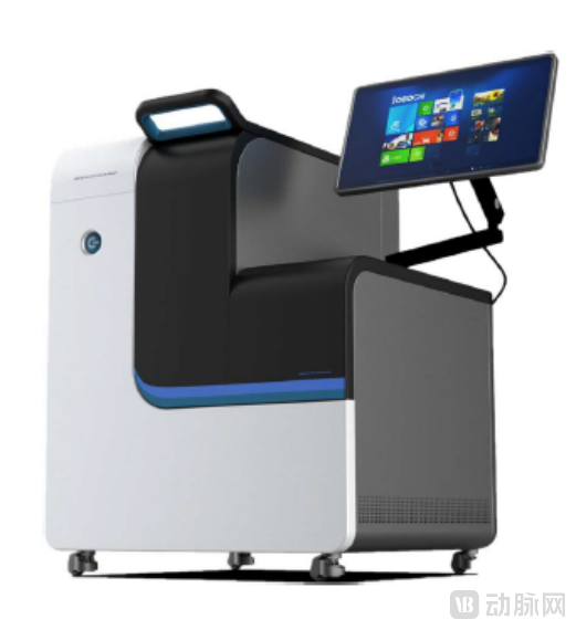
The FLI system developed by Xu Bingwei’s team utilizes more than five different modalities to simultaneously acquire independent signals from multiple channels, including two-photon autofluorescence, three-photon autofluorescence, second-harmonic generation, and third-harmonic generation. This enables imaging of a broader range of biomolecular markers, thereby allowing for more accurate identification of various tissue and cellular structures, such as tumor architecture and cellular atypia, as well as collagen fiber scaffolds, elastic fibers, blood vessels, lymphatic vessels, and extracellular vesicles. This provides robust technical support for the scientific detection and diagnosis of cancer and holds promise as a novel approach to tissue imaging.
The technical features of FLI confer multiple advantages.
Higher efficiency.For tissue samples of the same area, FLI imaging takes less than 3 minutes, whereas conventional pathological diagnosis often requires several days.
No sectioning required.By employing FLI technology, fresh tissue samples require no processing and can be directly placed on the scanning platform to complete the imaging process. With an imaging thickness of less than 1 micrometer, the system’s inherent “optical sectioning” effect and greater imaging depth readily enable single-cell layer imaging and three-dimensional tissue reconstruction.
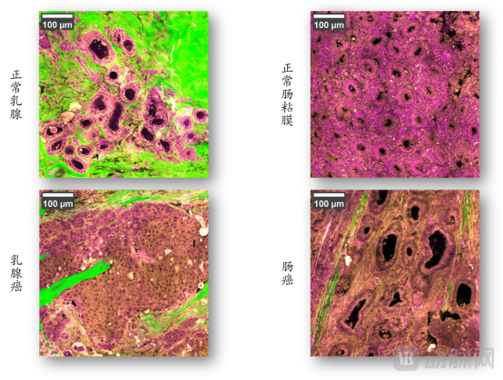
No staining (labeling) required.FLI technology captures nonlinear signals generated by endogenous marker molecules within human tissues under the action of femtosecond lasers, enabling label-free and stain-free imaging with a simple procedure and high reproducibility. This is a physical process that does not alter tissue cells, whereas conventional staining and labeling methods involve a series of chemical reactions that may cause changes to tissue samples and result in the loss of critical information.
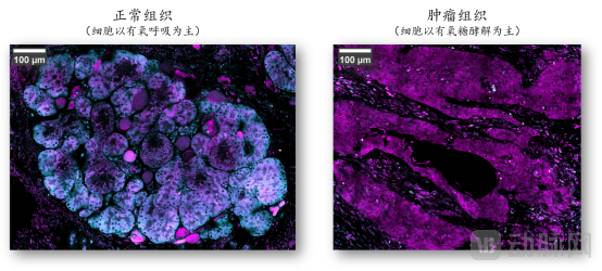
Information presentation is more comprehensive.FLI integrates multi-modal molecular imaging information to achieve multi-modal co-localization. A single scan can obtain basic information such as tissue structure and cell morphology, as well as more dimensional and richer tissue information including cellular energy metabolic status, proliferation and rearrangement of collagen fibers, and the density and distribution of exosomes, thereby facilitating rapid and precise personalized diagnosis. In contrast, conventional pathological diagnostic techniques often require multiple sectioning and different staining or labeling methods to acquire various types of information, resulting in a cumbersome, complex, time-consuming, and labor-intensive process.
Naturally integrated with AI.AI-based image analysis is an inevitable trend in the development of histopathology. Unlike current digital pathology, which requires slide scanning, color correction, image segmentation, and manual annotation, FLI directly generates standardized digital images. Its multimodal capabilities enable automatic recognition and annotation of tissue cells, seamlessly integrating with AI analysis.
These advantages make FLI a powerful tool for enhancing the diagnostic efficiency of pathologists and assisting clinicians in making rapid decisions.
Currently, pathological diagnostic instruments and consumables in China are predominantly imported, costing the country tens of billions of foreign exchange annually for updates. Accelerating the localization of high-end medical equipment and high-value consumables has become a major priority in China’s medical device sector.
In the “13th Five-Year Plan for Scientific and Technological Innovation in Medical Devices” issued by the General Office of the Ministry of Science and Technology, the “medical imaging field” was listed as the top priority among key directions for frontier technological development. The “multimodal molecular imaging” and “molecular pathology microscopic imaging technologies” to which the FLI system belongs are precisely the frontier technologies explicitly targeted for development in this field within the Plan. The “13th Five-Year” Special Plan emphasizes that strengthening basic and frontier research on medical devices, fostering “new theories, new methods, new materials, new tools, and new technologies” in medical devices, and leading transformative changes in medical paradigms through major original breakthroughs constitute the core pathway for advancing leapfrog development of China’s medical device industry.
Since its establishment, the Femtosecond Research Center has been successively recognized as a Technology-Based Small and Medium-sized Enterprise, a “Little Giant” Tech Enterprise, and a National High-Tech Enterprise. It has also received talent incentives and R&D support from competent authorities at the provincial, municipal, and district levels. Currently, core system patents for FLI technology have been granted in China, the United States, and Europe. The center has collaborated with more than 20 tumor hospitals in China on applied research, completed preclinical validation with over 1,000 samples, and is poised to launch a commercial prototype featuring fully independent intellectual property rights and autonomously controllable manufacturing.
“In our view, FLI is a world-leading innovative technology in cancer pathology, representing the trend of China’s medical device technological innovation shifting from primarily ‘following’ to ‘running alongside’ and even ‘leading.’ Leveraging China’s abundant clinical resources, our team has deeply integrated FLI with AI to effectively enhance the efficiency and accuracy of cancer pathology diagnosis, thereby alleviating the severe shortage of pathologists and technicians in China, where clinical demand is substantial,” said Xu Bingwei. “As a platform-based technology, FLI holds broad application prospects in the healthcare sector. Currently, our team is primarily focused on cancer-assisted diagnosis and surgical decision support. We aim to establish benchmarks for technology application through intraoperative decision-support scenarios, gradually open up the application interfaces of this platform technology, collaborate to develop solutions for various application scenarios, and join forces with medical and scientific research resources from all parties to co-create a new ecosystem for pathological diagnosis of cancer and other diseases.”
Tang Yinan, Investment Director at Yuanyi Capital, believes that the bottlenecks in efficiency and standardization of current pathological diagnosis lie in the upstream sample processing stage. Due to the need for human visual interpretation, obtaining pathological data from tissue samples requires steps such as slide preparation and staining, which are difficult to standardize. This severely constrains the timeliness and accuracy of downstream interpretation. The rapid, non-destructive imaging capability of FLI technology enables three-dimensional mapping of living tissue samples into the digital world, offering a novel approach to addressing the pain points of efficiency and quality in pathological diagnosis. The high-quality, standardized morphological and functional tissue data collected by the FLI system, combined with deep learning algorithms, can be used to train a series of new quantitative indicators. This facilitates the transition of pathological diagnosis from an empirical science to a quantitative science, opening up new application scenarios and untapped market opportunities. By leveraging China’s abundant biological sample resources, the company has established a dual advantage of world-class technology and massive clinical data. It is poised to build a globally competitive matrix of FLI systems and AI applications, leading industry development.
Other Supplementary Information
It is understood that FLI technology has broad application prospects in the following fields.
Basic Medical Research.Including tumor metabolism, tumor microenvironment, cancer stem cells, tumor heterogeneity, metastasis mechanisms, drug screening, and pharmacological research.
Translational Medicine Research.These applications include assisted diagnosis, screening of sensitive patients, prediction of therapeutic efficacy and prognosis, assessment and monitoring of treatment response, analysis of fibrotic diseases, as well as organoid-based companion diagnostics and selection of treatment regimens. In the context of organoid applications, traditional methods are unable to achieve continuous observation of the viability status of living cells. In contrast, FLI requires no specimen preparation and causes no damage to cells, enabling continuous monitoring of cellular states and changes under both normal culture conditions and drug exposure.
Clinical Diagnosis.FLI imaging of unsectioned, unstained fresh or fixed tissues, combined with AI-based analysis, can assist clinicians and pathologists in achieving accurate and rapid lesion diagnosis and clinical decision-making during and after surgery. This includes rapidly determining the benign or malignant nature of lesions, delineating the extent of suspicious areas, and providing probabilities for molecular subtyping and common gene mutations. It also enables rapid assessment of lymph node metastasis by visualizing the metastatic regions and sizes of positive lymph nodes and quantifying the number of positive lymph nodes.
Life Sciences Research.Including neural/brain imaging, in vivo animal/plant imaging, embryo imaging optimization, and live-cell observation and tracking.