DPM Company: Pioneering Molecular Imaging Technologies for Precision Surgery and Clinical Diagnostics
Since the discovery of X-rays by Röntgen in 1895, humanity has made tremendous progress in medical imaging technology over the past century. Its development has demonstrated a trend from two-dimensional to three-dimensional, from single-modality to multimodal fusion, and from morphological imaging to functional imaging, undergoing innovations through different technological stages including X-ray, ultrasound, CT, MRI, and PET. Throughout this process, the examination and diagnosis of diseases have become increasingly precise.
Chinese-made medical imaging equipment unfortunately missed the initial R&D phase of multiple imaging technologies; from an intellectual property perspective, the number of patents was on average five times lower, and development lagged by 20 years. However, it has caught up in the new wave of technological innovation.Molecular Imaging Technology (Molecular Imaging)train.
Molecular imaging technology employs imaging methods to visualize specific molecular events at the cellular level, reflecting changes at the cellular and molecular levels in vivo.Recognized internationally as one of the ten medical science frontiers with the greatest development potential, it is a cutting-edge medical imaging technology of the 21st century.
Based on Clinical Needs: Fluorescent Molecular Imaging for Precise Lesion Identification
Medical imaging technology has greatly facilitated precise preoperative diagnosis of human diseases and postoperative efficacy assessment in surgery. However, in many cases, physicians still struggle to visually discern subtle differences among various tissues. Clinical practice continues to face challenges in accurately delineating lesion boundaries, detecting minute lesions, and identifying critical anatomical structures.
Emerging optical molecular imaging technology holds promise for addressing this clinical challenge. This technique utilizes cells, reporter genes, or fluorescent dyes within living organisms, exciting these fluorophores with light of specific wavelengths to generate optical molecular images. It represents a novel approach for in vivo, real-time imaging.
Fluorescence Molecular Imaging Technology Applied in Clinical Practice Brings New Opportunities for Precision Surgery. In Recent Years, the Total Number of Articles in the Field of Global Fluorescence-Guided Intraoperative Navigation Has Grown Exponentially.Fluorescence molecular imaging-guided intraoperative navigation technology can visualize the in vivo distribution of fluorescent contrast agents within milliseconds, thereby assisting physicians in detecting minute lesions, identifying their location and morphology, and performing precise resection.
Among these, due to the absorption and scattering effects of biological tissues on different spectral bands, the penetration depth of photons in the visible light region is limited to the millimeter scale; whereasNear-infrared light can achieve a penetration depth on the centimeter scale.。
It has been successfully applied in intraoperative tumor imaging, lymph node biopsy, nerve imaging, angiography, and the diagnosis and treatment of tissue perfusion.
In 2009, at the World Molecular Imaging Congress (WMIC), Nobel Laureate in Chemistry Professor Roger Y. Tsien introduced a method for fluorescence molecular imaging-guided resection of tumor tissues in mice, pioneering the application of optical molecular imaging technology in surgical navigation. In 2011, European scientists developed a prototype system for molecular imaging-guided surgical navigation and applied it to clinical surgery for ovarian cancer in humans for the first time.
Although molecular imaging technology in China started slightly later than abroad, it has gained strong momentum, with certain fields now leading internationally.Represented by the team from the Institute of Automation, Chinese Academy of Sciences, which broke through the bottleneck of the “homogeneous algorithm” used in foreign products as early as 2008, innovatively proposed the “heterogeneous algorithm,” and successfully applied it to independently developed equipment, significantly improving the precision of tumor localization and causing a major sensation internationally.
Subsequently, the Key Laboratory of Molecular Imaging at the Center for Intelligent Medical Research, Institute of Automation, Chinese Academy of Sciences, has been leading the development of related theories and technologies in China in areas such as fundamental research and translational applications in molecular imaging. It is a laboratory with significant international academic influence in both basic research and clinical translation.
To promote the translation of scientific and technological achievements into practical applications, in 2016,The Key Laboratory of Molecular Imaging of the Chinese Academy of Sciences incubated DPM (including Beijing Digital Precision Medical Technology Co., Ltd. and Zhuhai Dipu Medical Technology Co., Ltd., hereinafter referred to as “DPM”)., DPM has become the vehicle for translating laboratory achievements into practical applications.
Among domestic fluorescence molecular imaging companies, DPM possesses a complete chain from theoretical research to clinical translation. The core technologies of its products have received support from national special programs, including the National 973 Program, the Major Scientific Instrument Special Project of the National Natural Science Foundation of China, and the Nanotechnology Special Project of the Ministry of Science and Technology of China.
Incremental Market: Fluorescence Endoscopy Equipment Remains a Blue Ocean
Near-infrared fluorescence imaging devices are a key element in navigation during fluorescence molecular image-guided surgery. So, what is the application mechanism of navigation in fluorescence molecular image-guided surgery? First, fluorescent contrast agents are administered via intravenous, local, or intraparenchymal injection; second, during open surgical procedures, using a device positioned above the field of viewOpen Imaging Devicevisualize the contrast agent; in minimally invasive surgery, useEndoscopic Imaging DeviceVisualization.
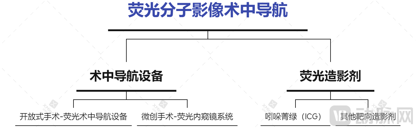
Major Products for Intraoperative Navigation with Fluorescent Molecular Imaging
In the past two years, open and minimally invasive intraoperative navigation products have successively received approval for market launch in China. So, how large is this market exactly?
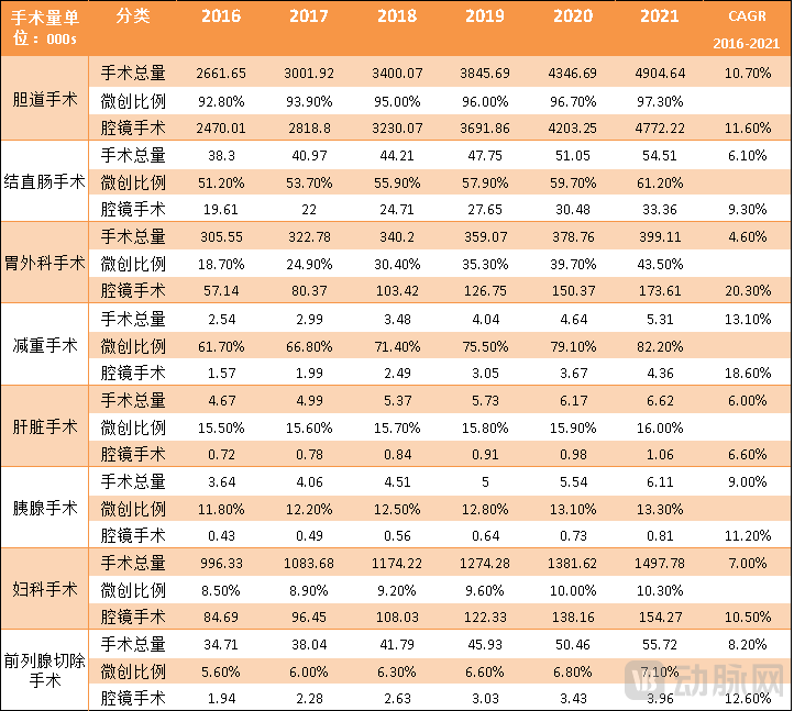
First, the total volume of surgical procedures in China has experienced rapid growth, and near-infrared fluorescence imaging navigation systems are widely applied in this field.
It is reported that from 2016 to 2021, the compound annual growth rate (CAGR) of overall surgical procedures in China, excluding obstetrics, ranged from 3.3% to 13%, indicating a trend of rapid development. Fluorescence imaging, leveraging its strong advantages for clinical translation—such as high sensitivity and specificity, absence of ionizing radiation, minimal side effects, and low equipment and usage costs—Exploratory applications have been implemented in clinical settings across departments including thoracic surgery, gynecology, gastrointestinal surgery, pediatrics, neurosurgery, and urology.
Furthermore, near-infrared fluorescence imaging enables real-time intraoperative in vivo imaging without interfering with the conventional surgical field and workflow. Based on these advantages,Near-infrared fluorescence imaging is poised to become the standard intraoperative navigation imaging tool in China’s 300,000 operating rooms.
Secondly, taking fluorescence endoscopy as an example, its clinical application is still in the early stages of development, and the market remains a blue ocean.
With continuous clinical exploration and innovations in equipment and consumable technologies, the indications for surgical procedures have been steadily expanding, while the proportion of minimally invasive surgeries has continued to rise. According to data from Life Science Intelligence, the share of minimally invasive procedures across various surgical subspecialties has increased year by year over the past three decades. As a growing number of patients benefit from these advances, an increasing number of outstanding domestic enterprises have entered the field to serve clinical practice.
Data Source: Life Science Intelligence
Minimally Invasive Surgery (MIS) primarily includes laparoscopic, thoracoscopic, hysteroscopic, and arthroscopic procedures. Owing to numerous advantages such as reduced trauma, fewer complications, lower risk of infection, and shorter hospital stays and recovery times, rigid endoscopic surgery is gradually replacing traditional open surgery, making minimally invasive approaches one of the key trends in surgical development in recent years.
As one of the revolutionary technologies in minimally invasive surgical equipment, fluorescence endoscopy systems have become a key competitive frontier for global endoscope manufacturers. However, within the multi-billion-dollar endoscopy market, the current penetration rate of fluorescence endoscopes remains low, indicating substantial room for future growth.
According to Frost & Sullivan data,China’s fluorescent rigid endoscope market is expected to enter a period of rapid growth, with a compound annual growth rate (CAGR) of 99.6% from 2019 to 2024. The market size for fluorescent rigid endoscopes is projected to reach RMB 3.52 billion in 2024, accounting for 32% of the overall rigid endoscope market.
Furthermore, in light of the current trend toward domestic substitution in China's endoscopy industry,Near-infrared fluorescence imaging, as a highly breakthrough technology in recent years, is expected to drive leapfrog development of domestically produced endoscopes from the perspective of technological innovation.
Product Portfolio Closure: Breakthroughs Achieved in Both Imaging Equipment and Contrast Agents
Overall, in the domestic market, DPM holds a position in the surgical molecular imaging marketThe Most Comprehensive Product Line. enterprises.Currently, we have established three major series of high-end optical molecular imaging surgical navigation systems: SuperEye, SmartEye, and WiseEye. In the field of surgery, we are also expanding our product lines to include ultra-high-definition endoscopes, 4K endoscopes, 3D endoscopes, 3D fluorescence endoscopes, and open fluorescence endoscope camera systems. Furthermore, we are strategically positioning contrast agents to create a closed-loop ecosystem of “equipment + consumables.”At the same time, it has made more far-reaching strategic arrangements in the commercialization of products based on cutting-edge imaging technologies.
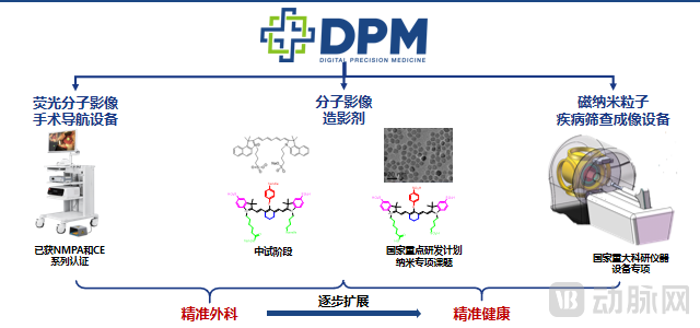
DPM’s Complete Product Portfolio Forms a Closed Loop Source: DPM
1. In-depth Advancement of Fluorescence Imaging Devices: Maturity in the First Near-Infrared Window and Breakthroughs in the Second Near-Infrared Window
Currently, the number of players in the near-infrared fluorescence imaging market is gradually increasing, and commercialized devices are being continuously innovated. To achieve high-quality visualization and imaging,Near-Infrared Fluorescence Imaging DeviceIt is necessary to have a sufficient near-infrared excitation light source, an optical acquisition system, filters, and a camera sensitive to near-infrared emitted light;Image results showIt is preferable to include color images, fluorescence images, and fused images of the surgical field simultaneously.
DPM’s Huiyan Series employs its proprietary 4CMOS technology, featuring high sensitivity, rapid image development, and real-time multispectral fused imaging.It is reported that DPM’s three major product series have been deployed for clinical trials and commercial sales in over 300 Grade A tertiary hospitals across China.
Imaging technology is one of DPM’s core technologies. Its team has independently developed key technologies, including multi-source data compensation and correction, rapid segmentation, precise registration, and real-time visualization, enabling multi-angle, high-throughput, and dynamic continuous acquisition of intraoperative images of tumors and other lesion tissues.
We have learned that DPM’s related research has received support from the National Key R&D Program on “Research and Development of Digital Diagnostic and Therapeutic Equipment” under the Ministry of Science and Technology of China;and took the lead in China to release group standards for near-infrared fluorescence imaging systems and near-infrared cold light sources in the industry.
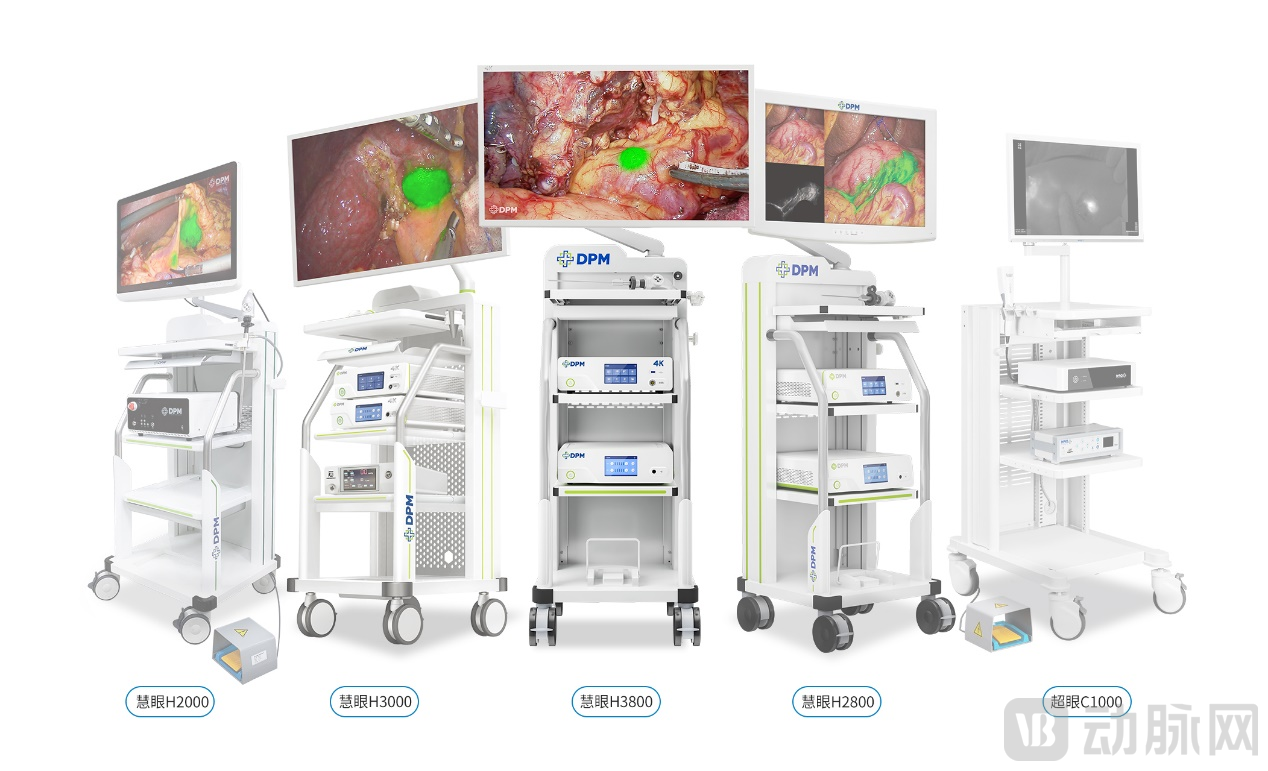
Near-Infrared Fluorescence Imaging Intraoperative Navigation Devices (Zhiyan and Chaoyan Series for Open Surgery; Huiyan Series for Minimally Invasive Surgery) Source: DPM
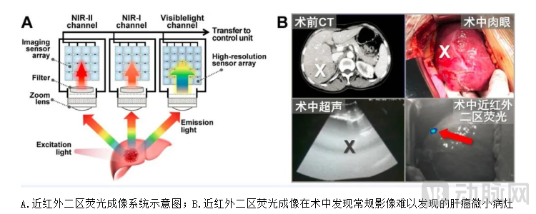
Currently, near-infrared fluorescence imaging-guided navigation worldwide primarily utilizes near-infrared I (NIR-I) imaging technology. However, scientists have discovered thatNear-infrared II (NIR-II) imaging (900–1700 nm wavelength range) offers deeper tissue penetration, higher signal-to-noise ratio, and superior spatiotemporal resolution compared to near-infrared I (NIR-I) imaging (700–900 nm wavelength range).。
The second near-infrared (NIR-II) window holds significant application value and potential in the development of novel contrast agents, cerebrovascular imaging, tumor detection, lymphatic system imaging, multimodal imaging, pharmacokinetic/pharmacodynamic studies, and clinical surgical navigation imaging.。Among the few manufacturers of near-infrared II (NIR-II) imaging systems, DPM has made extensive forward-looking investments and achieved initial results.
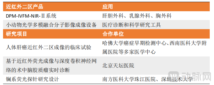
DPM's NIR-II Imaging Products and Related Research Source: DPM
According to reports, the DPM team has developed a novel near-infrared II (NIR-II) fluorescence imaging system, the DPM-IVFM-NIR-Ⅱ, along with surgical navigation technology. In combination with the fluorescent contrast agent indocyanine green (ICG), this system has been applied to human liver cancer imaging.
This application addresses the clinical challenges of intraoperative margin assessment and localization of microscopic tumor foci in liver cancer surgery, achieving the clinical translation of near-infrared II (NIR-II) fluorescence imaging, marking a “first-in-human” achievement.The related findings were published in *Nature Biomedical Engineering* and received positive acclaim from professors at Harvard Medical School.
The study results demonstrated that, compared with near-infrared I (NIR-I) imaging, near-infrared II (NIR-II) imaging offered higher sensitivity (100% vs. 90.63%) and a threefold higher signal-to-background ratio. This system not only assists surgeons in performing more precise hepatectomy for liver cancer, thereby reducing collateral damage to normal liver tissue, but also enables more thorough eradication of microscopic tumor foci.
Source: DPM
2. Fluorescent Contrast Agents (Fluorescent Molecular Targeting Probes)
Contrast agents can help imaging devices acquire high-contrast images, thereby enhancing diagnostic efficacy. Each contrast agent has specific excitation wavelengths, targeting properties, and clinical application scopes. Currently, optical contrast agents commonly used in clinical practice include sodium fluorescein, 5-aminolevulinic acid (5-ALA), methylene blue (MB), and cyanine dyes such as indocyanine green (ICG).
From an application perspective, the total global sales volume of contrast agents maintained an annual growth rate of over 30% from 2016 to 2020.Targeted contrast agents, as consumable products, represent a promising strategic direction for manufacturers of fluorescence navigation equipment. However, as they are essentially classified as pharmaceuticals, their development involves long cycles and substantial upfront investment. Consequently, there are few market participants, with DPM being one of the few.
For example, DPM, in collaboration with Beijing Tiantan Hospital and Peking Union Medical College Hospital, developed a novel GBM-targeted contrast agent to address clinical challenges such as the difficulty in determining glioblastoma (GBM) resection margins, thereby achieving high-sensitivity targeted imaging of gliomas during surgery. Trial results demonstrated that the median progression-free survival for patients undergoing fluorescence-guided surgery, compared to conventional surgery, increased from 3.6 months to 14.1 months, while overall survival improved from 13.5 months to 23.1 months.
3. MPI 3D Fusion Imaging Equipment with Magnetic Nanoparticles: A Global Leader
The development of the “In Vivo 3D Fusion Imaging Device Based on the Nonlinear Response of Magnetic Nanoparticles” (hereinafter referred to as MPI) is conducted under the support of the Major Scientific Instrument and Equipment Development Special Program of the National Natural Science Foundation of China, and it is also one of DPM’s future core products.
The primary advantages of this device are its imaging sensitivity, which is 1,000 times higher than that of existing magnetic resonance systems, and its temporal resolution, which is 2,000 times higher. It overcomes the depth limitations of optical imaging, enables real-time dynamic three-dimensional imaging, and facilitates non-invasive, precise early diagnosis in vivo for health screening applications.Currently, the "Specification for Image Quality Evaluation of Magnetic Particle Imaging Systems," co-drafted by DPM, has passed the group standard review organized by the Chinese Society for Graphics, a national-level academic society in China, and has been approved for establishment, laying the foundation for technical standards and specifications for MPI.
From the perspective of medical imaging technology development, fluorescence molecular imaging is undoubtedly a highly promising technology driving the advancement of precision surgical diagnosis and treatment. Extensive research indicates that methods for differentiating benign from malignant tissues based on molecular differences facilitate earlier and more accurate identification of pathological tissues, enable better determination of surgical margins during operations, and improve patient prognosis.
In recent years, the total number of publications in the field of fluorescence-guided intraoperative navigation has grown exponentially, with related imaging methods and novel fluorescent contrast agents becoming international research hotspots.. Meanwhile, related scientific research and commercial devices have also developed rapidly, especially in the field of tumor-assisted identification.
Currently, the number of domestic and international companies entering this field is continuously increasing.Backed by the Key Laboratory of Molecular Imaging of the Chinese Academy of Sciences, and leveraging leading theoretical research and extensive experience in the integration of medicine and engineering, DPM’s product portfolio not only covers multiple series of intraoperative optical molecular imaging surgical navigation systems and optical molecular imaging contrast agents, but also includes cutting-edge technological translations such as second near-infrared window (NIR-II) imaging devices and magnetic nanoparticle imaging systems. We look forward to and will continue to monitor DPM’s breakthroughs in molecular imaging technology.