Pair Intelligent Medical Image Annotation Platform Files IPO Prospectus
Manual annotation by medical experts serves as the cornerstone of AI research in medical imaging. Annotation software should aim to minimize the time physicians spend on manual labeling, alleviate the burden associated with this task, and help improve the quality and consistency of annotations. As the first domestically produced one-stop medical image annotation software, Pair features professionalism, convenience, universal ease of use, and intelligence. Since its public release in 2020, Pair has received substantial feedback, both positive and critical, driving continuous iterative optimization and intelligent upgrades. This article systematically summarizes and presents the key updates made to Pair in 2021. The core functional highlights of the Pair software are as follows:
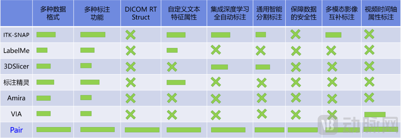
● Universally applicable to research projects, compatible with various imaging modalities and file formats
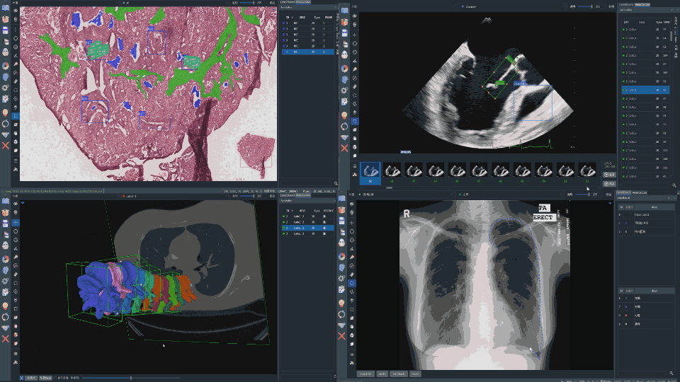
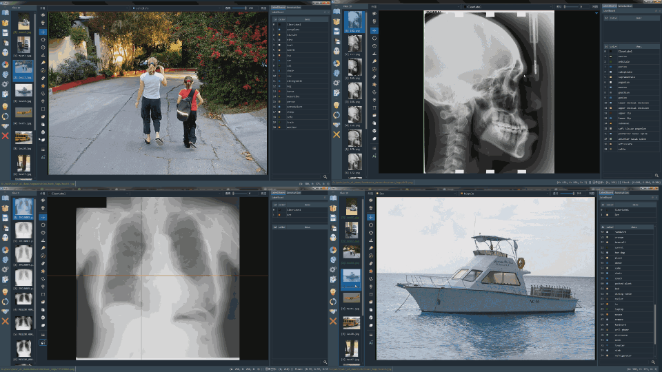
Pair is compatible with CT, X-ray, MRI, PET, ultrasound, histopathology slides, and cytology smears.Over 10 Imaging Modalities, compatible with 2D images, videos, 3D models, pathology slides, signals, radiotherapy contouring files, etc.20+ File Formats. All medical imaging AI research projects can be annotated via Pair, eliminating the need to frequently switch between different projects or undergo time-consuming and labor-intensive training on various annotation software platforms.
● Rich annotation features, covering the annotation needs of numerous medical imaging AI research projects
Pair provides measurement lines, angles, polygons, curves, surfaces, bounding boxes, keypoints, classification labels, text attributes, timelines, cross-sections, and more.Over 20 itemsAnnotation features encompass common annotation types required for AI-based image analysis, including measurement, segmentation, classification, object detection, keypoint localization, and action prediction. More importantly, Pair supports the hybrid use of various annotation functions and types, fully accommodating complex research and annotation requirements.
● Intelligent annotation assistance, freeing physicians from the burden of manual labeling
Pair provides multi-level intelligent assisted annotation features, significantly reducing the manual annotation workload for physicians. Pair offers automated assisted annotation functions such as threshold segmentation, magic wand, curve fitting, rectangular box tracking, and label memory/copying. Pair is uniquely equipped with proprietaryUser-Defined AI AnnotationandAutoSeg: Universal Intelligent Segmentation Annotation. Both features can be powered by deep learning models, enabling one-click intelligent annotation of anatomical structures using only a standard computer CPU.
● Standardized annotation workflows to support large-scale medical image annotation tasks
Pair provides configuration and management capabilities for annotation labels, effectively preventing label misuse and improper use of annotation functions caused by annotators’ operational errors or personal habits. This standardizes the annotation workflow and significantly reduces noise in annotation results. By optimizing memory management, Pair supports the simultaneous import and annotation of over 1,000 imaging files, annotation of ultra-large video files with up to 20,000 frames, and annotation of pathological image files exceeding 2 GB in size.
● Case privacy de-identification and image encryption provide security assurance for research projects
Patient case data and medical images are valuable resources for AI research, yet they face an escalating risk of privacy breaches. There remains a lack of user-friendly, general-purpose de-identification tools and annotation software with encryption capabilities. To address this, Pair provides comprehensive de-identification features and advanced annotation functions with multi-layered permission-based encryption and decryption, ensuring robust security for patient privacy, medical imaging, and annotation assets.
Concise and user-friendly workflows, with continuously improved and enriched tutorial use cases.
Pair features a concise and intuitive annotation logic design, enabling physicians to get started quickly and reducing the time spent on annotation interactions. Pair provides comprehensive documentation and video demonstrations to help users rapidly familiarize themselves with the software’s workflow, thereby saving training time for annotation. To date, Pair has released 15 well-produced video tutorials and will continue to invite multiple medical experts to share case studies on efficient annotation using Pair.
To date, Pair has largely completed the development of its core functional framework and is rapidly advancing toward comprehensive intelligence to further alleviate the burden of manual annotation for physicians. Pair maintains a high frequency of updates and optimizations, striving for utmost professionalism in the design of its annotation features and aiming to become essential foundational software. In 2021, Pair implemented numerous significant feature updates and optimizations, as detailed below:
● AutoSeg: Universal Intelligent Segmentation and Annotation
● AI-Assisted Annotation for User-Defined Models
● Video Timeline Event Annotation
● Multimodal Imaging Complementary Annotation
● Comprehensive support for DICOM RT Struct in radiotherapy contouring
● PairCode: Software for De-identifying Patient Privacy and Securing Medical Imaging Data
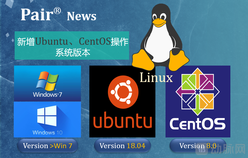
● Supports Windows, Ubuntu, and CentOS versions
Below, we will provide a detailed introduction to the major feature updates (Remarks: All medical images shown in this article have been de-identified to protect privacy):
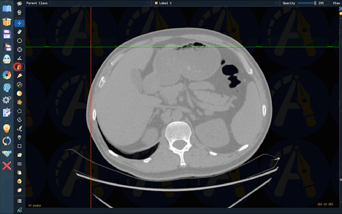
Fine-grained segmentation annotation is the most time-consuming task in medical imaging annotation. Different imaging modalities, anatomical structures, and lesions require heavily burdensome point-by-point segmentation annotations performed by physicians with varying specialties and qualifications. To address these critical challenges, the Pair team has developed a flagship intelligent feature.AutoSeg, aiming to achieve fine-grained segmentation and annotation of general anatomical structures, significantly reducing the time required for manual annotation, thereby accelerating the implementation and widespread adoption of AI imaging research.AutoSegThe advantages and characteristics are as follows:
● No configuration required, simple and straightforward, convenient and easy to use
● No distinction requiredImaging Modality, Anatomical Structure, Lesion Type
● Completed in 1 second, can be used on standard laptops
● I.Key Frame Selection Operation, to achieve fine-grained segmentation annotation
According to statistics, the use of PairAutoSegAutomatic segmentation and annotation features can save approximately70%the time required for segmentation annotation. Pair implements this functionality based on Huawei’s open-source deep learning platform, MindSpore, and completes the final deployment using MindSpore Lite. The efficient kernel operator implementation in MindSpore Lite fully leverages the computational power of standard CPUs, effectively balancing model computational demands with accuracy requirements. This reduces inference time to under one second, significantly enhancing the user experience of AutoSeg.
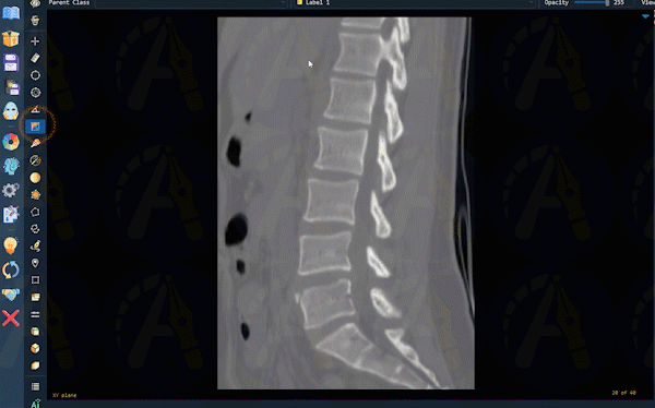
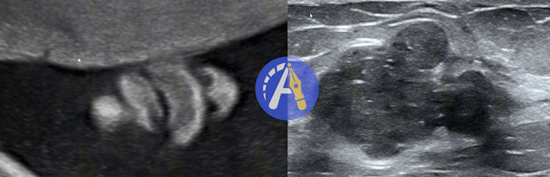
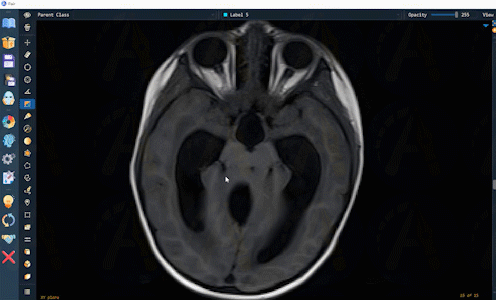
AutoSeg has been validated and utilized across a variety of anatomical structures and lesions in multiple imaging modalities, including CT, MRI, ultrasound, X-ray, PET, scanning electron microscopy, pathology, smears, and endoscopy. The annotation results generated by AutoSeg can be easily edited and modified to facilitate user fine-tuning and rapid confirmation. Below are examples demonstrating the application of AutoSeg across diverse imaging modalities and anatomical structures:


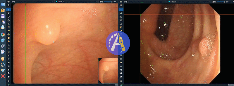
Display of AutoSeg Function Annotations on Colonoscopy Images
AIA is exclusive to Pair.One-Click AI Inference Feature, whether for classification, segmentation, keypoint localization, object detection, or multi-task learning, users need only provide a custom AI model and click the AI button to rapidly generate automated auxiliary annotations. Fine-tuning these annotation results completes the labeling process, saving annotation time and accelerating workflows. Currently, Pair’s AIA-assisted annotation feature supportsGoogle's TensorFlowandFacebook's PyTorchDeep Learning Platform, SupportingOver 50 Deep Learning Network Model Architectures. Pair has also recently added support for domestically producedHuawei MindSporeSupport for deep learning framework models.
Medical video imaging typically consists of image clips from different events arranged along a timeline, with varying annotation requirements for different attributes across segments. Examples include cardiac motions during different phases of the cardiac cycle and types of surgical instrument movements at various stages of a procedure. Currently, few annotation software solutions offer timeline-based annotation capabilities for medical video imaging. Pair’s timeline annotation feature, enabled by an intuitive interactive design, allows rapid multi-attribute annotation of arbitrary video segments, thereby efficiently advancing video annotation workflows. Specifically, within the timeline annotation interface, users can complete attribute annotation for a selected video segment by right-clicking and dragging the corresponding label to the appropriate frame on the timeline. Pair also provides a timeline-following function to facilitate user observation of the frames associated with each segment. To modify annotations, users can simply left-click and drag the endpoints on the timeline, ensuring convenient and efficient operation.

Single imaging modalities impose significant limitations on physicians’ ability to localize and annotate lesions. Synchronous observation and complementary use of multimodal information facilitate accurate lesion annotation by clinicians, such as combining T1- and T2-weighted MRI images, multimodal ultrasound images, PET and CT images, and cytology smear images with different fluorescent stains. Pair’s multimodal complementary annotation feature enables the simultaneous display of images from different modalities during the annotation process, reducing uncertainty in target region localization and contour delineation, thereby effectively assisting physicians in identification and annotation. Pair’s multimodal annotation functionality is compatible with 2D, video, 3D, and pathological images.
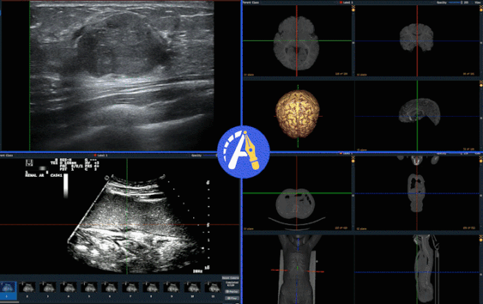
Multimodal Imaging Complementary Annotation Process
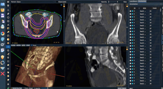
Given the significant demand for AI research in radiology, Pair has added comprehensive support for the DICOM RT Structure file format for radiological images. Radiologists willTPS(Treatment Planning System) The radiotherapy target contours delineated in the treatment planning system can be exported in RT Struct format and directly opened in Pair for visualization and further annotation optimization. Pair supports interactive annotation of over 1,000 RT contours. Furthermore, Pair can fill the contours within the RT Struct file and export them as segmentation annotation files, such as in .nii or .nii.gz formats, greatly facilitating radiologists' use of professional radiomics software, such asTexRAD、MaZda, etc., to conduct subsequent advanced analysis and research.
In the process of medical image annotation, it is crucial to effectively protect patient privacy and ensure the security of annotation resources. Existing annotation software often poses significant security risks in the annotation workflow:
● Images are not de-identified, leaving patient privacy information unprotected;
● There is a high risk of leakage for imaging resources and annotation result resources;
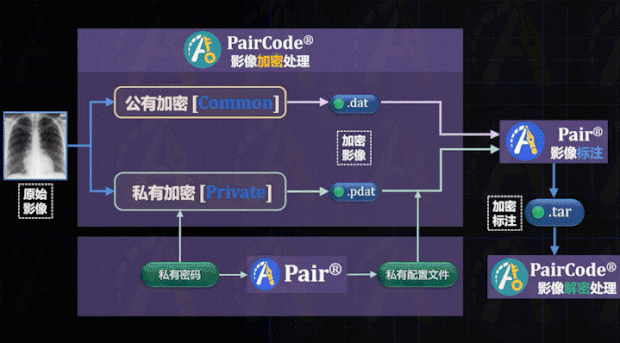
To meet the requirements for protecting patient privacy and ensuring the security of imaging resources, Pair provides PairCode, a secure auxiliary software that offers end-to-end encryption protection for image annotation tasks, thereby achieving dual-layer security for annotation resources. PairCode provides the following two functions:
DICOM Image De-identification
In medical imaging, DICOM data headers often contain sensitive patient privacy information. When raw data circulates as an annotation target, there is a high risk of patient privacy breaches. By leveraging PairCode’s data de-identification features, project managers can batch and customizably erase or modify sensitive entries—such as patient details and examination information within DICOM headers—to safeguard patient privacy.
Specifically, within the de-identification functionality of PairCode, privacy-related information in DICOM headers is categorized into five sections, allowing users to customize modifications or deletions based on project requirements. Meanwhile, PairCode provides a decryption key for the de-identified data, recording both the original and modified content of the header information to facilitate subsequent traceability. PairCode supports the processing of batch imaging files exceeding 10 GB in size.

PairCode De-identification Process Illustration
Annotation Resource Encryption and Decryption
To achieve secure, private deployment of annotation resources and reduce the risk of data leakage, PairCode provides encryption and decryption capabilities for medical images and annotations, ensuring secure data circulation:
● Project managers can customize the encryption and decryption of resources;
● Supports convenient encryption and decryption for over 20 professional medical imaging formats;
● Asymmetric high-order encryption ensures a zero-burden user annotation experience.
PairCode offers two encryption methods: public-key encryption and private-key encryption, enabling query and modification of encrypted images and annotation results.Can only be performed via the Pair software., thereby ensuring data security. After encrypting the data using PairCode, annotators only need to load the configuration file to annotate the encrypted images. The software will also automatically encrypt the annotation results, eliminating the need for additional manual encryption.
Currently, Pair offers versions for three different operating systems—Windows, Ubuntu, and CentOS—to meet the annotation needs of physicians, engineers, and cluster servers. Users can freely choose a version for installation and use. Annotation results generated on any of these operating system versions can be viewed and modified on other systems, facilitating the sharing of annotation outcomes among project team members. Additionally, Pair provides both Chinese and English language interfaces to accommodate users of different linguistic backgrounds.
、
● Professor Cai Yuexin, Department of Otolaryngology, Sun Yat-sen Memorial Hospital, Sun Yat-sen University
“Pair software truly achieves universal annotation support across scientific research projects, surpassing other annotation tools we have tried. Our team has adopted Pair for annotation in multiple AI research projects involving 3D medical imaging. Throughout the project implementation, we received timely responses and proactive support from the Pair team. We hope that Pair will continue to improve, further facilitating AI-related research and becoming a preferred tool for more physicians.”
● Dr. Yang Yi, Department of Orthopedics, West China Hospital, Sichuan University
“After adopting Pair software, the annotation of our 3D CT images has seen significant improvements in both speed and quality, allowing our research projects to progress rapidly. Its functionality is straightforward and intuitive, with virtually no learning curve, and its many intelligent annotation features are truly impressive. PairCode also effectively meets our professional needs for large-scale image de-identification. The Pair team consistently responds quickly to our customized requests, making them an excellent collaborative partner. We hope that Pair can help lower the barriers to AI research, enabling more physicians to complete their AI projects more quickly and effectively.”
● Zhang Zijian, Medical Physicist, Department of Oncology, Xiangya Hospital, Central South University
“Before adopting Pair, our data annotation workflow required switching between different software platforms depending on the data type. However, a chance encounter introduced our team to Pair, which provided an integrated platform for multimodal data annotation, particularly for radiotherapy contouring in CT-PET imaging. Pair is user-friendly and easy to learn, significantly boosting our team’s annotation efficiency. In certain project scenarios requiring customized features, we also received comprehensive support from the Pair team. We sincerely hope that the Pair team will continue to grow stronger and achieve greater success.”
To date, Pair has been adopted by over 2,000 users and more than 300 renowned research institutions, hospitals, and enterprises both in China and abroad, yielding substantial valuable feedback and functional suggestions. Backed by a stable and active R&D team, Pair will undergo continuous optimization and upgrades, with the commitment to establishing itself as a foundational, specialized domestic software solution and advancing the new AI infrastructure.

List of Selected Institutions Trialing the Pair Software

List of Hospitals Trialing Pair Software
The download link provides installation packages for Pair across different operating systems and versions, along with resources such as the Pair AIA model files and sample test images, enabling users to immediately experience the convenience of AI-assisted annotation. To access the aforementioned new features, please download Pair version 2.5.
https://www.jianguoyun.com/p/DUQd8gcQh-n1CBiLvcgD
Below are video tutorials on related features provided by Pair on Bilibili. You are welcome toLike, Coin, FavoriteOne-click triple action.
https://space.bilibili.com/616429598
For more detailed information, please follow Pair’s official WeChat and Bilibili accounts.

Pair was developed under the leadership of Professor Ni Dong and Dr. Yang Xin from the Medical Ultrasound Image Computing Lab (MUSIC) at Shenzhen University. The team consists of more than ten members. The Pair software has undergone nearly three years of development and continues to undergo significant optimization. Pair is positioned to address long-standing issues in medical image annotation software, striving to become the premier, most professional, and physician-centric domestic medical image annotation solution. Since its public debut at the MICS conference in July 2020, Pair has received valuable guidance and critical feedback from many senior experts, helping to identify additional problems worthy of resolution. Currently, Pair has basically completed the development of basic annotation functionalities for Phase I and is poised to enter Phase II, which focuses on comprehensive intelligence enhancement while further enriching the basic annotation features.
We extend our gratitude to physicians across various specialties for their affirmations and constructive criticism of Pair, and to experts in the field of medical image analysis for their attention and guidance. We also thank the Huawei Open Source Ecosystem and the MindSpore team for their prompt response and strong support in the development and optimization of the AutoSeg functionality. Finally, we appreciate the concerted efforts and unwavering perseverance of our team members, including but not limited to: Chen Chaoyu, Liu Wenxing, Hu Xindi, Shi Wenlong, Lin Mingrong, Luo Yiran, He Shuangchi, Liu Sijing, Liu Lian, Zhou Han, Wang Jian, Lv Fen, Gao Rui, and Wang Kaini.