Viz.ai Enters Its Fourth Year with FDA-Approved Imaging AI Platform Deployed Across 1,000 Hospitals and Millions of Patients Served

Viz.ai
Clinical Intelligent Medical Software Developer
In recent years, AI-powered medical imaging has emerged as a hot sector within the “AI + Healthcare” landscape.
According to data from Global Market Insights, the global market size for AI in healthcare was $4.2 billion in 2020, with the AI medical imaging segment accounting for $1 billion, representing over 24% of the global AI healthcare market. Lei Jun once remarked, “Even a pig can fly if it stands at the eye of the storm.” However, there is a second part to this saying: “If equipped with wings, it will soar even higher.”
Viz ContaCT Becomes the First AI Software to Enter NTAP, with a One-Time Reimbursement of $1,040.
Viz.ai is a medical imaging AI company headquartered in San Francisco, USA, founded in 2016. In 2018, the company launched its first product, Viz LVO (also known as Viz ContaCT). This platform leverages AI algorithms to analyze CT neuroimaging scans, detect indicators associated with stroke, and provide clinical decision support for stroke care to physicians. At that time, VCBeat also reported on this development.
Viz ContaCT Interface. Image source: Viz.ai official website
When the Viz ContaCT platform entered the market, Viz.ai was still in its seed round. Recently, the company announced the completion of a $100 million Series D financing round, led by Insight Partners and Tiger Global Management, with participation from Google Ventures and other investors.
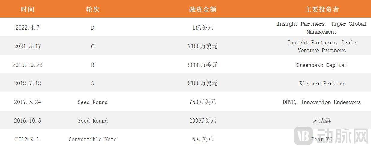
Viz.ai's Historical Funding Rounds Data Source: Crunchbase
In September 2020, the Centers for Medicare & Medicaid Services (CMS) included Viz ContaCT software in the New Technology Add-on Payment (NTAP) program, making it the first AI software to be included in NTAP. Under this program, up to an additional $1,040 can be reimbursed per patient who meets the NTAP criteria and uses Viz ContaCT.
What is NTAP? The U.S. Medicare system adopts the DRG-PPS model, which classifies patients into different diagnostic groups based on treatment methods and patient characteristics. Reimbursement is determined by these diagnostic groups rather than actual costs. However, since the reimbursable cost scope is updated only every two to three years, some new technologies cannot be incorporated in a timely manner. Therefore, the NTAP policy was introduced as an additional supplement.
There are certain criteria for inclusion in the NTAP: not covered by medical insurance, novelty, and substantial clinical improvement.
So how did the CMS take notice of Viz.ai’s ContaCT software?
In the United States, stroke is the fifth leading cause of death and the most common neurological cause of disability in adults. The therapeutic window for treatment is 6 to 24 hours; beyond 24 hours, the likelihood of sequelae increases dramatically. In particular, the therapeutic window for ischemic stroke is narrow, typically limited to 6 hours, as cerebral neurons begin to die after just five minutes of ischemia. Large vessel occlusion (LVO) is a common type of ischemic stroke.
According to the 2018 American Stroke Association Guidelines for the Early Management of Patients With Acute Ischemic Stroke, acute stroke care can be divided into three stages: prehospital, emergency department, and inpatient hospitalization.
When patients with suspected stroke are transported to primary care hospitals, CTA or MRI is performed first to determine whether the patient has large vessel occlusion.
If so, transfer to an advanced stroke center, and then assess for indications for mechanical thrombectomy;
If not, transfer to a primary stroke center for treatment.
From the moment a patient loses consciousness to the initiation of treatment, the process essentially constitutes continuous triage, where every second counts. However, numerous obstacles hinder this workflow. For instance, physicians rely on imaging reports to make diagnoses, yet there is typically a one-hour lag between completing CT angiography (CTA) and magnetic resonance imaging (MRI) scans and receiving the final reports. Additionally, transferring patients from primary care hospitals to stroke centers consumes valuable time, and significant information asymmetry exists between these two levels of care.
Direct Integration with Imaging Center for AI Analysis: Detection of Large Vessel Occlusion in 6 Minutes
How can this process be accelerated? Viz ContaCT software intervenes starting from the CTA and MRI scanning stage. Viz ContaCT integrates directly with the hospital’s DICOM Router, which serves as the network hub for storing and transmitting raw medical images, as well as the center for film printing and output.
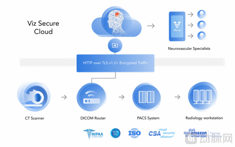
Viz ContaCT Workflow Logic. Image source: Viz.ai official website
Viz ContaCT directly retrieves data from the DICOM Router, leverages artificial intelligence to analyze imaging data, and automatically detects suspected large vessel occlusion (LVO) stroke (ContaCT). It reports to the on-call stroke center and sends real-time patient updates to stroke center physicians, including timestamps for emergency department arrival, CT scan completion, and issuance of the ContaCT assessment.
According to Viz.ai, the Viz ContaCT platform can determine whether a large vessel occlusion is present within six minutes after scanning, whereas traditional methods typically take about an hour.
Viz ContaCT also established a communication platform, Viz HUB, to facilitate connectivity between two hospitals. The platform supports voice, text, and video communications and is HIPAA-compliant, adhering to the official U.S. standards for healthcare information privacy and security.
All of the above can be referred to as pre-hospital emergency care for stroke. In essence, this approach parallelizes tasks that were previously performed sequentially from front to back, ensuring that by the time the patient arrives at the stroke center, the hospital has already received the patient’s data and is fully prepared.
If a patient arrives at the stroke center more than six hours after their last known time of being conscious, it is recommended to perform a CTP scan to assess the infarct core and ischemic penumbra. However, no matter how many tests are conducted, the principle remains that the timing for mechanical thrombectomy should not be delayed.
Viz.ai has developed the Viz. CTP platform for CT perfusion (CTP) examinations. This platform integrates directly with the hospital’s DICOM router, leveraging artificial intelligence to process CTP data and generate color-coded perfusion maps, which can be viewed by physicians and other relevant personnel within the software. According to Viz.ai’s official website, AI-generated images exhibit a 50% reduction in motion artifacts compared to traditional images.
By connecting to the hospital’s DICOM Router center, Viz ContaCT can also serve as a mobile image viewer.
Physicians can view a variety of high-quality images on this platform, including CT, CTA, MRI, X-ray, ECG, ultrasound, and 3D images. The platform features proprietary image compression technology, enabling image viewing even in areas with poor network connectivity. As a result, physicians are not confined to their offices and can review reports while on the go.
In December 2020, Shalitin et al. published a paper in the journal *Experimental Stroke & Translational Medicine*, stating that the Viz.ai software demonstrated 96% sensitivity and 94% specificity in analyzing CTA images from 2,544 patients across 139 hospitals.
Not limited to detecting large vessel occlusion, gradually adding multiple disease modules
Following the same logic as for ischemic stroke detection—accessing hospital DICOM routers to retrieve data and leveraging artificial intelligence to analyze imaging for alerts—Viz.ai’s Viz.ContaCT received FDA clearance for its hemorrhagic stroke module in 2020, for its pulmonary embolism and aortic dissection modules in 2021, and for its cerebral aneurysm module in early 2022. Its subdural hematoma module is currently under 510(k) review.
It is worth mentioning the modules for cerebral aneurysms and cryptogenic stroke.
Brain aneurysms are typically not detected during routine daily activities but are instead discovered incidentally during health check-ups or examinations for other conditions. Furthermore, even after a confirmed diagnosis, patients often remain uncertain about which specialist to consult for follow-up care. The Viz ANEURYSM module does not require manual activation; it automatically detects brain aneurysms that may be overlooked on CTA scans and informs patients of hospitals where treatment is available.
Cryptogenic stroke is not a standalone category; approximately 30% of acute ischemic strokes are cryptogenic, meaning their pathogenesis is unclear. In some cases, cryptogenic strokes are caused by cardiac issues. If a stroke patient is diagnosed with cryptogenic stroke upon hospital admission, the software includes a “Viz LINK” button to refer the patient to cardiology.
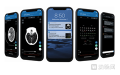
The Viz.ContaCT software is now accessible on various PC and mobile devices.
Viz.ai emphasizes that none of its modules can serve as standalone diagnostic tools; they merely identify specific images and alert physicians, enabling them to be prepared rather than caught off guard, with final determinations remaining dependent on clinical judgment.
At the 2020 International Stroke Conference, Viz.ai and researcher Hassan presented preliminary study data indicating that the use of Viz.ContaCT reduced patients’ average hospital stay by 2.5 days.
Recently, Viz.ai also published the results of a study from the University of California, San Diego (UCSD) on its website: at a Comprehensive Stroke Center (CSC), patients using Viz.ContaCT experienced an average reduction of 41 minutes in the time from hospital admission to groin puncture compared with traditional methods.
In 2019, Viz.ai partnered with Medtronic. Since then, the company has established collaborations with organizations and companies such as the American Heart Association and NICO. More than 1,000 hospitals, including the Mount Sinai Health System in New York, have adopted Viz.ContaCT, serving millions of patients.
In 2021, Viz.ai grew its team from 180 to over 350 employees and was named to Forbes’ 2021 Next Billion-Dollar Startups list.
Chris Mansi, co-founder and CEO of Viz.ai, is a neurosurgeon. He stated that the impetus for founding Viz.ai was witnessing the death of a female patient who had undergone successful surgery after a car accident but ultimately passed away due to a delayed diagnosis of cerebral vascular embolism. The other two co-founders, David Golan and Manoj Ramachandran, are experts in the fields of artificial intelligence and plastic surgery, respectively.
Viz.ai’s next steps include expanding its team to 550 employees and partnering with pharmaceutical companies to use AI for identifying patients eligible for clinical trials, thereby accelerating drug development.
Domestic Medical Imaging AI Products Are Being Deployed Successively, with AI + CT as the Mainstream
As of early March 2022, when the “Guiding Principles for the Registration and Review of Artificial Intelligence Medical Devices” was released, a total of 33 Class III AI medical device products had been approved by China’s National Medical Products Administration (NMPA), including approximately 20 imaging AI products. The first Class III certificate for an imaging AI product issued by the NMPA in China was granted in January 2020.
As early as 2018, it was claimed that the inaugural year for the clinical deployment of AI in medical imaging had arrived. In fact, this conclusion was not rash given the context of 2018, when investment in AI for medical imaging reached an all-time high with 31 financing deals. However, in 2019, the capital market remained fragile, and the number of financing deals dropped to 14.
In the second half of 2020, the AI medical imaging industry, having weathered the winter, began to recover and has grown increasingly popular year by year. According to Frost & Sullivan’s projections, the market size of China’s AI medical imaging sector is expected to increase from RMB 340 million in 2020 to RMB 92.31 billion in 2030.
In the first ten months of 2021, there were 140 investment deals in China's medical AI sector, including 39 deals in AI medical imaging, accounting for 27.86% of the total.
The range of diseases covered by AI in medical imaging is continuously expanding, now encompassing various regions including the head, chest, abdomen, and bones. Among these, lung-focused AI products—primarily for auxiliary detection of pulmonary nodules and triage support for pneumonia—have reached a high level of maturity, with multiple products having obtained Class III certification from the National Medical Products Administration (NMPA).
In terms of application modalities, CT-based AI imaging products have obtained the most regulatory approvals, followed by MR.