S-Fetus Phoenix Eye: KaiLi Medical Launches World's First AI-Powered Dynamic-Image-Based Prenatal Ultrasound Screening System
Ultrasound imaging screening holds significant value in the diagnosis of fetal congenital defects.
In the past, ultrasonic imaging techniques were limited to assessing the size and gross structural changes of intrauterine fetuses, with a narrow diagnostic scope that made it difficult to capture the detailed tissue architecture of the embryo or the subtle structural features of the developing fetus. Today, while ultrasonic diagnostic technology can reveal detailed structures across various fetal systems, it has also raised the threshold for physician interpretation.
Unlike the static images produced by CT and MRI, ultrasound images are dynamic and real-time. They are not only more difficult to interpret than static images, but also require physicians to acquire dynamic images from different planes along the same timeline and make real-time diagnoses, placing high demands on operators’ technical skills and image interpretation experience. This has indirectly led to the approximately 2 billion annual ultrasound examinations in China being heavily reliant on physician expertise. However, there is a scarcity of highly skilled sonographers in China; primary-care physicians, due to insufficient training and limited experience, often lack adequate diagnostic capabilities. Consequently, the accuracy of ultrasound diagnosis is difficult to guarantee, failing to meet the healthcare needs in regions with relatively scarce medical resources.
On May 21, SonoScape (Shenzhen SonoScape Medical Corp., Ltd.), a company that has been dedicated to ultrasound imaging research for 20 years, releasedFourth-Generation AI-Powered Prenatal Ultrasound Screening Product: “Fengyan S-Fetus”, providing solutions to the pain points in ultrasound diagnosis.
The breakthrough of this product stems fromCapable of automatically identifying and capturing images of 14 standard planes, including the thalamic plane and lateral ventricular plane, in real time; performing real-time image quality control; automatically measuring 12 parameters such as head circumference and biparietal diameter, as well as localizing the conus medullaris; and finally, automatically recording the results to generate a report."Implemented by doctors"Fully Intelligent Scenario-Based Mode for Hands-Free Imaging
With this technology, sonographers no longer need to rely on experience to locate standard planes for measurement; instead, they can directly obtain fetal growth parameters and ultrasound reports. This eliminates the need for frequent manual measurements via button operations and reduces error-prone data entry. The technology holds significant importance for effectively improving the efficiency and accuracy of prenatal ultrasound screening, addressing the uneven distribution of medical resources, and enhancing hospital quality control and management standards.
In fact, AI’s empowerment of ultrasound is similar to its applications in other medical specialties, primarily focusing on improving diagnostic accuracy and efficiency. However, unlike other modalities, ultrasound should serve as an inclusive diagnostic tool with broad applicability and a strong foundation in primary care settings. This endows AI-powered ultrasound with exceptional significance.
Driven by advantages such as being non-invasive, non-interventional, cost-effective, and widely adaptable, the market for ultrasound diagnostic equipment has experienced rapid growth. According to data from Fortune Business Insights, the global market size for ultrasound diagnostic equipment was USD 7.26 billion in 2020 and is projected to reach USD 12.93 billion by the end of 2028, representing a compound annual growth rate (CAGR) of 7.8%.
However, unlike static images, dynamic ultrasound footage is not easily archived. To conduct AI research on ultrasound imaging, it is essential to record and capture images of specific planes at specific moments. Meanwhile, challenges such as algorithm model development must be addressed to overcome objective factors including variations in scanning techniques, differences in color Doppler ultrasound equipment, and non-standardized data. The high technical requirements combined with the lack of standardized training data have resulted in very few enterprises focusing on AI-driven ultrasound research.
To truly develop valuable intelligent ultrasound products, companies must integrate ultrasound imaging with post-processing and combine traditional techniques with deep learning, all based on standardized data collection and processing, in order to gradually overcome these challenges.
SonoScape has 20 years of R&D experience in the ultrasound field and possesses profound expertise in core technologies such as artificial intelligence.“Phoenix Eye S-Fetus” is a brand-new technology further developed by SonoScape based on its first-generation intelligent applications.After three years of technological iterations, and through joint research by the R&D team and multiple intelligent obstetrics and gynecology centers,SonoScape has taken a further step forward, achieving an artificial intelligence breakthrough in the automatic capture of standard planes based on dynamic images.
From the first to the fourth generation of “S-Fetus,” SonoScape has achieved a leap from repetitive keystrokes to one-click acquisition, and from four views with four measurements to the automatic capture of 14 standard views and automated measurement of 12 parameters. This advancement provides greater possibilities for the development of prenatal ultrasound examination.
Reportedly, the “Phoenix Eye S-Fetus” technology is China’s first deep learning-based intelligent detection technology for obstetric ultrasound, and also the world’s first artificial intelligence technology capable of automatically capturing standard planes from dynamic images.Achieve fundamental breakthroughs by leveraging core algorithms, proprietary architecture, and cross-architecture optimization to unlock hardware potential, thereby addressing inherent limitations such as intelligence confined to isolated processes or partial stages, inevitable latency delays, cognitive and operational discontinuities for users when switching between intelligent and non-intelligent modes, and the continued need for manual user intervention.
Meanwhile, this technology can intelligently identify the imaging plane during real-time scanning by physicians, automatically capture standard planes throughout the process, perform automatic measurements, and accurately enter the measurement results into the corresponding fetal growth parameter fields in the report. Compared with conventional ultrasound techniques, the one-click fully automated measurement requires only 7.79% of the time needed by conventional methods, saving more than 90% of the time spent on measurements during the examination.
Among these features, the automatic recognition, image capture, and measurement of standard planes not only free sonographers from the need to constantly monitor the screen to locate standard views, allowing them more time to focus on assessing normal and abnormal fetal anatomical structures; but also enable physicians at primary care hospitals to acquire images of quality comparable to those obtained by sonographers at top-tier tertiary hospitals, thereby effectively addressing the imbalance in medical resource distribution.
In terms of growth parameter measurement, the “Phoenix Eye S-Fetus” system presents numerous fetal growth parameters to sonographers and conducts in-depth analysis of these parameters. This eliminates the need for physicians to frequently press buttons to measure growth parameters or consult growth parameter charts for interpretation, thereby reducing button presses by nearly 70%.
In ultrasound report generation, it assists physicians in accurately entering ultrasound parameters into the reports, thereby preventing data entry errors, eliminating repetitive manual tasks, and reducing physician workload.
“In recent years, some hospitals have introduced automated/semi-automated technologies into obstetric ultrasound image analysis; however, in practice, this approach has revealed issues such as low measurement accuracy, limited automation, and suboptimal clinical application outcomes.”
In the ‘Phoenix Eye S-Fetus’ workflow, the front-end generates multi-modal data according to scenario requirements, while back-end processing performs real-time reconstruction, processing, and optimization of this multi-modal data. For the reconstructed and optimized data, the AI module is required to perform real-time identification and tracking, along with high-precision analysis to extract standard planes. The standard plane decision and scheduling module then schedules the adaptive slice feature extraction module to extract quantitative features based on predefined strategies. Finally, quantitative analysis is conducted, followed by the completion of subsequent integrated automated workflows.Li Guanghao, Head of the Domestic Ultrasound Marketing Department at SonoScape, remarked.
SonoScape’s newly developed “Phoenix Eye S-Fetus” is built on a novel portable neural network model and emerging algorithmic mechanisms such as Transformer. Guided by clinical diagnostic and therapeutic needs, it achieves more comprehensive fusion, capture, and interpretation of image features in real-time sequential order, rather than relying solely on simple convolutional feature extraction.
A renowned hospital in China compared the clinical applications of “Phoenix Eye S-Fetus” and conventional ultrasound technology in acquiring standard fetal views, view recognition, and automated measurements for normal fetuses at 16–41 weeks of gestation. The comparative results showed that the S-Fetus technology achieved an automated view acquisition success rate of over 99%, with a single-attempt success rate exceeding 95%. For automated measurement of growth parameters, the accuracy reached over 99%, with a single-measurement accuracy exceeding 98%.
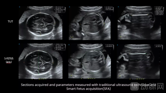
Obstetric Ultrasound Diagnosis Using the “Phoenix Eye S-Fetus” Technology (from left to right: transverse section of the thalamus, transverse section of the abdominal thalamus, longitudinal section of the femur)
It is reported that the “Fengyan S-Fetus” technology demonstrates good repeatability, enabling multiple automated measurements on the same imaging plane with negligible variation in results. For prenatal ultrasound practitioners with limited experience at primary-care hospitals, the use of “Fengyan S-Fetus” technology can improve the accuracy of obtaining standard imaging planes and measuring parameters, reduce errors during examinations, and enhance the overall quality of prenatal ultrasound assessments.
As can be seen, the “Phoenix Eye S-Fetus” technology can greatly simplify the operational process of prenatal ultrasound examinations, save examination time, and reduce the workload of sonographers, thereby improving the efficiency of prenatal ultrasound examination and diagnosis and facilitating the transition from conventional ultrasound to intelligent ultrasound.
By leveraging intelligent software for assisted diagnosis and treatment, the ultrasound examination workflow is streamlined, diagnostic efficiency is enhanced, and the precision of ultrasound-guided therapies is improved. This approach significantly elevates the diagnostic capabilities of primary care physicians and junior clinicians, thereby reducing the rates of misdiagnosis and missed diagnoses.
It is evident that intelligent ultrasound diagnosis represents a key application scenario of “AI + Healthcare” and constitutes a major trend in the future development of ultrasound diagnostics.
SonoScape’s recent technological breakthrough addresses the insufficient recognition of standard imaging planes, inadequate disease judgment capabilities, and urgent need for professional training among junior physicians; the heavy workload, excessive repetitive tasks encroaching on time for learning and professional development, and low efficiency among mid-career physicians; and the high psychological stress, diagnostic time consumed by routine tasks, and lack of time to address complex and refractory cases among senior physicians.
In addition to addressing the pain points of physicians, this technology enables junior doctors to quickly become proficient and helps mid- to senior-level doctors improve efficiency. It assists hospitals in reducing costs and increasing efficiency, while also lowering rates of missed and misdiagnoses and optimizing the allocation of medical resources. This leads to improved diagnostic accuracy, streamlined ultrasound diagnostic workflows, and a significant reduction in the threshold for performing ultrasound diagnostics, thereby aligning with the development of the “tiered diagnosis and treatment” policy.
It is reported that the “Phoenix Eye S-Fetus” system has already been deployed in some hospitals. Widespread adoption of obstetric ultrasound screening may be just around the corner.