Innovative Applications and Frontiers of Medical Imaging AI: A Vision by Professor Dinggang Shen
Medical imaging AI has experienced rapid development in recent years, with a plethora of imaging AI applications emerging continuously. What kind of medical imaging artificial intelligence constitutes truly clinically valuable innovative applications? What represents the cutting edge of medical imaging AI technology?
On June 17, Professor Shen Dinggang, Founding Dean of the School of Biomedical Engineering at ShanghaiTech University and Co-CEO of United Imaging Intelligence, was invited to participate in the “AI + Auxiliary Diagnosis Innovation and Development Forum” at VCBeat’s 6th Future Healthcare 100 Conference, where he delivered a keynote speech titled “Innovative Applications and Frontiers of Artificial Intelligence in Medical Imaging.”
Professor Shen Dinggang elaborated on three aspects: the development of medical AI, integrated diagnosis and treatment cases covering multiple scenarios, diseases, and entire care pathways, and prospects for frontier technologies.
In recent years, driven by continuous policy support, the practical application of technology, and the catalytic effect of the pandemic, the healthcare industry has entered a period of explosive growth in digitalization and intelligence. Medical imaging AI is considered the segment within artificial intelligence in healthcare most likely to achieve commercialization first. Consequently, many countries and regions worldwide, including the United States, China, and the European Union, are vigorously promoting the development of medical imaging AI and have successively introduced relevant action plans, such as the Brain Project.
Driven by global expectations and collective efforts, the market size for AI in medical imaging is projected to exceed RMB 280 billion over the next five years. The Asia-Pacific region is expected to witness the highest growth globally, with its market size forecast to surpass RMB 100 billion within this period, indicating substantial market potential. Undoubtedly, China remains the largest and most significant market in the Asia-Pacific region.
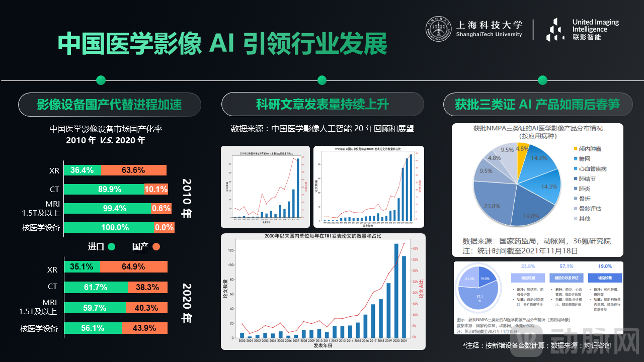
In China, this has been corroborated from the perspectives of industry, scientific research, and the approval process for Class III medical device certifications. On the industrial front, a decade ago, China relied entirely on imports for imaging equipment, particularly MRI systems above 1.5T and nuclear medicine devices. Over the past ten years, multiple domestic imaging equipment brands, represented by United Imaging Healthcare, have emerged, breaking the monopoly held by foreign manufacturers. The market share of domestically produced CT scanners has increased by nearly 30%, while high-end equipment has grown from zero to capturing nearly half of the market. Some domestic devices, such as United Imaging’s 2-meter PET scanner, have achieved “world-first” status, marking the beginning of overseas exports for China’s high-end imaging equipment.
In terms of scientific research, two decades ago, publications from China were rare in top-tier international journals and conferences. However, after 2014–2015, the number of published papers surged dramatically, accounting for a substantial proportion to date. An increasing number of leading scholars in the field are dedicating themselves to artificial intelligence in medical imaging, while more companies and hospitals are actively engaging in collaborative research in this area.
On the regulatory approval front, since 2020, various medical imaging AI products have successively obtained Class III certification from the National Medical Products Administration (NMPA), including solutions for pulmonary nodules, pneumonia, fractures, and cardiovascular conditions. This has laid the foundation for large-scale commercial deployment and accelerated the development of medical imaging AI in China.
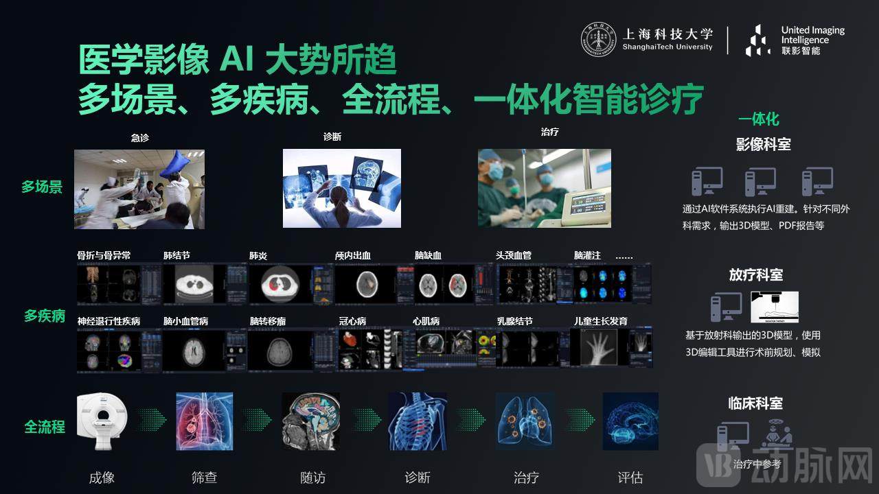
As AI products expand their coverage and become more closely integrated into physicians’ daily workflows, it has become evident that AI applications limited to a single anatomical site or clinical scenario fall far short of meeting hospitals’ real-world needs. Only by implementing multi-scenario, multi-disease, end-to-end, integrated AI-powered intelligent diagnosis and treatment can we usher in a new era of clinical care.
Taking stroke treatment as an example, the traditional stroke care pathway involves complex imaging examinations and requires extensive manual calculation of quantitative metrics, which is time-consuming and labor-intensive. Furthermore, stroke management necessitates multidisciplinary collaboration; however, the lack of an efficient tool for interdepartmental coordination results in low consultation efficiency.
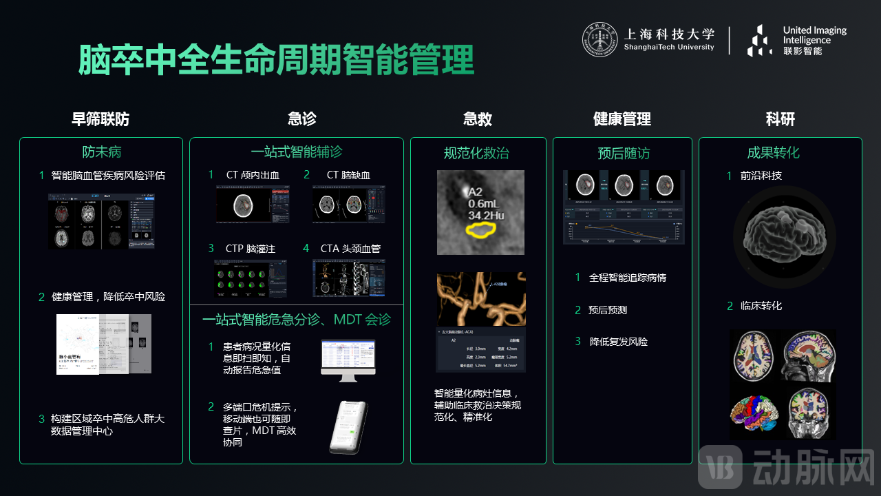
Our intelligent management system for the full lifecycle of stroke care provides support in early screening and collaborative prevention, emergency care, acute treatment, health management, and scientific research. In terms of early screening and collaborative prevention, the AI system helps assess the risk of cerebrovascular disease through early health checkups, identifies high-risk populations, and provides comprehensive brain health reports to help reduce stroke risk. Additionally, AI can be leveraged to establish a big data management center for regional high-risk stroke populations, enabling real-time monitoring and big data analytics.
In emergency settings, AI can provide one-stop intelligent diagnostic assistance by automatically detecting hemorrhage and ischemia based on CT images, and identifying the core infarct zone and culprit vessels from CTP and CTA examinations. Meanwhile, AI facilitates one-stop intelligent triage for critical cases and multidisciplinary team (MDT) consultations by automatically pushing quantified patient condition data to the MDT team, issuing multi-terminal critical alerts, and enabling physicians to review images anytime via mobile devices. As a result, the entire stroke diagnosis process can be completed in as little as five minutes, achieving efficient consultation.
In emergency settings, AI can also provide 3D localization and quantitative information on lesions to support standardized and precise clinical decision-making.
In health management, particularly during prognostic follow-up, AI can provide detailed curves comparing lesion changes, enable intelligent end-to-end tracking, and predict prognosis, thereby helping to reduce the risk of recurrence.
In terms of scientific research, the implementation of such a full-lifecycle intelligent stroke management system will unlock greater potential for frontier scientific exploration and clinical translation.
Apart from stroke, lung cancer is the leading cause of death among the Chinese population. Eighty-five percent of lung cancer cases are already at an advanced stage by the time they are diagnosed. If detected early, the 5-year survival rate for lung cancer patients can increase by at least 60%.
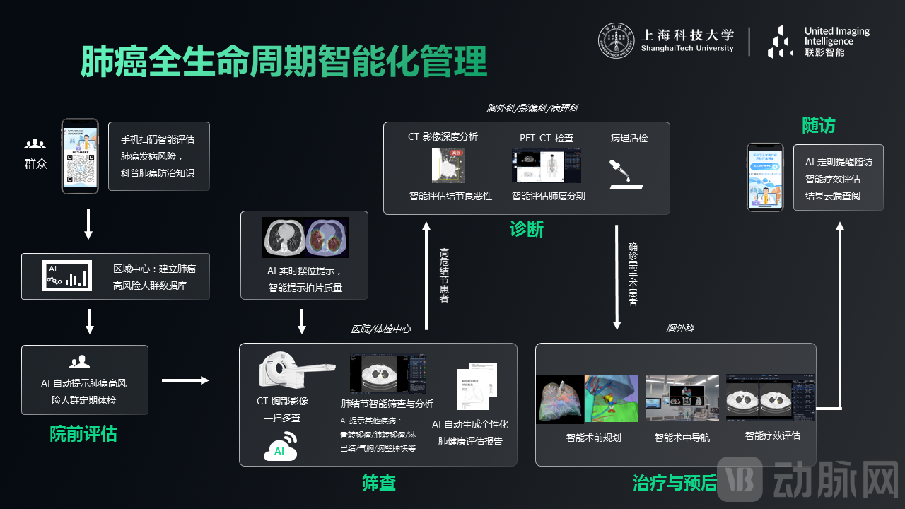
In the face of lung cancer, AI can also build an integrated intelligent health management system for lung cancer patients, covering pre-hospital assessment, screening, diagnosis, treatment and prognosis, and follow-up.
During the pre-hospital assessment phase, AI intelligently evaluates lung cancer risk based on self-reported information provided by users, while also helping to disseminate knowledge about lung cancer prevention and control. District centers can leverage this large-scale intelligent screening to establish a database of high-risk populations for lung cancer, thereby reminding these individuals to undergo regular health check-ups.
During the imaging process, AI can monitor image quality in real time, ensuring standardized imaging quality across different hospitals, health check-up centers, and regions. By leveraging CT scans for “one-scan, multiple-disease screening,” AI aids in detecting conditions other than lung cancer. Upon detection of pulmonary nodules, AI automatically assesses their benign or malignant nature and generates personalized, highly interpretable lung health assessment reports. This reduces communication costs between doctors and patients while encouraging individuals to take greater responsibility for their own health management. For high-risk nodules, AI evaluates lung cancer staging based on further PET-CT examinations.
During the surgical phase, physicians can leverage AI for preoperative simulation and planning, as well as intraoperative surgical navigation. Postoperatively, AI enables regular patient reminders for follow-up visits, intelligent follow-up management, and treatment efficacy assessment.
Collaboration among industry, academia, research, and healthcare is a crucial pathway for advancing cutting-edge technological development.

Since 2020, United Imaging Intelligence has collaborated with the Sun Yat-sen University Cancer Center to develop an AI-assisted detection system for brain metastases based on deep learning. In conjunction with multiple hospitals, a multicenter, multi-reader, prospective validation study was conducted. The AI model was trained and validated using data from 1,000 patients comprising over 10,000 metastatic lesions. The results demonstrated that, with no significant change in false-positive rates, AI assistance increased the average sensitivity of all reading physicians by 21% and reduced the average reading time per case by 40%. This study has been published online in the journal Neuro-Oncology, representing one of the few successful clinical-to-research translation cases globally in the field of brain metastases to date.
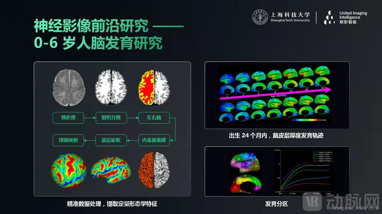
In the frontier of neuroimaging research, we have conducted studies on brain development in children aged 0–6 years, which is crucial for facilitating the early diagnosis of disorders such as autism. Given the substantial differences between infant and adult brain imaging, specialized algorithms are required. After years of research, we have developed a comprehensive suite of AI-based analytical algorithms for precise tissue segmentation, surface reconstruction, and spherical mapping. These tools enable the extraction of quantitative morphological features, such as cortical thickness, with the aim of characterizing the developmental trajectory of cortical thickness during the first 24 months after birth. Furthermore, based on these developmental trajectories, we parcellate brain regions to guide subsequent research on functional brain development.
In the field of brain atlases and neurodegenerative diseases, our team has undertaken the acquisition, quality control, and analysis of multimodal CT/MRI/PET data for the “Chinese Brain Atlas Research Science and Technology Innovation Platform,” contributing to the construction of a brain atlas covering individuals aged 20–80 years. Meanwhile, our team leads the Key Program of the National Natural Science Foundation of China titled “Research on Early Diagnostic Evaluation Models for Alzheimer’s Disease Based on Intelligent Fusion of Multimodal Medical Imaging.” This project leverages AI to accelerate MRI acquisition, learn the representational relationships between MRI, CT, and PET, and achieve effective multimodal fusion for the early diagnosis of Alzheimer’s disease. In clinical practice, by rapidly acquiring MRI scans and utilizing AI-trained models that capture the correlations between MRI and PET/CT, we can synthesize PET/CT images. These synthetic images are then processed by a pre-trained multimodal classifier to enable rapid diagnosis of Alzheimer’s disease, with the goal of completing the entire workflow—from image acquisition to final diagnosis—within minutes.
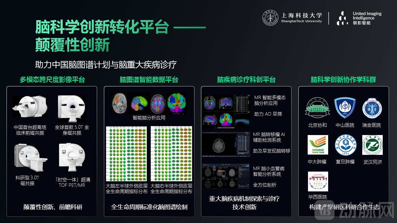
To construct a brain atlas of the Chinese population, we have partnered with multiple top-tier tertiary hospitals in China to establish an innovative translational platform for brain science. Our brain science platform exclusively utilizes high-end, research-oriented multimodal imaging equipment independently developed by United Imaging Healthcare, which has reached internationally leading standards. This equipment provides critical imaging technologies and reference data for animal studies, clinical research, and prospective scientific investigations. Building on high-quality imaging data, we have developed the “Brain Data Analysis Platform” and the “Brain Disease Diagnosis and Treatment Platform” to enable intelligent auxiliary diagnosis of Alzheimer’s disease, brain metastases, and cerebral small vessel disease. Through such collaboration among industry, academia, research, and healthcare institutions, we can effectively integrate the academic, medical, and industrial sectors, fostering an innovation ecosystem characterized by independent innovation and deep synergy.

Just as the universe holds infinite mysteries waiting to be explored, so does the brain. In the future, neuroscience may transcend research papers and computer screens, breaking through dimensional boundaries to realize a neural metaverse.
Currently, we already possess some of the technological foundations necessary to realize the Neuro-Metaverse. For instance, in the diagnosis of brain diseases, AI can automatically detect and segment lesions, perform qualitative and quantitative analyses, and provide reference data for clinical diagnosis. In terms of treatment, preoperatively, AI can offer 3D visualization for surgical planning, enabling one-click, diagnostic-grade 3D reconstruction of lesions and affected organs. It provides comprehensive 3D editing tools to assist physicians in surgical planning and simulation, offering quantitative metrics to help validate treatment strategies and predict prognosis. Intraoperatively, by integrating technologies such as AR and VR, real-time 3D analysis of intraoperative imaging, virtual remote surgical guidance, and remote operation of surgical robots can be achieved. Post-treatment, through AI-based structural quantification and network assessment, AI metabolomics, and AI radiomics, combined with AR and VR technologies, dynamic whole-body evaluations can be conducted. This helps physicians comprehensively monitor treatment outcomes, allows patients to immersively understand their health status, and facilitates better communication between doctors and patients.
With the continuous advancement of medical standards and modern technology, the future of medical AI will undoubtedly feature intelligent diagnosis and treatment that is multi-scenario, multi-disease, end-to-end, and integrated. We hope to see deeper collaboration among industry, academia, research, and clinical practice in the future, jointly driving the development of cutting-edge life sciences technologies.