Pulmongene Founder Dr. Nan Tang: How Foundational Research at NIBS Drives Therapeutic Innovation in Pulmonary Fibrosis
On the official website of the National Institute of Biological Sciences, Beijing, a video in the “Institute News” section highlights the major discoveries made by Dr. Tang Nan’s team in the field of idiopathic pulmonary fibrosis (IPF).
In 2019, the research team led by Dr. Tang Nan published a study online in the journal Cell, which not only established the world’s first mouse model that fully mimics the human disease course but also revealed a novel mechanism underlying the pathogenesis of idiopathic pulmonary fibrosis (IPF) based on this model, marking a milestone achievement in the history of IPF research and therapy.
Prior to the release of this landmark study, Tang Nan had served as a principal investigator at the Beijing Institute of Life Sciences for seven years. On this fertile ground for scientific research, which encourages originality and innovation and advocates the principle of “leading, not following,” Tang was able to fully leverage his expertise in his areas of greatest interest—lung development and lung regeneration—thereby laying a solid scientific foundation for the establishment of Pumu Biotechnology.
Now in its second year, Pumu Biotechnology, an innovative pharmaceutical R&D enterprise incubated by the Beijing Institute of Life Sciences, is dedicated to developing novel therapies for pulmonary diseases and organ fibrosis.

On the path of academic pursuit, Tang Nan has always followed his inner curiosity and passion for science.
Tang Nan pursued her undergraduate studies in clinical medicine. After graduation, she worked as a clinician in the Department of Internal Medicine at the First Affiliated Hospital of Xi’an Jiaotong University for four years—an experience that continues to significantly benefit her research. During her medical training, Tang developed a strong interest in basic scientific research and aspired to establish her own independent laboratory to explore numerous unanswered scientific questions.
Driven by a strong interest in basic scientific research, Tang Nan chose to pursue advanced studies in the United States, where he earned his Ph.D. at the University of California, San Diego (UCSD) under the supervision of Dr. Randall Johnson, focusing on angiogenesis and tumor vasculature.
During her doctoral studies at UCSD, Tang Nan became intrigued by the field of organ regeneration. Given that her dissertation research focused on blood vessels, she raised a critical question: while fabricating solid organs represents merely the first step in regenerative medicine, how can the vasculature that nourishes organ development and ensures proper function be successfully reconstructed? After careful consideration, Tang Nan chose to focus her research on the lungs, thereby embarking on her journey into the study of lung development and regeneration.
To study lung development, Tang Nan chose to pursue her postdoctoral research at the University of California, San Francisco, under the mentorship of Gail Martin. A towering figure in the field of embryonic stem cells, Gail Martin provided substantial support for Tang Nan’s research on lung regeneration and deepened her understanding of pulmonary development.
2012 was a landmark year for pulmonary research. In July of that year, The New England Journal of Medicine (NEJM) published a paper reporting on a British woman with lung cancer. Over the 15 years following surgical resection of one lung lobe, researchers used various imaging techniques to observe the alveoli in her remaining lung lobe and found that the number of alveoli had increased by 64%, accompanied by a significant improvement in lung function.
The emergence of this case has caused a sensation in the field of pulmonary research. Previously, the medical community generally believed that human alveoli were incapable of regeneration. However, for Tang Nan, the conclusion was entirely expected. In previous experimental observations, lung regeneration in mice and dogs had long been commonplace, yet these findings were consistently dismissed as “meaningless research” for human medicine.
Today, she has come to recognize the significance of this work and has clarified her research direction, which is:What Is the Underlying Mechanism That Enables Lung Regeneration After Partial Lobectomy in Humans?
It was in that year that Tang Nan, having completed his postdoctoral research, bid farewell to the University of California, San Francisco (UCSF) and returned to China to join the Beijing Institute of Life Sciences. There, he formally launched his research on alveolar regeneration—a line of inquiry that ultimately gave rise to Pumu Biotechnology.
At the inception of the research team at the Beijing Institute for Stem Cell and Regenerative Medicine, Tang Nan decided to establish a mouse model mimicking lung regeneration following pneumonectomy in humans, to investigate the mechanisms underlying lung regeneration. During this process, she and her team elucidated the origin of alveolar stem cells and their regenerative mechanisms, clarified the distinctions between type I alveolar epithelial cells (AT1) and type II alveolar epithelial cells (AT2), and identified the essential signaling pathways through which AT2 cells, acting as alveolar stem cells, undergo regenerative differentiation.
With a deeper understanding of alveolar regeneration, Tang Nan sought to block alveolar regeneration following lung injury to observe whether manifestations of certain pulmonary diseases would occur. They discovered that the cell cycle regulatory protein Cdc42 plays a crucial role in the process of alveolar regeneration. Alveolar stem cells lacking the Cdc42 gene failed to differentiate into type I alveolar epithelial (AT1) cells after lung injury. However, when they knocked out the Cdc42 gene, an unexpected outcome occurred: instead of the anticipated emphysema-like phenotype in the mouse lungs, a phenotype characteristic of idiopathic pulmonary fibrosis (IPF) emerged.
In the field of pulmonary fibrosis, the traditionally applied mouse model is the bleomycin model. This model requires mice to inhale bleomycin, triggering a severe immune response in the lungs that leads to pulmonary fibrosis. However,The bleomycin model does not represent a true IPF model.Prior to the release of Tang Nan’s research findings, she frequently heard colleagues in the industry complain about the limitations of the bleomycin model.
First, the bleomycin model exhibits strong immunogenicity. Intratracheal administration of bleomycin induces widespread abnormal cell death, triggering infiltration by lymphocytes, macrophages, and monocytes through the activation of a series of immune responses, along with the release of large amounts of cytokines, ultimately leading to pulmonary fibrosis. Its pathogenesis differs significantly from that of idiopathic pulmonary fibrosis (IPF) and does not replicate the characteristic pattern of IPF, which originates in the peripheral lung regions and progressively extends toward the central areas. Second, early pathological diagnosis of IPF reveals specific histopathological changes that are absent in the bleomycin model. Furthermore, the median survival time for humans after an IPF diagnosis is 2 to 4 years; once the disease manifests, it progresses rapidly, with continuous worsening of pulmonary fibrosis. In contrast, the bleomycin model reaches peak fibrosis on day 21, after which pulmonary fibrosis resolves spontaneously, a course inconsistent with the human disease progression.
Although a new method for constructing IPF mouse models has been discovered, it still requires scientific validation and refinement. Tang Nan and her team spent several years,By collaborating with clinicians and pathologists to discuss and compare pathogenic mechanisms, and by leveraging single-cell sequencing to contrast data from AT2 cells in mouse models with those from IPF patients, we ultimately confirmed and established a murine model of idiopathic pulmonary fibrosis (IPF) that faithfully recapitulates the human disease.
Tang Nan still recalls, “At that time, my students even compiled detailed comparative images of all upregulated factors in AT2 cells from IPF mice and those from IPF patients. Seeing such a high degree of similarity was truly exhilarating.”
This academic achievement represents a breakthrough in addressing the lack of animal models for idiopathic pulmonary fibrosis (IPF) and serves as a catalyst to stimulate further research across the field.
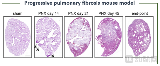
▲ Figure | The spontaneous, progressive pulmonary fibrosis in Pumu’s IPF mouse model closely mirrors the disease progression observed in IPF patients (Source: Wu et al., Cell, 2020; Pumu Biotechnology)
Developing a mouse model of IPF was only the first step. Building on this model, Tang Nan and his team conducted more in-depth research into the pathogenesis of IPF, with the aim of developing more effective therapeutic drugs for this disease.
Leveraging his extensive experience in translational medicine, Tang Nan decided to establish Pumu Biotechnology to embark on the path of new drug development.
Idiopathic pulmonary fibrosis (IPF) is referred to as “cancer that is not cancer.” The median survival time after diagnosis is 2–4 years, and approximately 80% of patients die within five years of diagnosis. Its five-year survival rate is only higher than that of pancreatic cancer and lung cancer. Globally, the incidence of this disease is rising. According to statistics from the World IPF Association, approximately 3.2 million people are living with IPF, with 1.22 million new cases diagnosed each year.
To date, no therapy has been proven effective in halting the progression of IPF or extending patient survival.
As the disease continues to progress, lung transplantation may be considered for suitable candidates. However, the 5-year survival rate after lung transplantation is only 47%–53%, and furthermore, many factors limit the feasibility of lung transplantation. Therefore,The ability to halt or reverse disease progression, extend life expectancy, improve quality of life, and reduce side effects remains the goal of drug development for IPF.
Previously, the pathogenesis of idiopathic pulmonary fibrosis (IPF) remained unclear, posing challenges to the development of therapeutic strategies. However, by analyzing AT2 cell states in IPF mouse models and in patients with IPF, Tang Nan’s team has elucidated its underlying pathogenic mechanism.
In healthy individuals, when lung injury occurs, alveolar type 2 (AT2) cells receive regenerative signals and undergo morphological changes, entering an intermediate state. Simultaneously, they begin to secrete a series of fibrotic factors that recruit downstream cells involved in tissue reconstruction to jointly facilitate injury repair. Once repair is complete, AT2 cells return to a quiescent state. However, in patients with idiopathic pulmonary fibrosis (IPF), the regenerative capacity of AT2 cells is impaired. Upon receiving signals of lung injury, these cells remain prolonged in the intermediate state, continuously secreting fibrotic factors and failing to revert to quiescence. These AT2 cells, persistently locked in the intermediate state, not only fail to achieve normal lung repair but also contribute to the development of IPF by excessively recruiting fibroblasts through overzealous repair efforts.
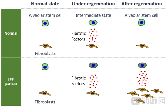
▲ Figure | Professor Tang Nan’s 2020 Cell paper on the pathogenesis of idiopathic pulmonary fibrosis (Source: Wu et al., Cell, 2020; Pumu Biotechnology)
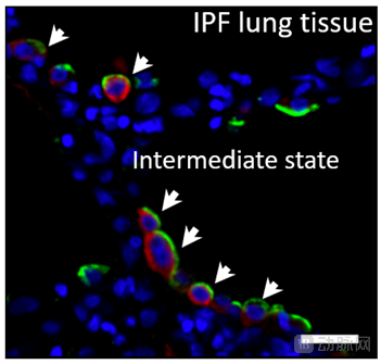
▲ Figure | Alveolar stem cells (AT2) in lung tissue samples from IPF patients are permanently arrested in an intermediate state (Source: Wu et al., Cell, 2020; Pumu Biotechnology)
Based on a clear understanding of the pathogenesis,Pumu Biopharma has developed PMG1015, a first-in-class innovative drug for idiopathic pulmonary fibrosis (IPF). This macromolecular drug features a novel target that is specifically expressed in fibrotic tissues and is virtually absent in healthy adults.
Currently, preclinical toxicology studies and the ongoing Phase I safety trial in healthy volunteers in Australia have both demonstrated the safety profile of PMG1015. Meanwhile, in preclinical pharmacodynamic studies, PMG1015 significantly prolonged survival in a mouse model of idiopathic pulmonary fibrosis (IPF), markedly attenuated weight loss, and reduced the extent of pulmonary fibrosis.
Furthermore, Pumu conducted head-to-head preclinical pharmacodynamic studies comparing PMG1015 with the marketed drug pirfenidone, demonstrating that PMG1015 was significantly superior to pirfenidone in terms of survival benefit and other efficacy endpoints.
Tang Nan revealed,PMG1015 has received implicit clinical trial approval from the CDE, and its clinical trials in China are set to commence shortly.
Entrepreneurship cannot succeed without the support of a team. One thing Tang Nan takes great pride in is the resilient perseverance of Pumu’s founding team.
Pumu Biotechnology has received strong support and guidance from Director Wang Xiaodong since its inception. From the very beginning, the company planned to take PMG1015 global and conduct clinical trials internationally. Due to the COVID-19 pandemic in the United States, Pumu decided to initiate Phase I clinical trials in Australia first. The entire team conducted comprehensive research in Australia, rapidly established an Australian subsidiary, and actively engaged with local CRO companies and investigative hospitals. Meanwhile, through close collaboration with local central laboratories, multiple laboratory testing methods were established. Even when the COVID-19 pandemic later swept through Australia, it did not affect patient enrollment or the progress of the clinical trials.
“In the future, Pumu Biotechnology will continue to focus on pulmonary diseases and fibrotic disorders, developing more innovative drugs that benefit patients. “The fields of pulmonary disease and organ fibrosis are broad enough to warrant in-depth exploration and development. We respect science, remain true to our original mission, and hope to bring truly original Chinese innovations to the international stage,” said Tang Nan.”