RapidAI Files IPO Prospectus After Transforming Stroke Care with AI-Driven Imaging Platform

RapidAI
Stroke and Cerebrovascular Imaging Technology and Product Developer
When AI-assisted diagnostic software first emerged, the expectation was that it would interpret data like a physician. Today, its development appears to have surpassed those initial expectations.
A Decade: From MRI Analysis to Covering the Entire Stroke Care Workflow
RapidAI is an artificial intelligence software that streamlines the stroke care workflow. From pre-hospital emergency services and imaging reports to assisted diagnosis, inter-hospital transfer, and post-operative review, RapidAI is involved in every step of stroke treatment.
Stroke, also known as cerebrovascular accident, is an acute cerebrovascular disease characterized by a group of conditions that cause brain tissue injury due to the sudden rupture or occlusion of cerebral blood vessels, which prevents blood from flowing into the brain. According to the Global Burden of Stroke report published in The Lancet, there were an estimated 12.2 million incident cases of stroke worldwide in 2019, resulting in 6.55 million deaths, accounting for 11.6% of total global deaths.
Stroke diagnosis and treatment guidelines in various countries all emphasize the importance of time. For instance, China’s guidelines repeatedly stress the need to “rapidly identify suspected stroke cases, transport patients to hospitals as soon as possible, and promptly administer thrombolytic therapy or endovascular thrombectomy to eligible patients with acute ischemic stroke.”
Accurate early diagnosis and treatment rely on various imaging technologies, while artificial intelligence excels at analyzing data and extracting image features. RapidAI’s founders, Albers and Bammer, likewise recognized that AI holds great promise in the field of medical imaging.
RapidAI originated from Stanford University. In 2008, Dr. Greg Albers, Roland Bammer, and software engineer Matus Straka established an artificial intelligence group at Stanford University. Funded by Don Valentine, the founder of Sequoia Capital, the group developed Rapid technology for the automated analysis of medical images.
In the second year, the team applied RapidAI technology to MRI image analysis for stroke, developing the RapidMRI software. Upon receiving scan files, the software provides quantitative, color-coded MR diffusion and perfusion maps.
In 2012, the multicenter DEFUSE 3 study related to this software published data indicating that RapidMRI can accurately identify the apparent diffusion coefficient (ADC), thereby helping physicians determine which patients could benefit from thrombolytic therapy within 12 hours of stroke onset. Also in that year, Greg Albers and Roland Bammer acquired the Rapid technology from Stanford University and established iSchemaView, Inc. in the United States, the predecessor of RapidAI.
In 2013, the RapidMRI software received FDA approval, and subsequently obtained CE certification in 2016, becoming the precursor to today’s RapidAI software.
Triage hemorrhagic vs. ischemic stroke; automatically assess ASPECTS score
Stroke is a collective term for a group of diseases, which are further classified into hemorrhagic stroke and ischemic stroke. Hemostasis is administered to patients with hemorrhagic stroke, while vascular recanalization therapy is provided to those with ischemic stroke.
Hemorrhagic stroke is further classified into subarachnoid hemorrhage, subdural hemorrhage, and epidural hemorrhage based on the bleeding location; ischemic stroke is categorized into large vessel occlusion (LVO) and non-large vessel occlusion. Currently, among AI-assisted diagnostic tools for stroke, the most rapidly advancing area is LVO ischemic stroke.
The management of stroke is, in essence, a continuous triage process; stroke centers in many countries are classified into primary and advanced levels, with referral mechanisms established between the two tiers.
According to the guidelines for stroke prevention and treatment, the first step in managing a patient with suspected stroke is to determine whether the condition is hemorrhagic or ischemic, which can be accomplished rapidly via non-contrast computed tomography (CT). Hemorrhagic lesions are particularly conspicuous on CT scans.
In 2020, RapidAI launched an ICH (hemorrhagic stroke) assisted diagnosis module. This platform can identify hemorrhagic stroke within minutes of receiving CT scan data, and precisely differentiate between subarachnoid hemorrhage, subdural hemorrhage, and epidural hemorrhage. According to RapidAI, the module has a sensitivity of 95%.
Non-contrast CT can also yield a key metric, the “Alberta Stroke Program Early CT Score” (ASPECTS), which reflects the extent of the core infarct and helps predict the efficacy of intravenous thrombolysis.
RapidAI Launches RapidASPECTS for Automated ASPECTS Scoring; According to RapidAI, the Software Improves Physicians’ Accuracy in Assessing ASPECTS Scores.
Accurate Assessment of the Tissue Window, 97% Diagnostic Accuracy for LVO
There is a saying in the stroke care industry: “time is brain.” Once it is determined whether a patient has hemorrhagic or ischemic stroke, they must be rushed to the appropriate department without delay.
RapidAI Develops Prehospital Communication Solution, Rapid for Prehospital, to Notify Team Members of Results and Patient Data Within Three Minutes for Rapid Response. Team Members Can Choose Alert Types—Receiving Notifications Only for Confirmed Diagnoses or for All Patients.
After excluding hemorrhagic stroke, ischemic stroke is further classified into transient ischemic attack (TIA), large vessel occlusion (LVO), and others. Among these, TIA is reversible, presents with mild symptoms, and has a low level of urgency, whereas LVO is highly urgent.
For patients with a high suspicion of large vessel occlusion, imaging should be performed as rapidly as possible.
China’s Guidelines for Stroke Prevention and Treatment strongly recommend the “one-stop CT imaging protocol” as the first-line imaging modality for acute ischemic stroke. This approach is not only rapid but also enables accurate assessment of the core infarct zone, the culprit vessel (including identification of the specific vessel and whether it is occluded), and the ischemic penumbra.
“One-Stop CT Protocol” consists of two examinations: CTA and CTP. CTA is the preferred method for identifying large vessel occlusion, while CTP facilitates “tissue window” assessment.
RapidAI has developed three platforms accordingly: RapidCTA, RapidCTP, and RapidLVO.
It is well known that reperfusion therapy for ischemic stroke has a "time window." Patients with indications for thrombolysis or thrombectomy within this time window should receive treatment as soon as possible, typically within 4.5 hours for intravenous thrombolysis and within 6 hours for mechanical thrombectomy. However, there is a group of patients who present beyond the time window but exhibit slow infarct progression, a small irreversible core infarct area, and a large ischemic penumbra with salvageable tissue. These patients can still benefit from reperfusion therapy, a concept referred to as the "tissue window."
In 2018, the results of the DAWN and DEFUSE 3 trials demonstrated that the use of tissue windows could extend the time window for mechanical thrombectomy in patients with anterior circulation large vessel occlusion to 16–24 hours. The American Heart Association and the American Stroke Association have also extended the treatment window for patients with acute stroke from 6 hours to 24 hours.
RapidAI introduces RapidCTP, the exclusive software designated post hoc in the DAWN and DEFUSE 3 trials, and one of the few software solutions available in China that enables automated imaging-based assessment of the tissue window.
Within two minutes of receiving raw data, RapidCTP delivers clearly color-coded CTP images and the ischemic penumbra area to the stroke team via PACS, email, and the Rapid mobile app, while assessing whether the patient is likely to benefit from reperfusion therapy.
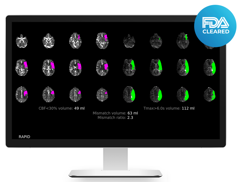
RapidCTP Interface
RapidCTA provides physicians with 2D and 3D CTA images by analyzing raw scan data. In fact, experienced physicians can identify patients with large vessel occlusion (LVO) based on CTA images alone; however, RapidAI has still developed the RapidLVO module. RapidLVO rapidly detects LVOs in the internal carotid artery (ICA) and the M1 segment of the middle cerebral artery (MCA), and highlights the relevant vascular segments.
According to RapidAI, this module has a sensitivity of 97% and a specificity of 96%.
Time from ER Arrival to Operating Room Reduced by 52 Minutes; Total Scans Exceed 2.8 Million
As mentioned above, stroke care systems in various countries are categorized into primary, advanced, and comprehensive stroke centers. When a patient is diagnosed with large vessel occlusion stroke, they are generally referred to a higher-level stroke center with mechanical thrombectomy capabilities.
At this point, the prehospital protocol, Rapid for Prehospital, will advise the physician on the optimal stroke center for patient referral and transmit the completed test results to the receiving physician.
After undergoing a series of imaging examinations, patients can initiate reperfusion therapy upon arrival at an Advanced Stroke Center. A new one-stop assessment tool is emerging abroad, whereby patients with high suspicion of large vessel occlusion bypass the traditional imaging workflow and are transferred directly to the catheterization laboratory for CTP assessment, including CBCTP.
RapidAI has developed the Rapid for Angio module for CBCTP examinations, providing color-coded CBCT perfusion imaging to help physicians identify the infarct core and areas at risk of progressing to infarction.
According to RapidAI, using the full suite of the RapidAI platform can reduce the time from a patient’s arrival in the emergency department to transfer to the operating room by up to 52 minutes.
RapidAI has also developed the auxiliary platforms Rapid Workflow for Stroke and RapidAI Insights. On the former platform, physicians can review all patient clinical data, communicate with referring physicians, and access summary reports after the patient undergoes surgery.
All RapidAI products are provided with access to RapidAI Insights, a dashboard that helps administrators control access and manage permissions, covering clinical imaging data, patient volume statistics, user logins, training, and more.
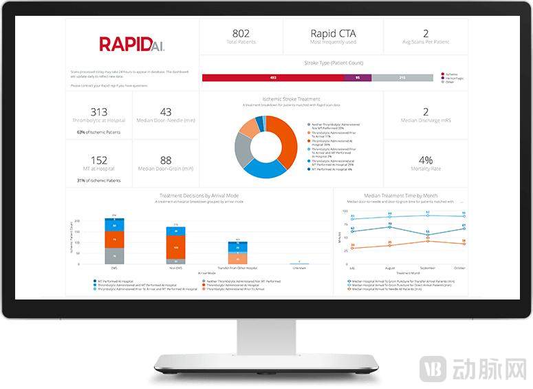
RapidAI Insights Interface
RapidAI has currently developed an intelligent assistance solution that covers the entire workflow of stroke care. According to RapidAI, the platform is used in more than 1,800 hospitals across over 60 countries and regions worldwide, including 18 of the top 20 hospitals in the United States, with a total scan volume exceeding 2.8 million.
RapidAI plans to expand this model to other cardiovascular diseases. In early 2020, RapidAI acquired Endovantage, a provider of aneurysm management products, and its auxiliary diagnostic modules for aneurysms and pulmonary embolism have now received FDA approval.
The Imaging AI Industry Is Developing Rapidly, with a Flourishing Landscape in the Stroke Field
With the advancement of medical and AI technologies, the “cross-sector integration” of artificial intelligence and healthcare is no longer novel, with numerous products already approved both in China and abroad. As of early March, coinciding with the release of the landmark AI review and approval policy, the Guiding Principles for Registration Review of Artificial Intelligence Medical Devices, a total of 33 Class III AI medical devices had received approval from the National Medical Products Administration (NMPA) of China.
Within the broader medical AI sector, imaging AI has seen the most rapid growth. Among the 33 approved certificates, approximately 20 are for imaging AI products. The first Class III medical device certificate for an imaging AI product issued by China’s National Medical Products Administration (NMPA) was granted in January 2020.
First, medical imaging databases are vast, with 80% of healthcare big data derived from imaging data; second, artificial intelligence excels at extracting image features. The global market size for AI in medical imaging is projected to exceed RMB 280 billion by 2027. Among AI-assisted diagnostic imaging applications, electrocardiogram (ECG) and computed tomography (CT) are the most prominent.
Stroke is the leading cause of death and disability among adults in China. In this field, imaging AI has evolved into various application types, including assisted diagnosis, lesion quantification analysis, and treatment efficacy assessment.