Centerline Biomedical Secures $33M Series B Funding to Advance AR-Guided, Low-Radiation Surgical Navigation Platform

Centerline Biomedical
Developer of Vascular Surgical Navigation Systems
The latest wave of AR/VR fever can be traced back to the metaverse.
The Metaverse, a neologism referring to a virtual world, denotes a parallel virtual realm created through technological means. In the second half of last year, Mark Zuckerberg, CEO of Facebook, announced the company’s entry into the metaverse, sparking widespread online discussion about this term borrowed from science fiction. Whether the metaverse represents mere conceptual hype or an actual trend remains debatable; however, the AR/VR industry, which serves as the foundational technology for the metaverse, has indeed seen a significant surge in popularity.
In the past few years, the AR/VR industry has seen a slump in capital market activity, but it “once enjoyed considerable prosperity.” In 2012, Google launched Google Glass, bringing AR/VR technology into the public spotlight. Subsequently, major tech companies such as Microsoft entered the fray. The sector continued to develop until it reached its first boom in 2016, with a total of 203 transactions recorded in the field that year.
But unexpectedly, it peaked at its debut. In 2017, the AR/VR market took a sharp downturn, and its prominence in the healthcare sector was surpassed by emerging technologies such as artificial intelligence and robotics.
Looking back at 2016, the hot projects in AR/VR healthcare were concentrated in four areas: personalized fitness, education and training, clinical assistance, and psychological disorders. With technological advancements and market changes, the surgical department has become the new demand driving the AR/VR healthcare market.
Traditional Interventional X-ray Radiation Is “Unavoidable”; Centerline Develops AR Surgical Navigation System
Centerline Biomedical is a medical technology company developing augmented reality (AR) imaging-guided interventional navigation solutions. Founded by Vikash Goel in 2014, the company is headquartered in Cleveland, Ohio, USA. Its flagship product, the IOPS System, received FDA approval in July 2019 for intraoperative AR navigation during endovascular aneurysm repair (EVAR) procedures.
Image guidance plays a crucial role in interventional procedures. Currently, the vast majority of interventional therapies rely on freehand puncture, akin to driving a car along an unfamiliar and complex route. Just as automobiles require GPS systems that display maps and vehicle positioning, interventional surgeries necessitate various methods to visualize patient anatomy and locate the position of surgical instruments.
Currently, the most frequently used image guidance modalities in interventional procedures include digital subtraction angiography (DSA), ultrasound, MRI, and CT.
Among these, digital subtraction angiography (DSA) is frequently used but involves the highest level of X-ray radiation exposure; ultrasound guidance is cost-effective and real-time, requiring no contrast agents and involving no radiation, but it offers lower image clarity and has blind spots in the field of view; magnetic resonance imaging (MRI) provides clear visualization but is expensive; computed tomography (CT) guidance is cost-effective and yields clear images, but it involves X-ray radiation and time delays.
X-ray radiation is a common occupational hazard for medical staff in interventional departments. Healthcare professionals in these departments are routinely exposed to excessive levels of X-ray radiation during their daily work. Short-term effects may include headaches, dizziness, and compromised immunity; prolonged exposure can lead to adverse impacts on hematopoietic, immune, and nervous systems. For instance, Wilhelm Röntgen, the discoverer of X-rays, died of cancer.
To reduce the “unavoidable” X-ray radiation exposure in traditional interventional procedures, Vikash Goel founded Centerline Biomedical to develop a low-radiation intraoperative navigation system—a venture rooted in a poignant story.
While studying in the Department of Computer Science at Cornell University, Vikash Goel met Dr. Roy Greenberg, the son of his advisor and an aortic repair specialist at the Cleveland Clinic. Sharing common interests, Dr. Greenberg invited Goel to participate in a Cleveland Clinic project focused on interventional image navigation using IOPS technology. The two collaborated for the next decade until Dr. Greenberg passed away from cancer, just as the IOPS technology was on the verge of its breakthrough.
Many people believe that Roy Greenberg developed cancer due to prolonged exposure to excessive X-rays. In 2014, Vikash founded Centerline Biomedical to carry on his late friend’s legacy, dedicating the company to reducing radiation exposure in interventional procedures and advancing intraoperative navigation imaging.
In 2016, Centerline Biomedical conducted its first preclinical study, demonstrating that internal structures could be visualized via a surgical navigation system even without fluoroscopy.
3D Color Imaging + Electromagnetic Positioning Technology for Low-Radiation, Contrast-Free Intraoperative Navigation
In 2019, the FDA approved the IOPS system for augmented reality (AR) navigation during endovascular aneurysm repair (EVAR) of aortic aneurysms.
Endovascular Aneurysm Repair (EVAR) is a commonly used method for treating abdominal aortic aneurysms (AAA). An abdominal aortic aneurysm is generally defined as an abdominal aortic diameter ≥ 3 cm. The main steps of EVAR involve inserting a catheter through the femoral or iliac artery, followed by the deployment of one or more collapsible stents into the AAA under digital subtraction angiography (DSA) guidance. The stent(s) span the aneurysm proximally and distally, thereby preventing blood flow into the aneurysmal sac and ultimately leading to its thrombosis and exclusion.
The X-ray radiation from a single interventional procedure has minimal impact on the average patient; however, an EVAR procedure typically requires approximately 150 mL of contrast agent, which can be life-threatening for patients with severe contrast allergy.
IOPS technology eliminates traditional DSA navigation by converting preoperative CT angiography images into 3D color images, which are then displayed on monitors in the operating room.
3D imaging reconstruction allows physicians to visualize a patient’s anatomical structures through the skin. But how is the position of surgical instruments determined? Current localization technologies include mechanical, optical, and electromagnetic tracking. Mechanical tracking, the earliest developed, offers limited accuracy. Optical tracking relies on surgical instruments equipped with markers and is widely used in surgery. Electromagnetic tracking, the most recently introduced technology, provides unique advantages in minimally invasive interventional procedures.
The IOPS® system employs electromagnetic tracking technology. An electromagnetic field generator is placed beneath the operating table, and surgical instruments are equipped with magnetic field positioning sensors. The magnetic field induces electrical signals in the sensors; by co-registering these signals with imaging data, the trajectory of the needle can be determined. To minimize errors, the IOPS® system utilizes a localization calibration patch placed on the patient’s lower back.
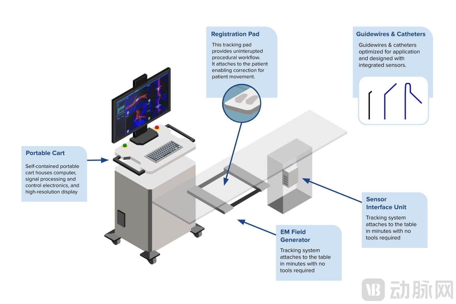 Schematic Diagram of the IOPS® System (Image Source: Official Website)
According to the Centerline Biomedical website, the IOPS® system reduces puncture time to just 53.4% of that required by traditional 2D navigation. Furthermore, the IOPS® system utilizes 3D color imaging with real-time needle tracking, offering superior performance compared to conventional 2D grayscale imaging.
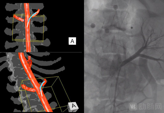
IOPS® System vs. Traditional 2D Imaging (Image source: official website)
In April 2020, IOPS® was used in clinics for the first time after receiving approval, marking the beginning of its commercialization journey. Centerline Biomedical offers three payment options: a fixed monthly fee, installment payments post-device usage, and customized payment plans.
Upcoming Launch of Two New LOPS Technology Applications: Peripheral Vascular Disease and Structural Heart Disease
In addition to EVAR, Centerline Biomedical has four R&D pipelines: peripheral vascular disease, structural heart disease, neurovascular disease, and endovascular & extravascular applications, with the first two already launched.
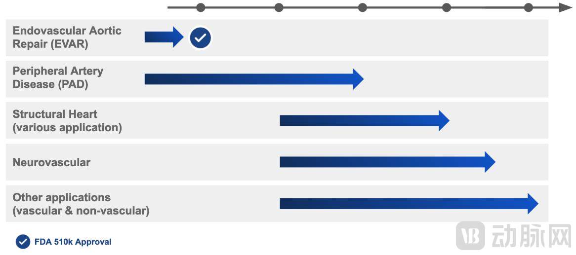
Centerline Biomedical’s R&D Pipeline (Image source: official website)
Centerline Biomedical recently completed a feasibility animal study on the use of AR technology for navigation in cardiac surgery. The company has named its new AR technology 4D-GNC, which builds upon its IOPS® product and utilizes four-dimensional structural mapping with augmented reality guidance through the heart.
In fact, Centerline Biomedical’s focus on structural heart disease dates back to 2018.
In early 2018, Centerline received an NIH/STTR Phase I grant to leverage HoloLens® AR technology for aortic aneurysm repair surgery (to date, HoloLens® has not been FDA-approved for use with the IOPS® system). In August 2018, it secured another NIH grant focused on researching the application of IOPS technology in heart valve replacement, while further developing its AR technology.
Founder Vikash Goel believes that structural heart disease represents a multi-billion-dollar supermarket and is one of the fastest-growing segments in healthcare. A surgical navigation system with low radiation exposure and precise imaging would simplify complex structural heart disease procedures.
Centerline Biomedical Announces Completion of $33 Million Series B FinancingRecently, Centerline Biomedical announced the completion of a $33 million Series B financing round. The round was led by Cleveland Clinic, with participation from GE Healthcare, RIK Enterprises, JobsOhio, Jumpstart Ventures, and G2 Group Ventures. Notably, GE Healthcare also participated in this round as part of its strategic expansion into medical AR/VR solutions.
According to Centerline Biomedical, this round of financing will be used for the development of new products, accelerate the commercial sales of approved products, and expand the clinical evidence database.
Rapid Development of AR Surgical Navigation with Multiple Subfields Emerging
AR technology initially existed only in people’s descriptions: images hovering in mid-air before an empty space, changing with a simple wave of the hand. As technology has advanced, AR has become a reality and is now applied across various industries. It is also thanks to the widespread adoption of AR/VR technologies that the concept of the metaverse no longer sounds far-fetched.
According to Global Market Insights, the global market size for the medical AR/VR industry exceeded USD 1.9 billion in 2020 and is projected to grow at a compound annual growth rate (CAGR) of over 35.8% in the coming years, with the surgical segment— including surgical navigation systems—serving as the primary growth driver.
Surgical navigation systems have undergone rapid development over the past two decades. Computed tomography (CT), ultrasound, X-ray, and magnetic resonance imaging (MRI) are all key technologies in surgical navigation systems, and augmented reality (AR) technology has also secured a place in these systems today.
Currently, AR surgical navigation display technologies fall into the following categories:
First, there is direct video display output, where anatomical structures are reconstructed in 3D to generate images that are overlaid onto other scenes and displayed within the video feed; this is currently the most mainstream AR display method. It is categorized into fixed displays and handheld mobile displays, with the IOPS® system introduced in this article being a typical example of a fixed display. Building upon standard video display, 3D video has also been developed, requiring physicians to wear 3D glasses for viewing. This approach generally necessitates a dimly lit operating room environment and is therefore commonly used in endoscopic surgeries.
Second, stereoscopic display. After all, no matter how clear a video is, it cannot match the clarity of human vision. Stereoscopic display technology directly overlays 3D reconstructed images onto the surgeon’s field of view. Stereoscopic displays fall into two categories: translucent screens and see-through headsets. For example, the surgical navigation system developed by HoloSurgical in the United States uses a translucent screen, allowing surgeons to view the surgical site with the naked eye through the handheld screen while 3D images are displayed on it. Microsoft’s HoloLens see-through headset is commonly used for AR surgical navigation; for instance, Novarad’s VisAR, an AR surgical navigation system recently approved by the FDA, utilizes the Microsoft HoloLens 2 headset to guide spinal surgeries.
Finally, there is direct-projection AR display, where 3D images are projected directly onto the human body.
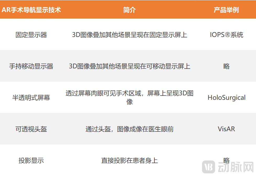 AR Surgical Navigation Display Technology
It is foreseeable that surgical navigation technology will continue to advance in the future, particularly in the field of interventional therapy, not only improving accuracy and work efficiency but also reducing X-ray radiation exposure and complications associated with contrast agents.