Digital Pathology: The Preferred Solution for High-Resolution Imaging and Multifunctional Demands in Scientific Research
Digital pathology is an emerging growth force in the field of pathological diagnosis.
According to the report from the 2020 World Congress of Pathology, the pathology market size is projected to grow from $30.3 billion in 2019 to $44.4 billion in 2024, representing a compound annual growth rate (CAGR) of nearly 6.1% from 2019 to 2024. Meanwhile, Grand View Research estimates that the global digital pathology market size was $767.6 million in 2019 and is expected to grow at a CAGR of 11.8% through 2027.
Currently, clinical pathology faces bottlenecks such as a significant shortage of pathologists, prolonged diagnostic turnaround times, and the need to improve diagnostic efficiency and accuracy. Digital pathology integrates digital imaging technology with traditional optical magnification devices by scanning physical slides into digital images. This transformation makes slide review more convenient and efficient, laying the foundation for remote consultations and digital storage of slides, thereby driving a leap forward in clinical pathological diagnosis. At present, the widespread application and importance of digital pathology in clinical practice have been fully recognized.
In fact, the applications of digital pathology extend far beyond clinical practice, with widespread use in scientific research, education, drug development, and disease control and prevention. Examples include pharmaceutical development and new drug safety assessment, toxicological and physiological studies, research on animals, plants, and minerals, disease control and epidemic prevention, and forensic pathology. Among these,Compared to clinical applications, digital pathology systems have generated relatively less discussion in the research sector, representing an untapped blue-ocean market.
Recently, VCBeat held discussions with Zhang Yueying, Director of Scientific Research in the Department of Pathophysiology at Shandong First Medical University, and Qiu Zhu, a researcher at the Molecular Oncology and Epigenetics Laboratory of the First Affiliated Hospital of Chongqing Medical University. Drawing on their practical research experiences, these conversations shed light on the robust and differentiated demands for digital pathology systems within the scientific research community.
Zhang Yueying, Associate Researcher and Associate Professor at the School of Clinical and Basic Medical Sciences, Shandong First Medical University; Director of Scientific Research in the Department of Pathophysiology; Master’s Supervisor in Pathology and Pathophysiology. She specializes in experimental pathology, with a focus on cancer stem cells and mechanisms of chemotherapy resistance, as well as experimental mesenchymal stem cell therapies for refractory pulmonary diseases. Dr. Zhang employs experimental pathological approaches to investigate the regulation of cancer stem cell stemness and the mechanisms underlying chemotherapy resistance from multiple perspectives, including exosomal miRNAs and the tumor microenvironment. She conducts experimental therapeutic studies using bone marrow-derived and umbilical cord-derived mesenchymal stem cells for radiation-induced lung injury caused by tumor radiotherapy, and carries out translational research targeting relevant molecular markers for disease diagnosis and treatment. Her major research findings have been published in internationally renowned journals in the field of stem cell research, such as Stem Cell Research & Therapy. She has undertaken 15 national-, provincial-, and ministerial-level scientific research projects, authored more than 30 academic papers, and received one Second Prize and one Third Prize of the Shandong Provincial Science and Technology Progress Award as a principal contributor.
Qiu Zhu, a faculty member at the Laboratory of Molecular Oncology and Epigenetics at the First Affiliated Hospital of Chongqing Medical University, has published numerous journal articles. He participated in the Chongqing Municipal Science and Technology Commission’s Special Project for Technological Innovation in Social Undertakings and People’s Livelihood Security, titled “Application and Promotion of Gut Microbiota Screening and Gene Microarray Analysis Technology in Liver Cancer Detection,” and was awarded the Second Prize of Chongqing Science and Technology Progress Award. The Laboratory of Molecular Oncology and Epigenetics at the First Affiliated Hospital of Chongqing Medical University, where Qiu Zhu works, was among the earliest institutions in Southwest China to establish a biobank. It currently stores more than 200,000 tumor samples. Leveraging this biobank, the laboratory publishes over 20 SCI-indexed papers annually. Around 2016, the laboratory began constructing a pathological sample bank, which now holds nearly 6,000 cases.
In scientific research, efficiency, convenience, and accuracy are paramount.
Previously, whether conducting research on cancer stem cells or mesenchymal stem cells, establishing pathological sample banks, or performing tumor pathology studies, all steps involving pathological experiments relied on the use of microscopes. Recalling this period, Professor Qiu Zhu stated, “In the past, building biobanks and carrying out pathological research were rather cumbersome processes. Taking immunohistochemistry as an example, observations could only be made under an optical microscope, after which researchers would use a camera to photograph the areas they deemed to meet experimental requirements.”
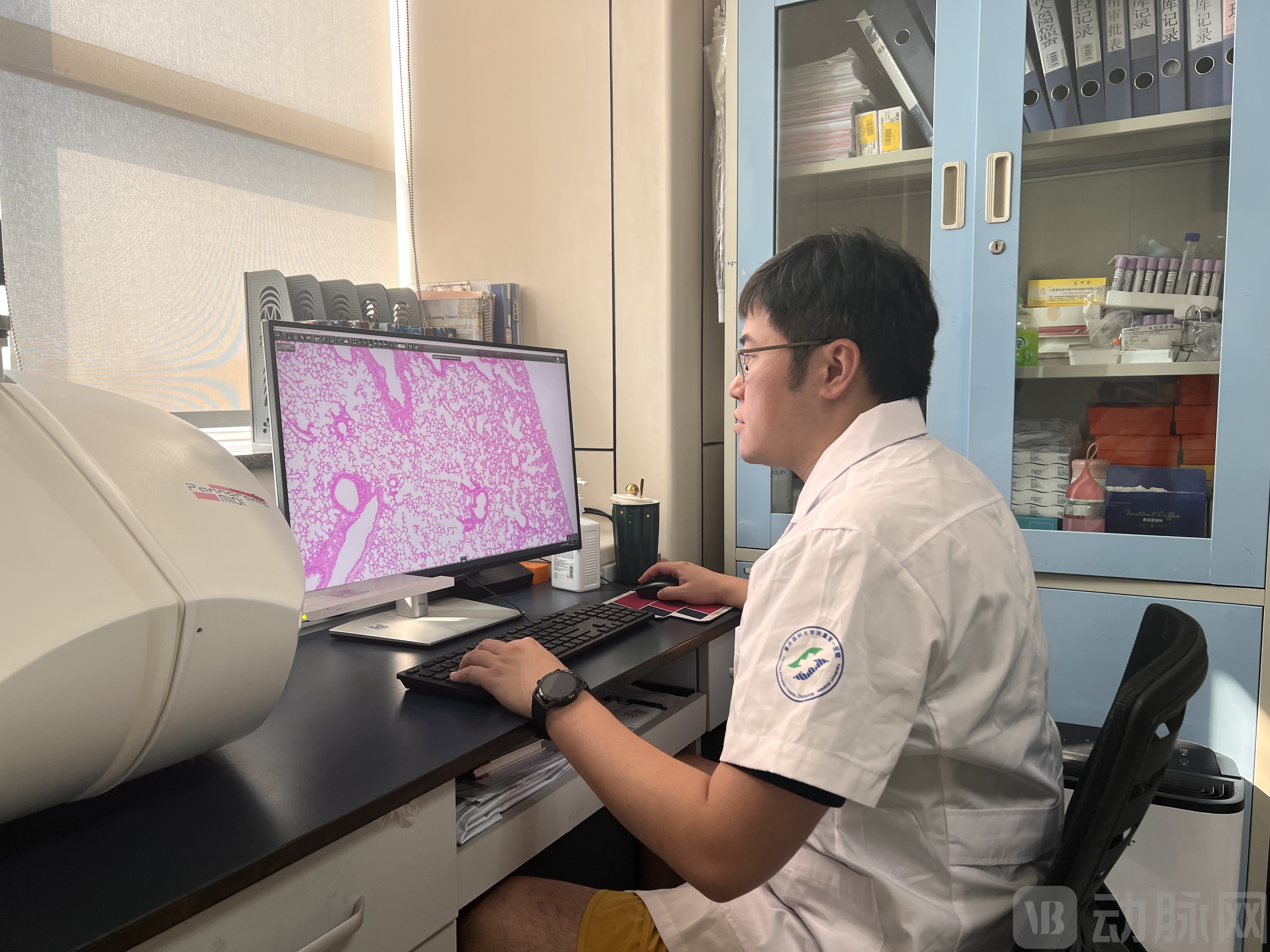
Teacher Qiu Zhu
“There are significant issues involved. For instance, not every student conducting pathological experiments specializes in pathology; they may struggle to accurately identify sites for tissue sampling and photography, easily confusing tumor tissues with normal ones. This leads to inaccurate imaging locations and low clarity, ultimately resulting in suboptimal experimental data.”
Director Zhang Yueying also stated, “In traditional formats, a single tissue section contains one tissue sample. To study 100 samples, 100 separate sections are required, followed by antibody application and validation for each of the 100 sections. This process is time-consuming and labor-intensive, and often leads to poor consistency in results.”
Digital pathology systems are a powerful tool for enhancing the efficiency and convenience of scientific research.
Leveraging digital pathology systems, slide review becomes more convenient and efficient with clearer images, while enabling the permanent digital preservation of massive datasets to facilitate subsequent retrospective or further analyses.“Previously, it was not uncommon for slides to be misplaced or damaged. Meanwhile, we frequently needed to transport tissue sections to the pathology department for analysis. In the past, this involved carrying multiple large boxes of slides and manually recording details in a notebook, resulting in a rather chaotic process.”Now, all it takes is bringing a computer to view high-resolution digital scanned slides and annotate them, significantly boosting work efficiency.“Teacher Qiu Zhu said.
Director Zhang Yueying employed the 3DHISTECH automated tissue microarray (TMA) system for experimental research using clinical samples, constructing tissue microarrays containing hundreds of specimens. By selecting representative cores with diameters ranging from 1 mm to 1.5 mm from each sample and arranging multiple samples on a single slide, she achieved high efficiency and exceptional convenience, while ensuring excellent consistency in results.

Director Zhang Yueying
Furthermore, performance is also a core focus of scientific research. Professor Qiu Zhu stated, “Since the introduction of the 3DHISTECH digital pathology slide scanner, the equipment has been in near-continuous operation. Some students need to scan hundreds or even thousands of slides during their experiments, leading to long queues; typically, reservations are booked up to one month in advance.” Under such high-workload conditions, the 3DHISTECH digital pathology slide scanner has demonstrated excellent performance.
Director Zhang Yueying and Teacher Qiu Zhu both mentioned,Compared to other application scenarios, scientific research imposes higher requirements on the resolution of digital pathology systems, necessitating a high-fidelity reproduction of the actual microscopic images.
Director Zhang Yueying noted that publishing articles is a critical task for researchers, and reputable journals impose stringent requirements on image quality. Professor Qiu Zhu also stated, “When submitting manuscripts, even if experimental results are successfully obtained, experts often question images with poor clarity from histological sections. High-resolution section images are akin to neat handwriting in a language exam—they significantly enhance the presentation of research findings.”
Optical microscopes are constrained by the diffraction limit of optical resolution, with a maximum imaging resolution of only around 200 nm. This limitation has become increasingly inadequate for advancing scientific research. Professor Qiu Zhu stated, “Previously, when using cameras for photography, we could only capture local regions, and the resulting images were two-dimensional with low clarity upon magnification. For instance, in tumor cell staining, unclear sections often led to misinterpretation of areas with faint or ambiguous coloration. In contrast, tissue sections scanned with the 3DHISTECH digital pathology slide scanner provide clear visualization of image details at both 20x and 40x magnifications. Furthermore, the accompanying software features automatic recognition capabilities, enabling precise identification.”
The secret to the 3DHISTECH digital pathology slide scanner’s ability to maintain high resolution lies in its incorporation of two APO objectives: a 20× objective with an NA of 0.8 and a 40× objective with an NA of 0.95.All are at the highest level within their respective tiers., ensuring that the scanned images are consistent with the color and authenticity observed under the microscope, thereby meeting the requirements for scientific research and subsequent publication.
In addition to high resolution, another specific requirement of scientific research for digital pathology systems is reflected in the complexity of required functionalities.
If a digital pathology system offers only basic digital slide scanning capabilities, it will fall far short of meeting the complex demands of scientific research. For instance, in the process of establishing a pathology sample bank and conducting pathological research, Professor Qiu Zhu requires the scanning of a large volume of brightfield H&E-stained slides and fluorescence immunohistochemistry slides.
Director Zhang Yueying also stated, “In research on cancer stem cells and mesenchymal stem cells, we need to perform complex analyses such as immunofluorescence and multicolor fluorescence, as well as quantitative analysis of certain markers. This requires digital pathology systems to offer additional functionalities, including bright-field H&E staining, immunohistochemistry, immunofluorescence, and FISH fluorescence scanning.”
3DHISTECH Digital Pathology Slide Scanners can scan various types of slides, meeting the diverse needs of scientific research., the diversity and customizability of its analytical functions have significantly expanded the breadth of product applications. In 2021, 3DHISTECH obtained an import registration certificate for pathological slide scanners from the National Medical Products Administration (NMPA). This marks the world’s first digital pathology product with fluorescence scanning capabilities approved by a national regulatory authority, enabling integrated brightfield and fluorescence scanning.
Overall, 3DHISTECH’s digital pathology system has left a deep impression on many researchers with its performance in precision, resolution, clarity, scanning speed, after-sales service, and digital analysis software.
Director Zhang Yueying concluded, “The era of rapid development in digital pathology has arrived. The application of digital pathology in the research field is extensive, not limited to pathological research laboratories.”Digital pathology is like a soldier’s firearms; it has become standard equipment for any major research group or scientific platform."Against this backdrop, Tangier will deepen and broaden its strategic layout in the field of scientific research."
Regarding the further application of digital pathology in scientific research, Director Zhang Yueying pointed out that AI-assisted diagnosis represents a promising direction, one that will bring revolutionary innovation to pathological research and teaching by more intelligently supporting both research and clinical auxiliary diagnosis. She also expressed hope that future digital pathology systems will feature lower costs, higher image quality, faster scanning speeds, and greater intelligence and user-friendliness.
Professor Qiu Zhu expressed his expectations for the hospital’s digital pathology local area network (LAN). “The hospital where I work is divided into three sectors: scientific research, clinical practice, and pathology. We hope to integrate digital pathology with the hospital’s LAN to strengthen in-depth communication among these three departments regarding pathological issues. For instance, rare and complex diseases have long posed challenges in clinical diagnosis and treatment. By leveraging the digital pathology LAN, researchers, clinicians, and pathologists can collaboratively analyze such cases online. Through close cooperation among the three departments, we can issue more professional diagnostic reports and improve the diagnosis and treatment of rare and complex diseases.”