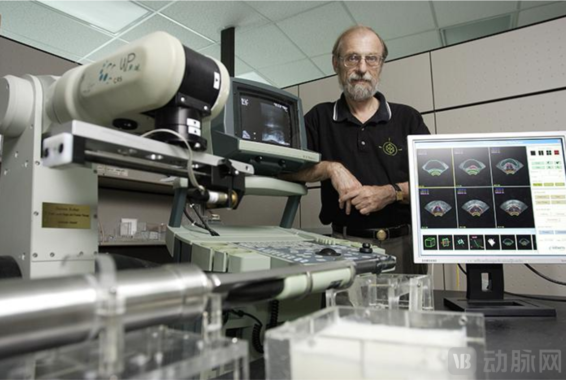
Exclusive Interview with Dr. Aaron Fenster, Fellow of the Canadian Academy of Health Sciences: How His Cost-Effective and Precise 3D Ultrasound Systems Entered a Billion-Dollar Market — 42 Patents, 13 Licensees, and Recently Filed IPO Prospectus
In 1942, K. T. Dussik of Austria first used an A-mode ultrasound imaging system to perform transcranial penetration detection, officially inaugurating the application of ultrasound imaging technology in the medical field.
Today, ultrasound medical imaging has traversed an 80-year journey, progressing through A-mode, B-mode, M-mode, and Doppler ultrasound phases. Although the field has entered a mature stage, the market remains highly dynamic, with ultrasound imaging advancing toward new technologies such as three-dimensional (3D) imaging, CT/MR image fusion, and deep learning.
According to Grand View Research, the global market size for ultrasound equipment was $7.9 billion in 2021 and is projected to grow from 2022 to 2030Growing at a compound annual growth rate of 4.5%。
In fact, the continuous expansion of the ultrasound market is attributable to the rising adoption rate of ultrasound equipment for diagnosis and treatment, coupled with the growing number of patients with chronic diseases. Furthermore, the increasing demand for minimally invasive procedures and advancements in ultrasound imaging technology are also key drivers propelling the development of the ultrasound market.
As a new technology in the field of ultrasound—The Inventor of 3D UltrasoundandThe Pioneer of Ultrasound-Guided Surgical Navigation Technology, Professor Aaron Fenster has been deeply engaged in the field of 3D ultrasound for over three decades, having published 3Over 100 SCI papers, with42 Invention PatentsAmong them,Patent rights were assigned to 13 companies,and independently founded three companies。
In recognition of his contributions to medical imaging and three-dimensional ultrasound guidance, Professor Fenster was named by the International Organization for Medical Physics as one of the world’s top 50 medical physicists of the past 50 years in 2013.
So, what prompted Professor Fenster to choose 3D ultrasound technology? What are the advantages of the 3D ultrasound imaging system he developed, and which clinical challenges can it address? Furthermore, regarding the commercialization of research findings, what insights and strategic choices does he share?
With these questions in mind, atConsulate General of Canada in Guangzhouat the recommendation of,VCBeat Orange BureauandProf. FensterA deep conversation was held.
Shift in R&D Focus: From X-ray to 3D Ultrasound
3D ultrasound technology was not the starting point of Fenster’s R&D.
Fenster’s initial research focus was on X-rays and computed tomography (CT), and he had already developed numerous imaging solutions utilizing X-rays and CT by the 1970s and 1980s.
However, in the early 1990s, several clinicians approached Fenster to highlight the substantial clinical demand for ultrasound-guided interventional procedures (biopsy and therapy), urging him to advance research in this area.
Ultrasound-guided interventions impose higher requirements on the quality of ultrasound medical images and information presentation. At that time, few researchers or companies focused on optimizing ultrasound imaging solutions, and the development of three-dimensional ultrasound technology remained largely unexplored.
Drawing on his years of in-depth experience in medical imaging, Fenster believes that the application of 3D ultrasound technology can bring revolutionary changes to clinical diagnosis and treatment. He quickly adjusted his research focus, deciding to become a “pioneer” in the development of 3D ultrasound technology.
And so it has been for more than thirty years.
Compared with traditional two-dimensional ultrasound, three-dimensional ultrasound provides vivid and intuitive stereoscopic images that reveal the three-dimensional morphology, internal structure, and spatial relationships of the region of interest, while enabling precise volume measurement. This facilitates accurate localization, characterization, and quantitative assessment in disease diagnosis and treatment.
Furthermore, various three-dimensional ultrasound imaging techniques can be integrated, such as power Doppler, color Doppler, surface rendering, and transparent rendering. This fusion enables comprehensive one-pass scanning and imaging of large solid organs, visualizing their internal tissue architecture and the three-dimensional morphology of the vascular tree, thereby paving the way for ultrasound-guided surgical navigation.
Fenster told VCBeat that various devices and features for 3D ultrasound imaging have already been developed by ultrasound vendors and researchers in the current market.
Some of these systems generate three-dimensional images based on electromagnetic or optical tracking methods; however, they require the addition of expensive tracking devices, are susceptible to environmental interference, and may introduce additional cumbersome steps in clinical practice. Others employ array-based two-dimensional piezoelectric ultrasound detectors to produce real-time three-dimensional images. Although this approach ensures image quality, it has significant limitations in terms of cost and operational flexibility.
Fenster’s developed 3D ultrasound system integrates 3D ultrasound machines, robotic navigation, and advanced 3D image processing technologies. By incorporating external mechanical devices, it enables precise control and recording of the movement of conventional ultrasound transducers.
Furthermore, as this system is compatible with all ultrasound transducers available on the market,Not only can it reduce operating costs, but it also offers high flexibility.。
During this period, Fenster also spearheaded the establishment of the Robarts Research Institute, whose medical imaging research has maintained a significant international lead. The institute currently engages in scientific collaborations with scholars from more than 20 countries, holds over 80 patents, has executed 15 licensing agreements, spun off eight companies, and raised nearly RMB 2.4 billion in funding.
Throughout his journey, Fenster has received countless awards and titles, yet he remains humble and low-key, consistently emphasizing that his achievements are primarily attributable to his team. Upon being awarded the Order of Ontario, he frankly stated, “Although I have received this honor, my team and students are even more deserving of this award.”
Equipped with External Mechanical Devices for 3D Image Reconstruction
It is reported that Fenster's team's 3D ultrasound system is primarily used in clinical diagnosis and image-guided interventions.
AtIn clinical diagnosis,Three-Dimensional Ultrasound Imaging of the Entire BreastPrecise and efficient cancer screening for women with dense breasts;3D Ultrasound of the Carotid ArteryFor monitoring changes in carotid artery plaques and the increased risk of stroke due to plaque rupture;Three-Dimensional Ultrasound Screening for Thyroid TumorsCan be performed by non-experts, alleviating the pressure on higher-level medical care;Observation of Arthritic Changes in the Knee and Wrist Joints Using Three-Dimensional Ultrasound, to assist physicians in evaluating specific treatment modalities.
Taking Carotid Ultrasound Imaging as an Example to Assess the Risk of Stroke. The traditional method for diagnosing carotid atherosclerosis is Doppler two-dimensional ultrasound, and non-invasive medical imaging provides good soft-tissue contrast.
This method was once regarded as the clinical standard for assessing the severity of disease at the carotid bifurcation and served as a key factor in determining patient eligibility for carotid endarterectomy. However, its high cost and demanding technical requirements make it ill-suited for the large number of patients requiring long-term monitoring of symptom changes.
Recent studies have shown that three-dimensional-based measurements of carotid atherosclerosis, withTotal Wall Volume and Total Plaque Volume Representation, more sensitive than luminal stenosis or carotid artery intima-media thickness (IMT) testing.
The Fenster team acquired carotid artery images using a conventional two-dimensional transducer, then employed a motorized mechanism to linearly translate the conventional 2D ultrasound transducer along the carotid artery, achieving a scanning length of at least 5 centimeters along the vessel. Subsequently, a series of two-dimensional ultrasound images were acquired at specific intervals.
If 2D ultrasound images are acquired at intervals of 0.25 mm, with a frame rate of 30 frames per second, then a length of 5 cm would require 200 ultrasound images. These images will be captured by a digital image acquisition device within 6 seconds and reconstructed into a 3D image by a computer.
Subsequently, deep learning tools are employed to segment the acquired three-dimensional images, generating volumetric measurements of the vessel wall that include plaque and the vascular wall itself, thereby facilitating further assessment and monitoring of changes in the patient’s carotid arteries. Additionally, these measurements can serve as indicators for evaluating stroke risk.
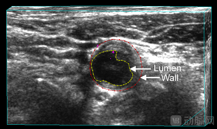
Top Panel: Lumen and Outer Vessel Wall Segmented
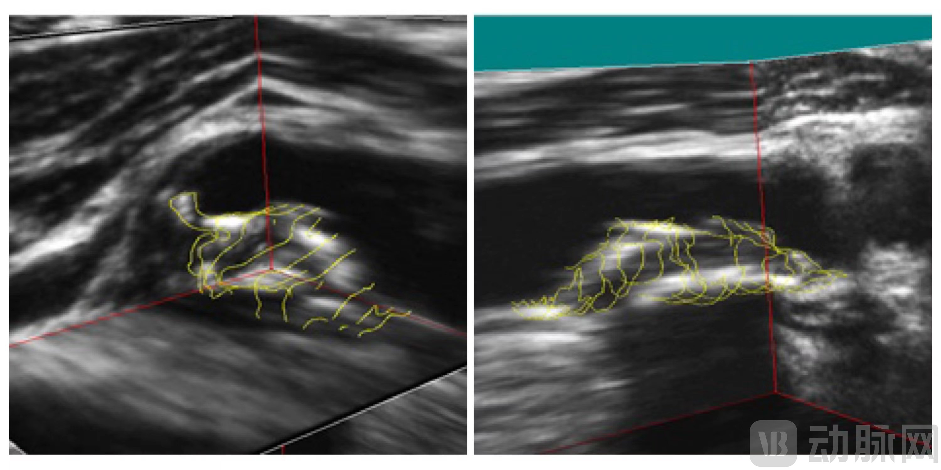
Bottom Panel: Segmented Carotid Atherosclerotic Plaque
In addition to optimizing clinical diagnosis, three-dimensional ultrasound can alsoIntegrated into Image-Guided Interventional Systems, for exampleThree-Dimensional Ultrasound-Guided Prostate Biopsy、Prostate and Gynecological Brachytherapy、Ablation of Focal Liver Tumorsetc.
Take ultrasound-guided prostate biopsy, for example. The established definitive method for diagnosing prostate cancer is transrectal ultrasound (TRUS)-guided 12-core needle biopsy of the prostate. Although this approach is widely used, it is suboptimal, with a false-negative rate as high as 25%.
To this end, Fenster developed a 3D ultrasound-guided prostate biopsy system. This system utilizes pre-operative MR images to identify suspicious lesions for biopsy and fuses these MR images with intraoperative TRUS images, thereby guiding the biopsy needle to the MR-defined suspicious targets under 3D ultrasound guidance.
The physician inserts the TRUS transducer into the patient’s rectum and rotates it 200 degrees around its longitudinal axis to generate a three-dimensional TRUS image. After identifying biopsy targets through fusion with MR images, the system guides and deploys the biopsy needle to obtain core samples, while automatically tracking and recording changes in biopsy location in real time.
Fenster stated that this ultrasound-guided system includes a hinged multi-joint stabilizer and a transducer tracking mechanism, which can support all end-fire TRUS transducers for prostate biopsy available on the market.
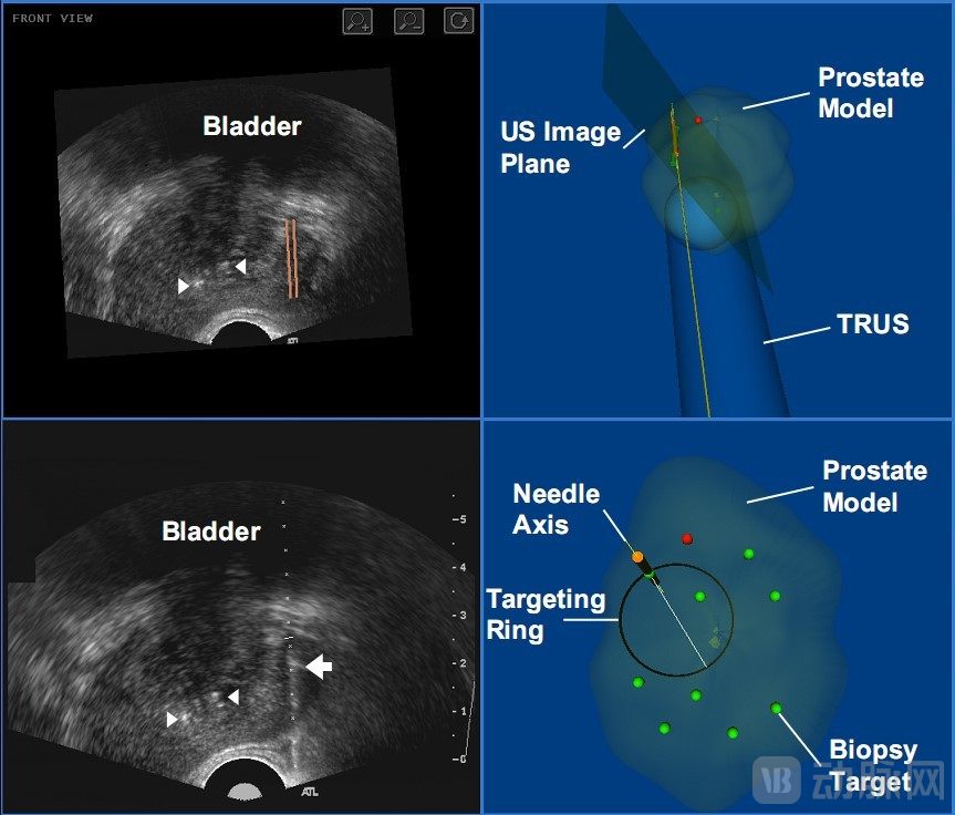
The figure above displays the four-window view of the user interface for a 3D TRUS-guided prostate biopsy system: (top left) automatic slicing of the 3D TRUS image to real-time match the orientation of the 2D TRUS transducer; (bottom left) real-time 2D TRUS image; (right) two panels showing the locations of biopsies within the 3D prostate model—green dots indicate the target sites for biopsy, and red dots indicate the next site to be biopsied.
13 Companies Have Been Licensed: How Should Future Technology Transfer Be Structured?
Fenster is one of the few scientists who are enthusiastic about technology transfer.
In the 1990s, Fenster first transferred the technology to a company free of charge, which then commercialized it and launched it in multiple national markets.
This seemingly “money-losing” deal, however, offered Fenster a positive insight:By leveraging enterprises to facilitate technology transfer, scientific research can benefit a broader population; furthermore, continued collaboration with enterprises post-transfer can further advance scientific research.。
It is worth mentioning that Western University, where Fenster is affiliated, implements “The inventor holds patent ownership.“Policy, which allows inventors to file patents without university involvement and leaves each technology collaboration entirely under the inventor’s responsibility, undoubtedly grants Fenster an inherent advantage in translating research outcomes into practical applications.”
Fenster told VCBeat that their partners include non-profit organizations, startups, and multinational corporations. Additionally, they maintain close ties with the departments of Medical Imaging (Radiology), Radiation Oncology, Rheumatology, and Neurology at London Health Sciences Centre and St. Joseph's Health Care London.
After clearly defining the content and ownership of each invention patent, the Fenster team will collaborate with institutions of varying natures based on different types of inventions and patents.
“Typically, we license our technology to help companies integrate 3D ultrasound systems into their products, and occasionally engage in joint development of additional applications based on customer needs,” Fenster continued. “CurrentlySome patents have been licensed to multiple small and medium-sized enterprises (SMEs) as well as multinational corporations., many products have been launched for global sales, such as prostate biopsy systems。”
Today, in addition to his research work, Professor Fenster continues to enthusiastically share his technology with various companies, seeking potential collaboration opportunities.
When discussing future models for translating research outcomes into practical applications, Fenster said, “In the past, we primarily facilitated technology transfer by founding companies and licensing patents, while maintaining long-term collaborations with licensee companies to provide them with further innovations and recommendations. I hope to continue this model.”
Fenster warmly welcomes Chinese companies interested in collaboration. In fact, he has long shared an indissoluble bond with China.
The Roberts Institute, where Fenster is based, continues to attract numerous visiting scholars and researchers from China. Moreover, most of them maintain contact with Fenster after returning to China, jointly applying for collaborative funding projects and continuing mutual visits and exchanges.
Prior to the COVID-19 pandemic, Fenster visited China almost every year to deliver lectures and engage in academic exchanges at the institutions of his former students (now collaborators), and to give invited speeches at academic forums.
Collaborating with Chinese enterprises to promote the application of 3D ultrasound navigation systems in the Chinese market would also serve as another way for Fenster to continue its engagement with China.