China's Original AI Platform for Structural Heart Disease, Tavigator™, Debuts at China Structural Week 2022
On a summer day in 1929, when German physician Werner Forssmann blindly inserted a rubber ureteral catheter into his own blood vessel, he likely did not anticipate that, over the next century, cardiac catheterization would be widely employed not only for diagnostic purposes but also for the treatment of heart and vascular diseases, benefiting tens of millions of patients annually.
From hypothesis formulation to animal experiments and then to human clinical trials, the development of transcatheter aortic valve replacement (TAVR) technology has undergone a lengthy process over the past two decades. With Professor Alain Cribier successfully performing the first human TAVR procedure in 2002, this technology has ultimately evolved into a mature therapeutic option that can significantly improve quality of life.
CT imaging is an indispensable diagnostic and therapeutic modality in the preoperative workup for transcatheter aortic valve replacement (TAVR). The 2022 “Expert Consensus on Process Optimization of Transcatheter Aortic Valve Replacement (TAVR)” also explicitly underscores the importance of preoperative imaging assessment. Preoperative CT evaluation for TAVR plays a pivotal role in selecting anatomical indications, formulating intraoperative strategies, anticipating and mitigating risks, and ensuring patient safety.[1]. Its novel examination and diagnostic techniques enable operators to fully understand the patient’s three-dimensional anatomical structure before performing surgical procedures guided by two-dimensional imaging. This approach facilitates the assessment of annular anatomical characteristics, coronary artery height, ventricular and vascular anatomy, and even the identification of adjacent structures, thereby providing essential information for preoperative selection of valve size, formulation of deployment strategies, intraoperative complication risk identification, and prognostic evaluation, ultimately making the surgical procedure safer and more efficient.[2]。
Artificial Intelligence (AI) is a field within computer science. AI-based algorithms can perform mapping tasks traditionally carried out by humans in a faster and more precise manner, presenting the results with greater multidimensionality and enhanced clarity and intuitiveness.
By leveraging AI-driven algorithms for fully automated segmentation, reconstruction, mapping, and quantification of anatomical structures, the system can complete TAVR procedural mapping tasks—such as delineating the aortic annulus and other key anatomical structures—within seconds, while ensuring high consistency and stability in measurement accuracy.[3]。
AI can also minimize measurement bias caused by operator experience and misjudgment of risk factors for surgical complications, while playing a crucial role in helping surgeons predict prognostic outcomes of TAVR procedures.[4]. It should be emphasized that AI can facilitate the development and refinement of such surgical planning protocols, thereby promoting broader adoption of these procedures in clinical practice. This is particularly important for medical centers that are in the early stages of implementation, have low procedural volumes, or lack experienced operators.
The prerequisite for establishing a patient-centered, safe, and efficient preoperative assessment is to accurately identify patients at high risk of adverse events versus those at lower risk, and to conduct more detailed analyses on the former, who will become the focus of more rigorous, time-consuming, and resource-intensive subsequent monitoring. In predicting prognoses for patients undergoing transcatheter aortic valve replacement (TAVR) or those with congenital heart disease, coronary artery disease, and heart failure, artificial intelligence (AI) has been proven superior to classical statistical methods.[5-8]. Meanwhile, AI’s high computational efficiency enables the simultaneous integration of numerous predictors, making it a powerful tool for modeling large numbers of variables and their interactions. This capability may lead to the “unexpected” discovery of novel variables and potential new predictors that extend beyond the current scope of clinical treatment decision-making. As the indications for transcatheter aortic valve replacement (TAVR) expand, AI can better support clinicians in assessing TAVR prognosis, which is crucial for aiding patient selection and accurately predicting surgical outcomes.[9-10]。
At the recently concluded 6th China Structural Heart Disease Week (China Structural Week 2022), Professor Wu Yongjian from Fuwai Hospital, Chinese Academy of Medical Sciences (National Center for Cardiovascular Diseases), led an imaging analysis research team that dedicated five years to in-depth study.Achieved five major technological breakthroughs, including multi-target, small-scale, pixel-level image annotation and anatomical structure mapping of the aortic root; research on high-precision automatic segmentation algorithms for aortic root structures using three-dimensional convolutional neural networks; development of fully automated, high-precision algorithms for aortic root target localization and measurement; research on intelligent multi-planar measurement algorithms; and research on intelligent algorithms for predicting TAVR surgical risks.
Professor Wu Yongjian’s team, in collaboration with Tuowei Moxin Data Technology (Nanjing) Co., Ltd., completed the algorithm research, engineering development, and cloud service deployment of Tavigator™, a China-originated preoperative decision-support platform for transcatheter diagnosis and treatment of structural heart disease.Leveraging AI Technology to Advance the Development of TAVR in China, Paving New Pathways for Novel Clinical Diagnosis and Treatment Models with Modern Smart Healthcare Characteristics.
Based on the Tavigator™ system, physicians need only upload CT images of the aortic root to achieve fully automated segmentation and 3D reconstruction of the aortic root anatomy, leaflet typing, annulus and multi-planar localization measurements, automatic localization and measurement of coronary ostia height, annular angle analysis, calculation of optimal projection angles, quantitative calcium analysis, left ventricular volume analysis, anatomical risk alerts, automated valve sizing and selection recommendations, and generation of dynamic structured reports—all without manual intervention. The entire process for detailed TAVR aortic root analysis takes just one minute, significantly faster than the tens of minutes currently required by clinicians for aortic root image analysis. Powered by a high-performance parallel computing system, this solution substantially enhances imaging analysis efficiency, offering significant scientific value and clinical relevance.
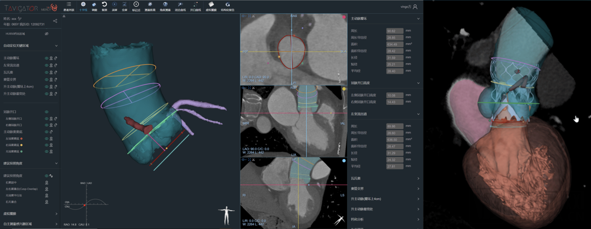
Fusion Display of 2D CT Images and 3D Aortic Root Models
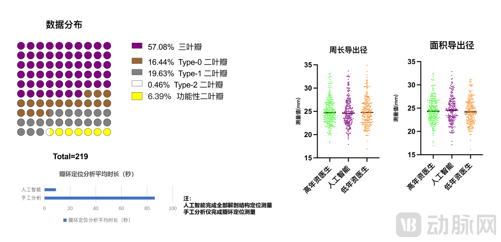
At the China Structural Heart Disease Week, preliminary results from a retrospective data analysis led by Professor Wu Yongjian and his team were simultaneously released. The study included preoperative CT imaging data from 219 patients who had undergone transcatheter aortic valve replacement (TAVR) at Fuwai Hospital. The data covered various leaflet morphologies, including tricuspid aortic valves (TAV), Type-0 bicuspid aortic valves (BAV), Type-1 BAV, Type-2 BAV, and functionally bicuspid aortic valves (FBAV). An artificial intelligence (AI) system and junior physicians in the imaging core laboratory independently analyzed the enrolled imaging sequences. Using the preoperative measurement assessment reports prepared by senior physicians in the imaging core laboratory as the gold standard, a comparative analysis was conducted with the perimeter-derived diameter and area-derived diameter of the annular plane as primary endpoints to validate human–AI concordance. Furthermore, subgroup analyses were performed based on different leaflet morphologies and degrees of calcification to evaluate the impact of leaflet morphology and calcification volume on AI-based measurement and assessment outcomes. Additionally, a comparative analysis was conducted between manual measurements by clinicians and the AI system regarding the time required for aortic annulus localization and measurement.
The statistical analysis results show that the AI system, in terms of accuracy of core measurement indicators,All demonstrated a high correlation with measurements by senior physicians at the core imaging laboratory, and outperformed those by junior physicians at the same laboratory;The artificial intelligence system can accurately classify valve leaflets. Preoperative imaging of different leaflet types demonstrates high correlation and consistency with the core measurements obtained by senior physicians at the imaging core laboratory. Meanwhile, the AI system exhibits strong robustness and good data generalization capability. Furthermore,Artificial intelligence significantly outperforms manual analysis in efficiency while ensuring analytical accuracy.
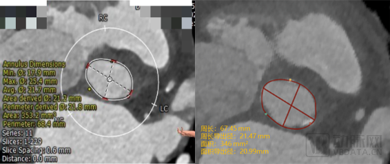
Results of annulus localization and measurement by senior physicians (left)AI-based localization and measurement of the annulus (right)
 Human-Machine Concordance Study—Based on Preoperative CT Imaging Samples from 219 TAVR Patients
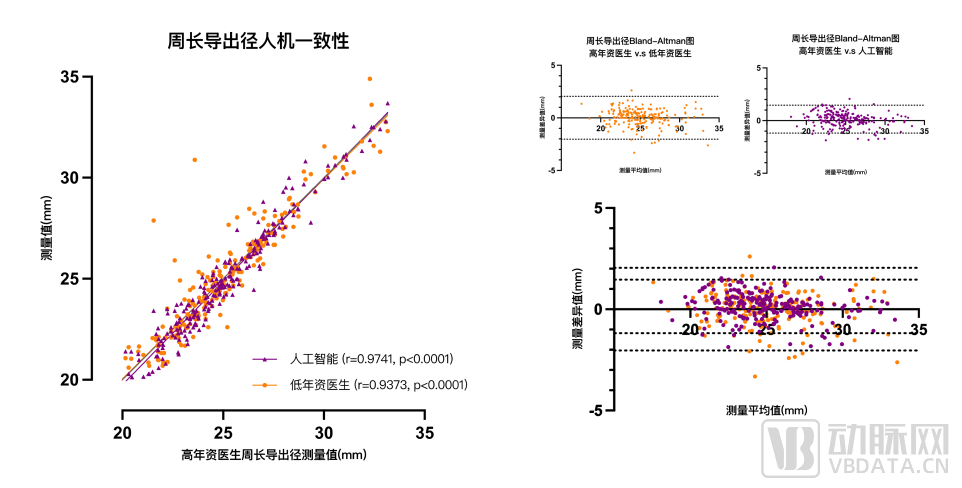
Study Results on Perimeter-Derived Diameter: AI Measurements Outperform Junior Physicians
(Circumference-derived diameter r: 0.9741 > 0.9373)
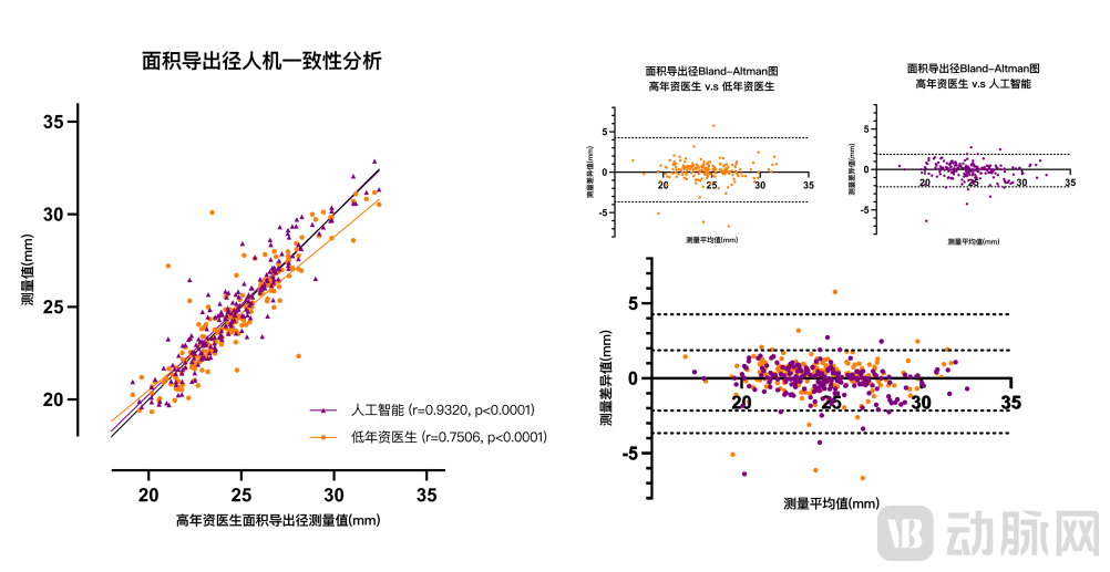 Study Results on Area-Derived Diameter: AI Measurements Outperform Junior Physicians
(Area-derived radius r: 0.9320 > 0.7506)
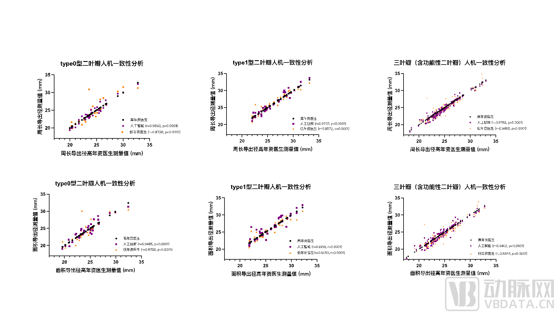 The AI system demonstrated extremely high correlation and stability with measurements obtained by senior physicians across cases with different valve leaflet morphologies.
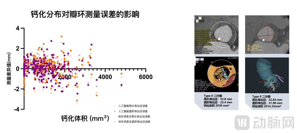 Artificial intelligence systems demonstrate high correlation and stability with senior physicians in the analysis of highly calcified cases
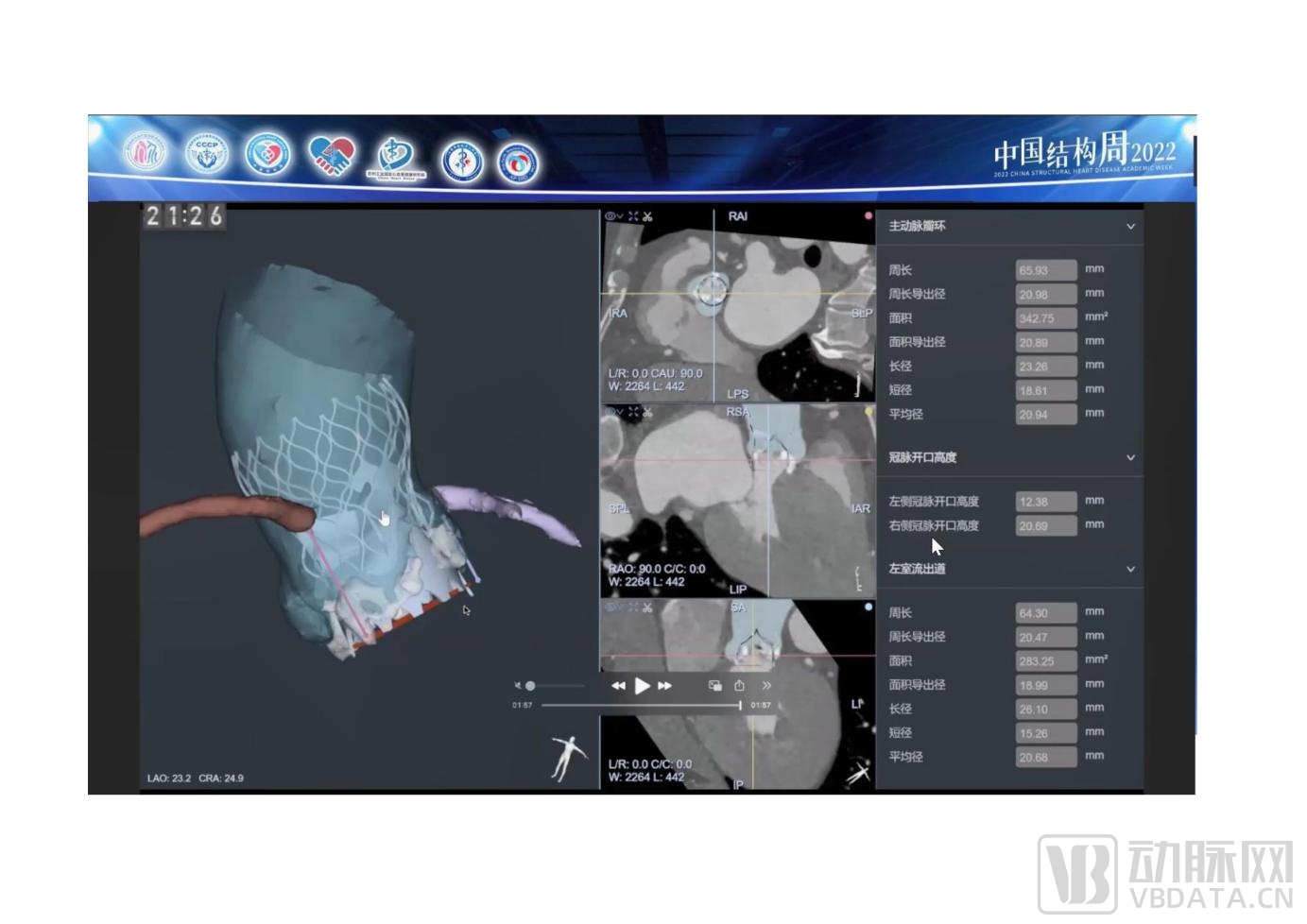
During China Structural Heart Week, researchers conducted a comparative pre-TAVR assessment using the Tavigator™ system in a patient with functionally bicuspid aortic valve deformity at intermediate surgical risk.The Tavigator™ system completed the measurement and assessment of the aortic root in just one minute. In contrast, using traditional specialized software, physicians at an imaging core laboratory would need to perform approximately 1,500 mouse clicks, over 700 keystrokes, and move the mouse a cumulative distance of more than 100 meters, with total processing time approaching 40 minutes. Taking the annulus perimeter-derived diameter—the most critical basis for valve sizing—as an example, the AI-measured result was 20.98 mm, showing only a slight difference from the expert-measured value of 20.8 mm. By analyzing measurements of aortic root anatomy via AI, and combining raw CT images with operator experience to build datasets for training artificial intelligence models, risk prediction results nearly comparable to those of experienced operators can be achieved. This case not only demonstrates the reliability of Tavigator™ data measurements in clinical practice but also fully highlights the potential for risk prediction based on anatomical structures. For many centers currently performing or planning to perform TAVR procedures, this technology not only saves substantial physician work time but also holds significant value in enhancing understanding of TAVR procedures, identifying potential risks embedded within anatomical structures, planning surgeries, and improving surgical outcomes.
The application of AI in TAVR aims to streamline workflows, enhance analytical efficiency, improve accuracy and consistency, and present the data underlying CT images from more diverse and comprehensive perspectives.It frees up more time for clinicians to focus on tasks that are better suited to human capabilities and carry greater meaning and value. The steady integration of AI into clinical decision support systems requires first establishing and thoroughly validating the robustness of algorithms, along with implementing specific regulations and standardization for the validation of novel AI algorithms. Continuous training, education, and outreach for all healthcare professionals are also essential to overcome skepticism and distrust, enabling healthcare providers to utilize these algorithms in real time without imposing cumbersome changes to their daily workflows. Meanwhile, research demonstrating the advantages and cost-effectiveness of AI algorithms compared to “traditional” clinical practices is urgently needed.The advent of Tavigator™ will undoubtedly drive significant advancements in China’s TAVR technology, substantial progress in TAVR clinical practice, major upgrades in the TAVR industry, and considerable benefits for TAVR patients.

Professor Wu Yongjian, Fuwai Hospital, Chinese Academy of Medical Sciences:As a new round of global technological revolution and industrial transformation takes shape, AI and digital technologies are rapidly evolving, becoming a key driving force behind this change. As a significant attempt at localized innovation in applying artificial intelligence to the clinical diagnosis and treatment of structural heart disease, Tavigator™ still has a long journey ahead. While optimizing accuracy, enhancing functionality, and improving performance, greater emphasis must be placed on conducting large-scale prospective cohort studies based on this technology to validate the reliability and validity of the model. Building on these efforts, a novel clinical pathway for TAVR characterized by modern smart healthcare will gradually evolve. Driven by the “four wheels” of clinical experience, big data, artificial intelligence, and high-performance computing, comprehensive integration across clinical practice, technology, and industry will ultimately be achieved. In the future, beyond TAVR, surgical planning standards will be established for mitral valve, tricuspid valve, and other structural heart diseases, with the ultimate goal of sharing Chinese expertise globally.

Professor Guo Yingqiang, West China Hospital of Sichuan University:In recent years, thanks to breakthroughs in algorithms such as deep learning, continuous improvements in computing power, and the ongoing accumulation of massive datasets, artificial intelligence has truly transitioned from laboratory research to widespread industrial and clinical application. AI has had a significant impact on cardiovascular research and practice. In the field of structural heart disease, mastery of cardiac anatomy has always been the most critical task for surgeons, including interventional cardiologists. Tavigator™ represents the direction of new technology applications in medicine, offering new possibilities for the processing and mining of medical big data. It enables experienced physicians to make more accurate judgments and helps novice physicians learn more rapidly, thereby facilitating the global promotion and adoption of TAVR technology.

Professor Yang Jian, Xijing Hospital of Air Force Medical University:AI technology can decipher highly complex intrinsic patterns from vast amounts of clinical data, offering broad application prospects in the healthcare industry. The field of structural heart disease involves numerous clinical data parameters; 3D structural visualization often fails to meet standards, and 4D physiological functions exhibit significant variations among patients of different ages, sexes, and ethnicities. AI technology can effectively address these challenges, thereby improving the quality of patient care while reducing medical costs. Applications such as AI simulation, the use of hyperrealism in medical device training, and the printing of personalized anatomical models will all promote the development of precision medicine and may serve as the foundation for the future flourishing of new technologies.

Professor Ruediger Lange, German Heart Center:It is gratifying to witness the seamless integration of Chinese artificial intelligence technology with clinical applications. To date, most relevant evidence has stemmed from single-center studies lacking validation across diverse populations, and issues regarding algorithm reproducibility warrant serious attention. We eagerly anticipate the day when Tavigator™ will be deployed in Germany and worldwide.
1. Expert Consensus on Clinical Pathways for Transcatheter Aortic Valve Replacement in China (2021 Edition). Chinese Circulation Journal, January 2022, Vol. 37, No. 1 (Serial No. 283). Article ID: 1000-3614(2022)01-0012-11 DOI: 10.3969/j.issn.1000-3614.2022.01.003
2. Guo Shuai, Zhang Bin, Wu Yongjian. Recent Advances in Transcatheter Aortic Valve Replacement[J]. Chinese Journal of Medicine. 2020,(1). DOI:10.3969/j.issn.1008-1070.2020.01.002.
3. Astudillo P , Mortier P , Bosmans J , De Backer O , de Jaegere P , Iannaccone F , et al. Automatic detection of the aortic annular plane and coronary Ostia from multidetector computed tomography. Interv Cardiol 2020;2020:9843275.
4. El Faquir N , De Backer O , Bosmans J , Rudolph T , Buzzatti N , Bieliauskas G , et al. Patient-specific computer simulation in TAVR with the self-expanding Evolut R valve. JACC Cardiovasc Interv 2020;13(15):1803–12.
5. Hernandez-Suarez DF , Kim Y , Villablanca P , Gupta T , Wiley J , Nieves-Ro- driguez BG , et al. Machine learning prediction models for in-hospital mor- tality after transcatheter aortic valve replacement. JACC Cardiovasc Interv 2019;12(14):1328–38.
6. Diller GP , Kempny A , Babu-Narayan SV , Henrichs M , Brida M , Uebing A , et al. Machine learning algorithms estimating prognosis and guiding therapy in adult congenital heart disease: data from a single centre including 10 019 patients. Eur Heart J 2019;40(13):1069–77.
7. Zack CJ , Senecal C , Kinar Y , Metzger Y , Bar-Sinai Y , Widmer RJ , et al. Leveraging machine learning techniques to forecast patient prognosis after percutaneous coronary intervention. JACC Cardiovasc Interv 2019;12(14):1304–11.
8. Angraal S , Mortazavi BJ , Gupta A , Khera R , Ahmad T , Desai NR , et al. Machine learning prediction of mortality and hospitalization in heart failure with pre- served ejection fraction. JACC Heart Fail 2019 pii: S2213-1779(19)30541-4.
9. Lopes, R.R.; van Mourik, M.S.; Schaft, E.V.; Ramos, L.A.; Baan, J.; Vendrik, J.; et al. Value of machine learning in predicting TAVI outcomes. Neth. Heart J. 2019, 27, 443–450.
10. Penso M, Pepi M, Fusini L, Muratori M, Cefalù C, Mantegazza V, et al. Predicting Long-Term Mortality in TAVI Patients Using Machine Learning Techniques. J Cardiovasc Dev Dis. 2021 Apr 16;8(4):44. doi: 10.3390/jcdd8040044. PMID: 33923465; PMCID: PMC8072967.
11. Key Points of the 2021 Annual Report on Interventional Therapy for Structural Heart Disease Chinese Journal of Interventional Cardiology, January 2022, Vol. 30, No. 1 DOI: 10.3969/j.issn.1004-8812.2022.01.003