Exclusive Interview with Dr. Zhao Chan of Peking Union Medical College Hospital: Pioneering International First-in-Class Precision Diagnosis and Treatment Systems for Ocular Diseases, Multiple Products Finalized and IPO Filing Submitted
Last year, Novartis announced the acquisition of UK-based ocular gene therapy company Gyroscope Therapeutics for $1.5 billion.
Subchoroidal Retinal InjectionIt is one of the optimal routes of administration for ocular gene therapy. Gyroscope Therapeutics employs a sclerotomy approach to position a microcatheter in the suprachoroidal space, followed by the use of a microneedle to inject the gene therapy agent into the subretinal space, thereby achieving drug delivery via the transchoroidal and subretinal routes.
Compared with the traditional transvitreal subretinal injection approach, this drug delivery route offers simpler operation, higher safety, and lower dependence on surgical equipment and skills, holding significant promise for clinical application.
Novartis’ high-profile acquisition has heightened industry awareness and emphasis on this route of administration. However, Gyroscope’s approach requires incising the sclera, which is only 1 mm thick, followed by suturing upon closure, entailing certain surgical thresholds and risks. Is there a more minimally invasive and safer method to enable drug delivery via this route?
It is reported that the team led by Zhao Chan from Peking Union Medical College Hospital, after years of research and development, has achieved this by employing simple yet ingenious mechanical principles.Precise localization of the suprachoroidal space, upon which a minimally invasive drug delivery and interventional system for suprachoroidal and transchoroidal subretinal administration was designed, represents the first international innovation to apply minimally invasive interventional technology systems to ophthalmic clinical practice.
Under the influence of "“InnoChina China” 2nd Biomedical High-Value Patent Project Selection Activityat the invitation of, Artery Orange Fruit Bureau had the honor of interviewingDr. Zhao Chan, Associate Chief Physician in the Department of Ophthalmology at Peking Union Medical College Hospital; Assistant to the Director of the Department of Ophthalmology (member of the leadership team, overseeing scientific research and laboratory operations); Principal Investigator (PI) at the Key Laboratory of Fundus Diseases, Chinese Academy of Medical Sciences。
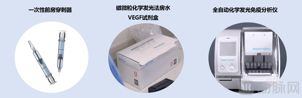
In fact, in addition to the suprachoroidal space targeted therapy system, Dr. Zhao Chan’s team has also developed an aqueous humor-based companion diagnostic system to provide diagnostic evidence for precision treatment. To date, the disposable anterior chamber paracentesis device has been granted three Chinese invention patents, four utility model patents, and three international patents (in the United States, Germany, and Japan). The novel resistance-sensing suprachoroidal injection therapy system has had four invention patent applications filed, four utility model patents granted, and two PCT international patent applications submitted.
Aqueous Humor Companion Diagnostic System: Enhancing the Precision of Intraocular Targeted Therapy
Fundus vascular diseases are common causes of blindness, including diabetic retinopathy, neovascular age-related macular degeneration, and retinal vein occlusion.Intravitreal injection of targeted therapeutic agents is currently a major treatment modality for retinal vascular diseases., among which anti-vascular endothelial growth factor (VEGF) targeted drugs were the first to achieve industrialization and large-scale clinical application.
“There is significant individual variability in the efficacy of intraocular targeted therapy, with both overtreatment and undertreatment coexisting,” said Zhao Chan. “VEGF inhibitors are based on population-level data and represent a generalized treatment approach; however, when substantial individual differences exist, they fail to yield homogeneous prognostic outcomes.”
For intraocular targeted therapy, in addition to anti-VEGF agents, there are also hormonal medications (such as glucocorticoids).
To determine which targeted therapy is more suitable for the patient, Zhao Chan’s teamThe First International Proposal of an Aqueous Humor Companion Diagnostic System, by collecting a small amount of aqueous humor to evaluate its response to the drug, this system includesDisposable anterior chamber paracentesis device, intraocular fluid detection kit (magnetic particle chemiluminescence method), chemiluminescence immunoassay analyzer, and other products。
Zhao Chan pointed out that aqueous humor companion diagnostics face two challenges.
First, aqueous humor collection is challenging. As the eyeball is a small organ, only trace amounts of aqueous humor can be collected. However, most existing syringes are calibrated in milliliters, making it impractical to aspirate 50–100 μL of aqueous humor. Furthermore, standardization, safety, and precision during the sampling process are difficult to ensure, which hinders the widespread adoption of intraocular fluid collection and subsequent diagnostic testing in outpatient ophthalmology clinics.
In this regard, Zhao Chan's teamDisposable Anterior Chamber Paracentesis Device, employing precise negative pressure control technology to enhance the accuracy of the negative pressure tube by 100-fold, enablingPrecise Collection of 50 μL Aqueous Humor, In addition, aqueous humor collection can be performed in general settings such as outpatient clinics and treatment rooms, thereby reducing the risks of infection and puncture-related injuries. Currently,This technology has been granted patents in China, the United States, Japan, and Germany, has completed clinical trials, and has obtained registration certification as an innovative medical device.。
Second, aqueous humor testing is challenging. Test results are correlated with the volume of fluid obtained; therefore, examining micro-volume samples imposes higher technical requirements on the assay.
Based on this, the team led by Zhao Chan developed a magnetic particle chemiluminescence assay kit for intraocular fluid detection (VEGF, IL-6, and IL-10 in vitro diagnostic kits) and an automated point-of-care testing device for aqueous humor chemiluminescence assays.Only 15–20 μL of sample is required to deliver test results within approximately one hour. Currently, both products have been finalized and are being prepared for registration and clinical trials.。
Aqueous Humor Companion Diagnostic System Series Products
Zhao Chan told VCBeat’s Orange Bureau that while numerous current studies attempt to assess treatment responses through imaging of fundus vascular diseases, their accuracy remains insufficient, and there are no mature individualized treatment plans based on such imaging. “Aqueous humor companion diagnostics hold the promise of establishing a more precise and refined multidimensional diagnostic system.”
Suprachoroidal Therapy System: Precise Targeting of the Choroid/Retina
If aqueous humor testing is an important tool for the precise diagnosis of ophthalmic diseases, then the suprachoroidal space therapeutic system represents a disruptive achievement in the field of ocular injection drug therapy.
As one of the organs with the highest water content in the human body, the eyeball is essentially a fluid-filled cavity system. The retina, choroid, and other major functional tissues constitute the thin inner wall of the eyeball, which is no more than 0.5 mm thick and serves as the primary site affected by intraocular diseases.
Conventional treatment approaches often involve direct intravitreal injection of the drug solution into the vitreous cavity (the space within the eyeball), relying on free diffusion of the medication through the vitreous humor to deliver it to the affected tissues.
Although this administration method is simple, it suffers from poor tissue targeting, drug clearance via aqueous humor drainage, and a short half-life. Consequently, frequent dosing is typically required in clinical practice to maintain therapeutic efficacy. This not only consumes substantial medical resources but also increases potential risks such as intraocular infection and needle-related injury. More critically, intravitreal injection of glucocorticoids is highly likely to cause complications such as glaucoma and cataracts.
In response to the aforementioned challenges, the ophthalmology community has gradually recognized over the past decade that drug delivery via suprachoroidal injection may represent a superior route for intraocular administration.
The so-called suprachoroidal space (SCS) refers to the potential space between the choroid and the sclera. Injection into the SCS allows the drug to diffuse throughout the entire SCS and penetrate through the choroid into the retina, thereby achieving a treatment modality with superior tissue targeting and fewer side effects, thus enhancing safety and efficacy.
However, the sclera is generally only about one millimeter thick. How can one precisely puncture this one-millimeter-thick dense tissue without damaging the underlying choroid and retina, which are merely a few tenths of a millimeter thick? This presents another highly challenging technical issue.
To this end, Harvard and MIT jointly designed a highly unique syringe in which the needle is connected to the plunger. During puncture, when the needle tip is positioned within the dense scleral tissue, the medication inside the syringe cannot enter the tissue, allowing the needle to continue penetrating the tissue under hydraulic pressure.
When the needle tip reaches the loose connective tissue cavity, fluid is released from the tip. At this point, advancing the rear plunger does not cause the needle tip to penetrate further; precise localization of the suprachoroidal space is achieved through pressure differential sensing.
It is reported that a paper on this study was published in a Nature subsidiary journal, but it failed to achieve commercialization because the needle was not securely fixed, making it prone to lateral deviation or slippage, which could lead to serious complications.
To address the aforementioned risks, Zhao Chan’s team employed a spring to maintain stable positive pressure. The injection needle penetrates the piston with its distal end sealed, achieving dual-layer axial fixation to prevent axial deviation during puncture. Furthermore, a novel differential resistance sensing device was utilized to enable fluid delivery into the target tissue through the side ports of the puncture needle.
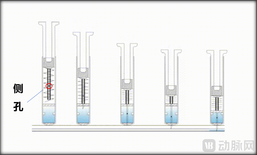
Schematic Diagram of Injection Needle
Zhao Chan revealed to VCBeat,The injection needle has been finalized after multiple iterations, achieving precise suprachoroidal space injection in ex vivo porcine eyes and successfully demonstrating its capability in live rabbit eyes with a scleral thickness of only 0.3 mm.。
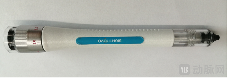
Injection Needle Shaping Product
Meanwhile, using a novel suprachoroidal space injection device, viscoelastic agents are injected into the SCS to dilate the scleral puncture site, thereby enabling catheter placement in the suprachoroidal space and completing subretinal injection via microneedle.Achieving minimally invasive transchoroidal and subretinal injection without sclerotomy, which is significantly superior to the Gyroscope solution mentioned at the beginning of the article.。
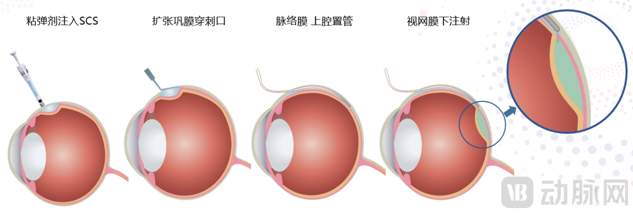 Minimally Invasive Transchoroidal and Subretinal Injection
“This technology enables precise, targeted treatment of fundus diseases, including gene therapy, stem cell therapy, small-molecule chemical drugs, and biologics. We are currently in partnership discussions with five leading gene therapy companies both in China and abroad,” said Zhao Chan.
Platform-level minimally invasive ophthalmic technology with significant future scalability
Actually,Suprachoroidal Space Injection System: A Highly Scalable Platform Technology, thereby expanding the possibilities for minimally invasive implantation and interventional therapies in ophthalmology based on this anatomical space.
For example,Development of a Novel Suprachoroidal Aqueous Humor Drainage Device for Glaucoma Treatment, is a novel MIGS technique involving minimally invasive suprachoroidal shunt implantation; andEpidural Anesthesia, in labor analgesia, this technology can be utilized to more precisely and safely administer anesthetic into the lumbar epidural space, blocking the spinal nerve roots and reducing the risks associated with inaccurate drug placement during childbirth.
It is reported that this series of precision diagnosis and treatment systems for fundus vascular diseases was jointly developed by Zhao Chan’s team and Beijing Huashi Nuowei Medical Technology Co., Ltd. through a medical-engineering collaboration, with intellectual property rights co-owned by both parties.
In Zhao Chan’s view, both the “medical” and “engineering” components are crucial to the translation of medical-engineering integrated products. Medical experts must possess sufficient professional sensitivity, while the engineering side requires technical personnel to develop the product. Additionally, an intellectual property management team and regulatory testing specialists are needed, followed by product promotion. The translation process involves a long and complex chain.
Zhao Chan does not advocate for technical professionals to start their own businesses. “Based on my experience, once medical personnel leave clinical practice, their innovative ideas become constrained; after transitioning into the commercial sector, they no longer focus solely on product innovation.”
Zhao Chan, meanwhile, has focused primarily on technology, continuously optimizing and jointly improving the platform based on recommendations from collaborative R&D partners. “Our next step is to develop derivative products leveraging the foundational suprachoroidal space therapy technology. For instance, the novel suprachoroidal aqueous humor drainage device for glaucoma treatment mentioned earlier, as well as applications in epidural anesthesia, have both completed product prototyping and entered the animal testing phase.”