Nanovision's Fuyan-24 Static CT Enters Clinical Trials, Paving the Way for Mesoscopic Imaging with Cellular-Level Resolution

Nanovision
X-ray Detector and Static CT Product R&D Provider
VCBeat has learned at the earliest opportunity,On November 23 at 13:00, the clinical launch ceremony for the world’s first whole-body multi-source static CT system—the Compound Eye 24 Static CT System—was successfully held at the 86th China International Medical Equipment Fair.

According to reports, the Fuyan 24 Static CT was independently developed by Nanovision (Nanjing) Technology Group Co., Ltd. (hereinafter referred to as “Nanovision”). It is the world’s first next-generation whole-body medical CT without slip rings. The system adopts a dual-ring structure composed of “64 photon-flux detector arrays and 24 scanning X-ray source arrays.” Since the X-ray tube and detectors do not need to rotate during scanning, trailing artifacts are eliminated, fundamentally achieving a significant improvement in spatial resolution. Its voxel resolution is 64 times superior to that of spiral CT technology, and it features multi-energy spectral imaging capabilities.
Meanwhile, leveraging innovations in underlying technologies, Nanovision’s first static CT product has achieved a scan slice thickness of 0.165 mm with isotropic resolution, and its next-generation product is expected to break through the 0.100 mm (100 micrometers) threshold, truly enabling mesoscopic imaging.
More precise scanning can yield more valuable diagnostic information and enable earlier detection of lesions, thereby facilitating more accurate diagnoses and ultra-early screening for diseases. Based on such ultra-early screening, static CT has the potential to truly advance the early detection, diagnosis, and treatment of various conditions. For instance, it could shift the diagnosis of diseases like chronic obstructive pulmonary disease (COPD)—which are often identified only at end-stage—from late to early stages, transforming their management from irreversible to reversible interventions, and ultimately holding promise for altering the disease spectrum.
Notably, core components of the static CT, such as the array-based integrated X-ray source and photon-flux detector, are independently developed by Nanovision. Previously, both the static CT system and its core components were included in the “List of Key Core Technologies and Critical Components for CT Equipment in China’s Medical Industry,” receiving strong national support.
Meanwhile, this static CT product has also garnered significant attention from top-tier medical institutions worldwide, such as the Mayo Clinic, Cleveland Clinic, Massachusetts General Hospital of Harvard University, Washington University School of Medicine, and MD Anderson Cancer Center.
In clinical practice, imaging at the millimeter scale and above is generally considered macroscopic imaging, such as imaging of organs and tissues like visceral organs and blood vessels; such imaging is typically measured in centimeters or millimeters.
Imaging at the micrometer scale is considered mesoscopic imaging, such as that of tissues and cells, typically measured in micrometers. Among these, 100 micrometers is regarded as the boundary between macroscopic and mesoscopic imaging.
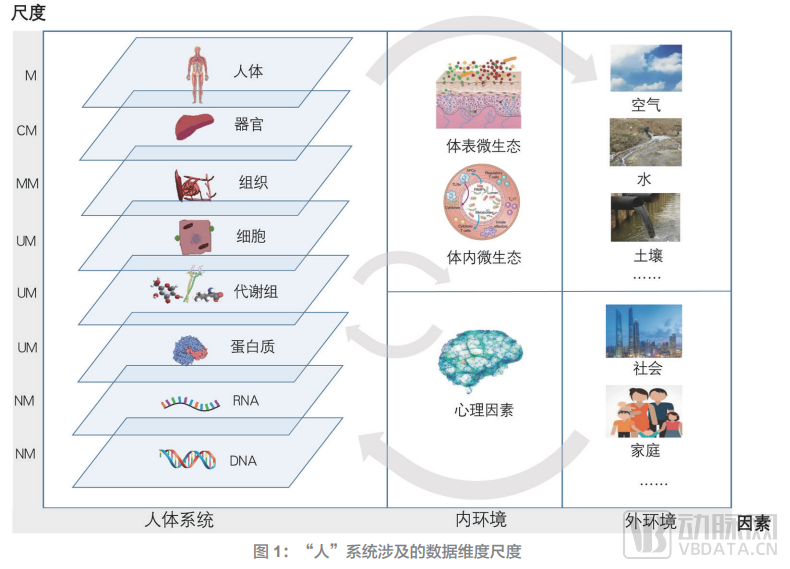
(Image source: White Paper on Computational Medicine)
Data indicates that macroscopic imaging primarily visualizes the anatomical structures of organs and tissues, enabling clinicians to make diagnoses based on morphological and anatomical changes. In contrast, mesoscopic imaging facilitates diagnosis by assessing alterations in biological characteristics at the tissue and cellular levels.
Based on this, mesoscopic imaging enables earlier identification and screening of diseases, whereas macroscopic imaging requires the disease to progress to an intermediate stage with gross morphological changes before a diagnosis can be made.
Currently, after decades of development, medical CT technology has seen increasingly refined image quality and clinical capabilities. However, in terms of imaging precision, it remains confined to the realm of macroscopic imaging. Furthermore, with advances in technologies such as cell therapy and gene therapy, clinical treatment approaches have progressed to the cellular and even molecular levels. Existing macroscopic imaging modalities are insufficient to meet the demands of clinical diagnosis and treatment, let alone the broader advancement of medicine. There is an urgent clinical need for mesoscopic imaging techniques capable of precisely visualizing cells and tissues. This strong clinical demand will inevitably serve as the primary driving force propelling medical CT toward more refined mesoscopic imaging.
For example, since their inception, spiral CT scanners—the most widely used modality in the market—have been engaged in a race to increase the rotation speed of the X-ray tube–detector assembly and to widen the detector array. Specifically, spiral CT acquires attenuation data of X-rays passing through the human body by rotating the gantry via slip rings, thereby enabling scanning from multiple angles. Throughout their development, manufacturers have continuously increased the rotation speed of the X-ray tube–detector assembly and expanded detector width to obtain richer information and achieve more accurate diagnoses.
However, spiral CT has now approached two limits but failed to break through the boundary of mesoscopic imaging.。
First, the high-speed rotation of spiral CT scanners around the human body generates substantial centrifugal force. The maximum centrifugal force they can withstand is 75G, corresponding to a fastest rotation speed of 0.25 seconds per revolution. Currently, existing spiral CT scanners on the market are approaching this speed limit.
Second, the pixel size of the detector and the amount of effective information obtained per pixel determine the spatial resolution of each CT scan. Currently, the most advanced spiral CT scanners worldwide have pushed the physical segmentation of detectors to its limit, with their spatial resolution approaching the theoretical ceiling of spiral CT technology.
Unlike spiral CT, Nanovision's first product, which has initiated clinical trials this time“Compound Eye 24” static CT adopts a unique technical approach, achieving a scan slice thickness of 165 micrometers, which approaches the 100-micrometer mesoscopic imaging threshold recognized by experts. Its next-generation product is expected to truly realize mesoscopic imaging.。
While top-tier spiral CT products struggle to break through into mesoscopic imaging, the first-generation static CT product already approaches this capability, owing to its disruptive technological principles.
According to the introduction, static CT eliminates the slip ring design. It adopts a dual-ring structure composed of an electronic scanning X-ray source and photon-counting detectors, and utilizes multiple fixed X-ray sources distributed in a ring to sequentially expose and complete the CT scan.
Specifically, the X-ray sources arranged in a ring structure emit X-rays sequentially under exposure control timing and are received by corresponding detectors, producing an effect similar to the rotational projection of X-ray sources in spiral CT scanners. This decouples the temporal resolution of CT equipment from mechanical rotation speed. As a result, static CT can easily overcome the centrifugal force limitations inherent to spiral CT, thereby significantly enhancing temporal resolution. For instance, thanks to innovations based on this technology, the temporal resolution of static CT is ten times higher than that of conventional spiral CT, with the fastest scanning speed reaching 0.08 seconds per rotation.
Meanwhile, for each X-ray source in static CT, the exposure time and exposure energy can be configured according to clinical requirements, enabling static CT to be suitable for imaging of all body parts.
In terms of spatial resolution, the physical pixel size of static CT detectors has been reduced from 1 mm to below 0.25 mm, achieving a reconstructed resolution of 0.165 mm. Compared with conventional spiral CT, static CT has achieved a leapforward breakthrough in imaging fine anatomical structures, with spatial resolution64-Fold IncreaseIn addition, static CT features multi-energy spectral imaging capabilities, offering clearer image acquisition and lower scan radiation, with radiation doses reduced to 40–50% of those associated with conventional CT.
Overall, spiral CT has approached its limits and is unlikely to achieve disruptive breakthroughs; in contrast, static CT is an emerging technology on the rise. Its first-generation product already approaches mesoscopic imaging, and the next generation is expected to truly realize mesoscopic imaging, with the potential to achieve nanoscale imaging as technology advances.
Nanovision’s static CT product, “Compound Eye 24,” has initiated clinical trials, marking not only the first clinical application of a static CT system globally but also the transition of mesoscopic imaging from concept to reality.
Based on Disruptive Innovation, Static CT Has Significant Clinical Advantages.
First, the "Compound Eye 24" static CT enables high-precision imaging, with a spatial resolution of 21 LP/cm at 10% MTF and a scan slice thickness of 0.165 mm (165 micrometers). Compared to conventional CT used for anatomical imaging of tissues and organs, static CT makes biological imaging of tissue cells possible due to its higher spatial resolution.
Through high-precision imaging, static CT can detect smaller lesions at an earlier stage, advancing disease detection and diagnosis. This holds promise for achieving ultra-early diagnosis of tumors and other diseases, thereby facilitating the clinical goals of “early screening, early diagnosis, and early treatment” and improving survival rates for malignancies such as cancer.
Second, the "Compound Eye 24" static CT enables ultra-large matrix scanning and reconstruction at 0.165 mm with a 2048×2048 resolution. Compared with conventional CT, static CT increases pixel information by 16-fold, thereby more accurately reflecting the true nature of diseases and lesions.
Third, the “Compound Eye 24” static CT enables low-dose imaging and silent scanning. Compared with conventional CT, static CT can reduce the radiation dose by more than 40% while maintaining the same image quality. Meanwhile, during scanning, the noise level in the scanner room is below 60 decibels, significantly reducing noise interference during examinations.
Fourth, the "Compound Eye 24" static CT features higher temporal resolution, with a maximum scanning speed of 0.08 seconds per rotation. This effectively minimizes motion artifacts and delivers more precise clinical images.
Clinical experts stated: “Given the high-precision imaging, large matrix imaging, and high temporal resolution of static CT, it may enable ultra-early cancer screening and precise disease diagnosis.”
For example, numerous clinical cases have demonstrated that pulmonary nodules smaller than 5 mm still carry a risk of malignancy. However, many clinical guidelines clearly state that pulmonary nodules less than 5 mm require only follow-up and no clinical intervention. This is because the imaging resolution of current CT equipment reaches only 0.5–0.6 mm, with scanning/reconstruction matrices of merely 512×512, enabling detection but not precise characterization of lesions smaller than 5 mm.
Encouragingly, static CT scanners, which are poised for clinical application, leverage high-precision imaging capabilities to clearly visualize details of lesions smaller than 5 mm, thereby facilitating accurate diagnosis and differential diagnosis of such small lesions. Consequently, static CT is expected to further advance the timeline for lung cancer diagnosis and treatment, enabling ultra-early screening and helping patients with lung cancer and other tumors achieve “early detection, early treatment, and early recovery.”
Throughout human history, the disease spectrum has continuously evolved alongside advancements in medical technology. For instance, smallpox, which once ravaged the globe, was eradicated with the rise of vaccine technology, while cardiovascular and cerebrovascular diseases have been curbed by the maturation of endovascular intervention techniques. Accordingly, we believe that static CT, as a disruptive innovation, will also drive changes in the disease spectrum.
Taking chronic obstructive pulmonary disease (COPD) as an example, the disease has typically progressed to the mid-stage by the time it is currently diagnosed, causing irreversible damage to the lungs. Static CT, characterized by high-precision imaging and accurate diagnosis, enables precise diagnosis in the early stages of COPD (before irreversible damage occurs). This allows physicians to intervene earlier in treatment, thereby significantly reducing COPD mortality rates and ultimately driving a shift in the disease spectrum.
Beyond the aforementioned applications, static CT holds further potential yet to be explored. Just as the evolution from 2G to 4G revolutionized communications, the transition from macroscopic to mesoscopic imaging has opened a new frontier in medical CT. Static CT and mesoscopic imaging are expected to exert a disruptive impact on clinical practice.