RSNA2022: Charting the Path to Redefine Radiology Through Patient-Centric Imaging and AI Empowerment
GE Hangwei
Medical Device R&D and Manufacturer
As the world’s top footballers converged on scorching Doha, biting winds in Chicago brought together leading radiology experts from around the globe.
Early Sunday morning, the Arie Crown Theater was already packed to capacity. Bruce Haffty, President of the Radiological Society of North America (RSNA) Annual Meeting, took center stage to kick off the conference with a presentation titled “Diagnostic Imaging: Value from the Lens of the Patient.”
This year’s content can be seen as an extension of last year’s theme, “Redefining Radiology.” He believes that medical imaging can eliminate the uncertainty faced by patients, alleviate their anxiety, and instill hope in overcoming disease. Therefore, through this mechanism,Imaging can deliver greater value in helping patients enhance their sense of control over their disease.。
Professor Bruce Haffty did not provide a comprehensive answer on how to realize this vision throughout his lecture. However, in light of this year’s conference theme, “Empowering Patients and Partners in Care,” empowering imaging in healthcare from the patient’s perspective may represent a potential pathway for the future development of this discipline.
William Kissick, a professor at Yale University in the United States, proposed the famous “Impossible Trinity”: under given constraints, it is difficult for a country’s healthcare system to simultaneously achieve “improved quality of medical services, increased accessibility to medical services, and reduced prices for medical services,” thereby employing an economic model to conduct a macro-level analysis of the operational logic of healthcare systems.
Reality often fails to meet the idealized assumptions of economic models; beyond balancing economic benefits,It is also necessary to be more empathetic, standing from the patient’s perspective to understand what they truly desire during the diagnosis and treatment process.
What Do Patients Want Most During Their Diagnosis and Treatment? A High-Quality Image? A Meticulously Detailed Treatment Plan? Or Rational Imaging Data Analysis?
Each of the aforementioned questions is evidently crucial for both physicians and patients; however, in Professor Bruce Haffty’s view, beyond economic benefits, some “Patient Care"Go in."
“From the physician’s perspective, medical imaging serves as a decision-making tool to determine next steps, such as whether to adjust the radiotherapy plan or administer additional chemotherapy. In contrast, for patients, beyond using imaging to understand the physician’s recommendations, it is more important to gain certainty about treatment outcomes, alleviate anxiety, and even foster hope.” These intangible benefits may hold greater significance than merely understanding clinical decisions.
To further elucidate the significance of imaging for patients, Professor Bruce Haffty cited an ongoing prospective trial—the TRIM study. In this study, patients with cutaneous melanoma undergoing surgical treatment are randomly assigned after surgery to either routine clinical follow-up or regular PET/CT scan follow-up, to compare the differences in therapeutic outcomes between the two follow-up modalities.
Similar to conventional trials, the primary endpoint of this trial is to determine whether regular imaging examinations can improve patient survival rates. However, the design incorporates minor adjustments to past traditional practices, specifically regarding “Health-Related Quality of Life”Incorporate the trial results as a secondary focus. In other words, the researchers aimed to verify thatPatients’ experiences may stem not from the reports and scans, but from the act of undergoing the examination itself—even if regular follow-up does not improve survival rates, might it nonetheless provide greater peace of mind?
Currently, the trial has not yet provided an answer to this inference; however, from the perspective of Professor Bruce Haffty’s clinical experience,Imaging examinations are inherently highly significant.
“For a 50-year-old woman at average risk, mammography holds significant value. However, by the time she reaches 80, her primary care physician may inform her that the benefits of mammography are limited for women in their eighties and recommend discontinuing screening. Yet I can tell you that many women aged 80 to 90 whom I have encountered derive substantial reassurance and well-being from this screening. The test remains meaningful.”
Therefore, value is subjective and depends on your perspective; it may differ from the viewpoints of physicians or insurance companies, prevailing societal perceptions, or discussions among patients. However, as patients, we all have ways to assess the value of medical imaging. For example, when spraining an ankle in daily life, imaging results often reveal a fracture. Although this is not the outcome we hope for, such examinations enable us, as patients, to understand the diagnosis and know what steps to take next, as well as inform family and friends. This provides us with effective comfort and peace of mind.
Extended Analysis Based on Professor Bruce Haffty’s Perspective. As the most intuitive representation of a patient’s understanding of their own disease, medical imaging encompasses the patient’s reflections, planning, hopes, and even deeper existential considerations regarding the future. Therefore, medical imaging should provide enhanced support for patient treatment throughout all stages of interventional diagnosis and therapy.
However, in procedure-oriented healthcare workflows, the value of radiology departments is severely underestimated due to their incomplete information processing, thereby making it difficult to translate Professor Bruce Haffty’s vision into reality.Such incomplete information processing often stems from the relatively inefficient information transmission systems in hospitals.
In most countries, radiology departments and clinical departments are not closely integrated. Part of the reason is that various departments emphasize the tool-like role of radiology while overlooking its clinical significance; another part is due to a lack of interoperability, preventing imaging data from being seamlessly transferred across multiple systems.
Fragmented connectivity has led to a series of adverse consequences for patients. In China, for instance, the lack of interoperability among medical images often forces physicians to request repeated imaging studies, which not only consumes valuable time for both doctors and patients but also exacerbates the financial burden of healthcare on patients.
Professor Bruce Haffty’s discussion on empowering nursing care also highlights an area where radiology departments should excel but currently fall short. In an ideal healthcare delivery system, patients should receive continuous, real-time, high-quality, and highly efficient care upon arriving at the hospital. However, current practices tend to prioritize quality and efficiency while overlooking assessment from the patient’s perspective.
To achieve whole-course care, medical imaging must not only be integrated into the process but also permeate every stage of diagnosis and treatment. By providing accurate and comprehensive information to alleviate patient anxiety, establishing an outcome-oriented evaluation system, and truly empowering both physicians and patients, we can redefine the roles, values, and interactive dynamics between radiologists and patients.
Redefining medical imaging through such a pathway may require external support.First, the development of interoperability, hospitals need not only to integrate various information systems with PACS, but also to establish connections between in-hospital data and external mobile terminals, enabling patients and their family doctors to more easily access complete historical treatment information.
Secondly, AI-driven intelligent construction, which has been a recurring topic at the RSNA over the years.
On the one hand, continuous, real-time diagnostic and therapeutic support cannot rely solely on radiologists; therefore, hospitals need intelligent tools to provide foundational support for patients, thereby safeguarding the intrinsic value of physicians.
On the other hand, as radiology departments begin to participate in the whole-course management of patients, despite being supported by intelligent auxiliary diagnostic and treatment tools, they will still consume time and effort due to events outside of imaging examinations to varying degrees. This poses a severe challenge for doctors who are already short on time. Therefore, the empowerment of imaging technology and artificial intelligence may be a necessary choice for modern radiology to enter the next era.
At the 2018 RSNA annual meeting, Professor Vijay M. Rao, the Chair, explicitly articulated the Radiological Society’s support for and endorsement of artificial intelligence technology in an interview with the Daily Bulletin.
In an interview with the Daily Bulletin, he stated: “AI and machine learning applications have effectively demonstrated their value in radiology, but we have only scratched the surface of these technologies. Today’s AI applications liberate physicians from repetitive tasks, enabling them to work with greater efficiency; patients no longer face prolonged waits due to technological advancements, making the entire field of radiology more transparent.”
As Vijay M. Rao had envisioned, the integration of artificial intelligence with medical imaging deepened over the subsequent four years, with AI increasingly embedded in workstations and spanning a wide range of diseases and clinical workflows.
At this year’s RSNA, artificial intelligence technologies showcased in previous years have undergone further iteration. At the largest exhibition booth of this year’s RSNA, GE HealthCare introduced SIGNA Experience for magnetic resonance imaging (MRI), progressively integrating AI technology with its imaging software platform and workflow solutions. Among these innovations, AIR Recon DL, an AI-based reconstruction technique, improves signal-to-noise ratio (SNR) and image clarity while reducing scan time. To date, approximately 5.5 million patients worldwide have undergone scans using AIR Recon DL.
According to Xue Jie, President and CEO of GE Healthcare’s Global MRI business, “SIGNA Experience is designed to alleviate the immense pressure currently facing the healthcare industry, helping medical institutions achieve efficient and streamlined examination processes. Due to the surge in imaging demands from the COVID-19 pandemic, a shortage of radiologists, staff burnout, and the increasingly heavy disease burden, clinicians are confronted with a high volume of scanning tasks. These user-friendly operations and efficient workflows,”Whether for novices or experienced technicians, scanning tasks can be easily completed without compromising image quality or output volume.”
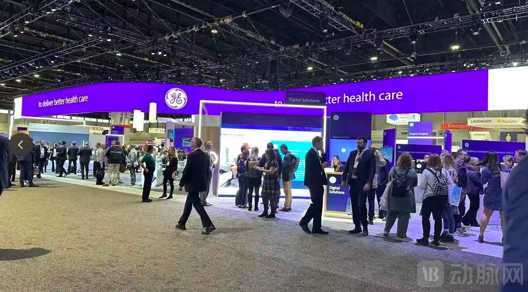 RNSA GE Healthcare Booth
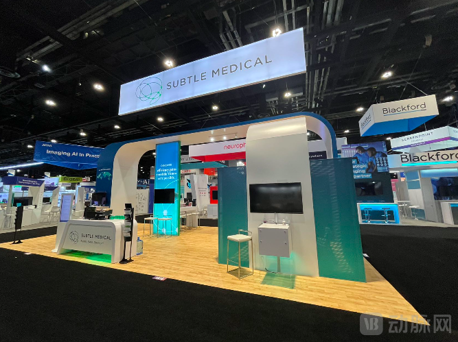
GE Healthcare’s philosophy appears to help radiology unlock value by enhancing efficiency and expanding physician resources. However, some startups have taken a different approach, focusing on accelerating the medical imaging process. For instance, Subtle Medical, founded by a Stanford team, has concentrated exclusively on this niche segment.
Subtle Medical’s SubtlePET and SubtleMR have been showcased at the RSNA annual meetings over the years. Empowered by AI, Subtle Medical accelerates MRI and PET imaging processes by 4–10 times while ensuring diagnostic-grade accuracy; it also leverages AI to enhance MRI images, reducing contrast agent usage by a factor of 10.
This year, Subtle Medical introduced its SubtleSynth product line, which has just received FDA approval. It leverages deep learning to synthesize STIR-like contrast from existing T1- and T2-weighted images, enabling a leap forward in the speed and enhancement of spinal imaging.
 RNSA Deep Medical Booth
AI-powered auxiliary diagnostic products on display permeated the entire exhibition hall. CT, MR, XR, ultrasound—each modality had its corresponding AI solutions. However, unlike in China, foreign manufacturers tend to focus their efforts on specific domains, such as stroke, thoracic, and cardiac applications, with few companies achieving the broad coverage of digital human solutions seen among Chinese enterprises.
Certainly, the development of intelligent software technology does not mean that cutting-edge medical imaging technology is reaching new heights.
GE Healthcare’s energy CT, Philips’ spectral CT, and Siemens’ photon-counting CT were all exhibited in the showroom and garnered widespread attention from leading scholars in this year’s published papers and conference lectures. Various new MR and molecular diagnostic equipment from different manufacturers also made their debut; further details are not provided here.
Riding the wave of AI’s ascent, vendors from outside the traditional healthcare sector have also been drawn in. At this year’s RSNA, NVIDIA was also present, unveiling multiple tools aimed at accelerating AI development for hospitals and research institutions.
NVIDIA’s MONAI, an open-source AI framework for medical imaging accelerated by NVIDIA technology, is designed to reduce the complexity of workflows from research and development to clinical practice.
With MONAI, developers can easily build and deploy AI applications, streamline the model development workflow, create models ready for clinical integration, and help physicians interpret medical imaging results more easily while gaining deeper insights into patient conditions.
Haris Shuaib, Head of AI Transformation at the AI Centre, stated, “Researchers, hospitals, and startups across the broader healthcare ecosystem are beginning to recognize the benefits of integrating streamlined AI workflows into their operations. The open-source MONAI ecosystem is dedicated to maximizing interoperability and impact by standardizing hundreds of AI algorithms, while reducing deployment timelines from three to six months down to just a few weeks.”
Currently, the AI Centre has developed algorithms that improve diagnostic accuracy for conditions such as COVID-19, breast cancer, brain tumors, stroke, and dementia. AIDE enables seamless and secure integration of approved AI algorithms with patient cases, allowing data to be processed without leaving the hospital.
Sebastien Ourselin, Deputy Director of the AI Centre, stated, “The AI Centre has played a pivotal role in the process of integrating AI into national healthcare. We are committed to implementing safe, robotic AI innovations in clinical practice, and the deployment of MONAI marks a significant milestone in this journey. This goal can only be achieved through robust collaboration between academia and industry leaders such as NVIDIA.”
In recent years, Chinese radiology has been steadily converging with the world’s leading nations, both in terms of hardware and software infrastructure and in the fundamental understanding of the discipline itself. Despite the ongoing impact of the COVID-19 pandemic, 32 domestic imaging-related manufacturers participated in the RSNA Annual Meeting, sharing the strength of Chinese radiology with medical industry professionals from around the globe.
As the only domestic medical imaging AI company exhibiting on-site at this conference, Shukun Technology presented its “Digital Doctor” product portfolio, which comprises more than 30 products. These include independently developed, comprehensive healthcare solutions covering the entire workflow of disease screening, assisted diagnosis, and treatment decision-making for major common and chronic conditions such as cardiovascular and cerebrovascular diseases and cancer.
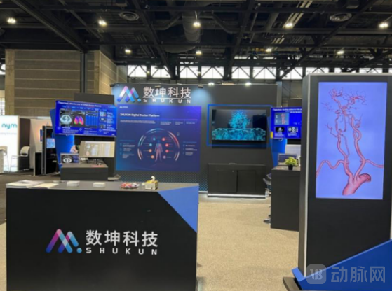 RSNA Shukun Technology Booth
In terms of imaging equipment, industry giants such as Neusoft Medical, Mindray, Anke, and Jusha Medical set up exhibitions on-site in Chicago. Taking Neusoft Medical as an example, its flagship products, including the NeuViz Epoch CT, NeuMR 1.5T MRI, and NeuWise PET/CT, were all showcased. Amid the pandemic, Neusoft Medical’s overseas market expansion is expected to accelerate further.
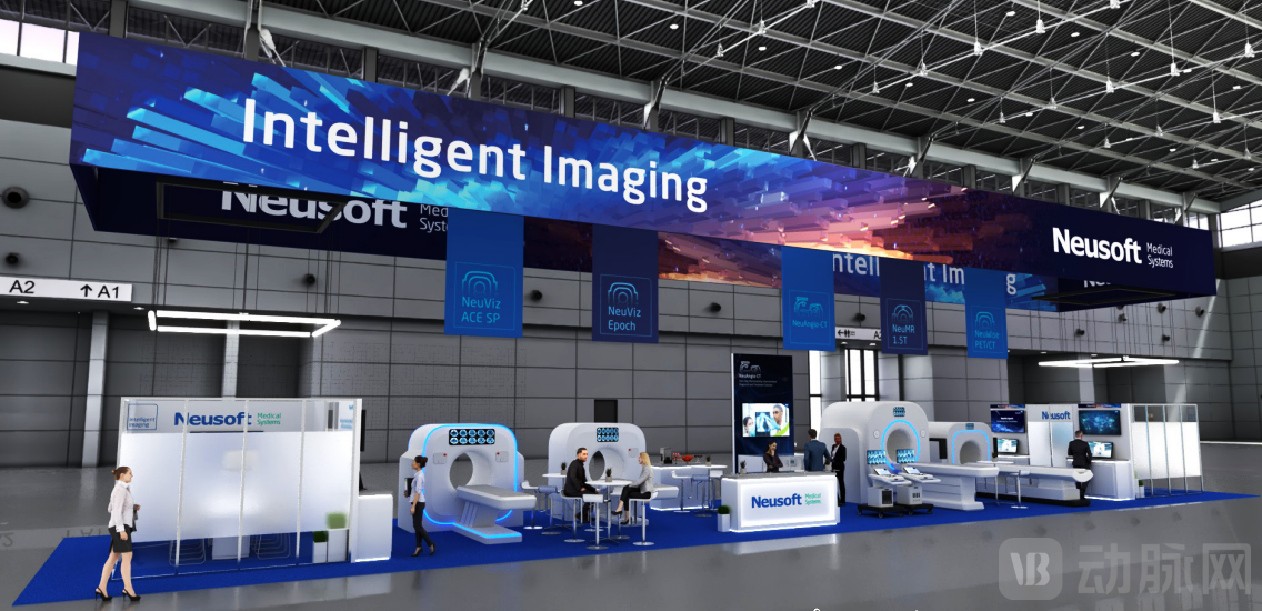
Neusoft Medical Booth at RSNA
Although United Imaging Healthcare did not have an exhibition booth at this year’s RSNA, it rented three conference rooms to showcase its full product portfolio. Meanwhile, Wandong Medical and NanoVision presented their products online, highlighting the disruptive innovations of Chinese-made CT systems to a global audience.
As we witness the deepening expansion of Chinese radiology into overseas markets, we also hope that leading medical imaging hardware and software enterprises will effectively integrate their cutting-edge insights—beyond mere technology—with radiological healthcare in China. Redefining the value of medical imaging and delivering it to patients is equally crucial for the future development of Chinese radiology.