DPM Advances NIR-II Imaging with Breakthroughs in Methods, Systems, and Clinical Translation
In the era of clinical medicine advancing toward “precision,” the rapidly emerging optical molecular imaging technology from DPM companies has become a powerful tool driving precision diagnosis and treatment. Leveraging technical advantages such as intuitive visualization, high accuracy, and rapid imaging, optical molecular imaging can accurately detect minute lesions, residual disease, nerves, lymph nodes, and other tissues, thereby efficiently assisting clinicians in decision-making and helping to improve patient outcomes.
Currently, DPM’s optical molecular imaging technology has been applied in the development of novel pharmaceutical agents, cerebrovascular imaging, tumor detection, lymphatic system imaging, multimodal imaging, pharmacokinetic/pharmacodynamic (PK/PD) studies, and intraoperative navigation, demonstrating broad prospects for future development.
NIR-II Molecular Imaging Technology
Development Potential and Application Value
Current optical molecular imaging primarily utilizes light signals within the visible spectrum (400–700 nm) and the near-infrared I window (NIR-I, 700–900 nm). In recent years, the emerging near-infrared II (NIR-II) molecular imaging technology, which leverages light signals in the 900–1700 nm wavelength range, has demonstrated significant potential and application value in biomedical fields such as in vivo imaging and disease detection. This is attributed to its ability to acquire images with lower background noise, higher resolution, improved contrast, and greater penetration depth.
In this highly promising emerging technology sector, industry players are racing to develop imaging system products. Among them,DPM Company possesses comprehensive advantages spanning imaging methodologies, imaging systems, fundamental research, and clinical translation.DPM was incubated by the Key Laboratory of Molecular Imaging of the Chinese Academy of Sciences, with Beijing Digital Precision Medical Technology Co., Ltd. serving as its R&D center and Zhuhai Dipu Medical Technology Co., Ltd. as its production base. Its scientific research product portfolio covers near-infrared II small animal in vivo imaging systems, multimodal small animal in vivo imaging systems, and magnetic particle imaging (MPI) systems. Its core technologies have received numerous accolades, including the Gold Medal at the Geneva International Exhibition of Inventions, the Gold Medal at the China National Invention Exhibition, and the Patent Excellence Award from the China National Intellectual Property Administration. The “Optical Molecular Imaging Surgical Navigation System” won first prize in the China Medical Device Innovation and Entrepreneurship Competition; the “Near-Infrared Fluorescence Imaging Intraoperative Navigation System” received support from the Ministry of Science and Technology’s Key Special Project on “Research and Development of Digital Diagnostic and Therapeutic Equipment”; and the “Broad-Spectrum Super-Resolution Intelligent Endoscopic Molecular Imaging System” was selected for the Ministry of Industry and Information Technology’s “Open Competition Mechanism” project for AI medical device innovation. Related core technologies have been published multiple times in Nature and Science subsidiary journals, as well as in journals such as the Journal of Clinical Oncology (JCO).

The Key Laboratory of Molecular Imaging of the Chinese Academy of Sciences, incubated by the Intelligent Medicine Research Center of the Institute of Automation, Chinese Academy of Sciences (CASIA), was officially established in 2013. With the laboratory’s rapid development and continuous accumulation of scientific achievements in recent years, its positioning within CASIA’s “One-Three-Five” strategic plan has evolved. During the 12th Five-Year Plan period, the laboratory was designated as one of the “Five Key Cultivation Directions,” specifically under “Intelligent Medicine and Novel Medical Devices.” During the 13th Five-Year Plan period, CASIA elevated the laboratory’s strategic status to one of the “Major Breakthrough Directions,” namely “Multimodal Medical Imaging and Intelligent Medical Equipment.” The Institute has set higher strategic requirements for the laboratory’s development in the new five-year period and provided strong support.The laboratory is generally positioned to address challenging scientific and technical issues in the field of biomedical engineering. It brings together core researchers from CASIA engaged in basic and clinical translational research in molecular imaging, big data analysis in biology and medicine, and the development of intelligent medical equipment. Leveraging information science and technology as its foundation and tools, the laboratory focuses on early detection, precise diagnosis, and treatment and rehabilitation of major diseases at the molecular and cellular levels. Aimed at leading the development of related theories and technologies in China in areas such as fundamental molecular imaging research, platform construction, equipment development, and application translation, the laboratory strives to become a renowned research and translational center with significant international academic influence.
DPM Near-Infrared II In Vivo Small Animal Imaging
Multispectral, Multifunctional Imaging System
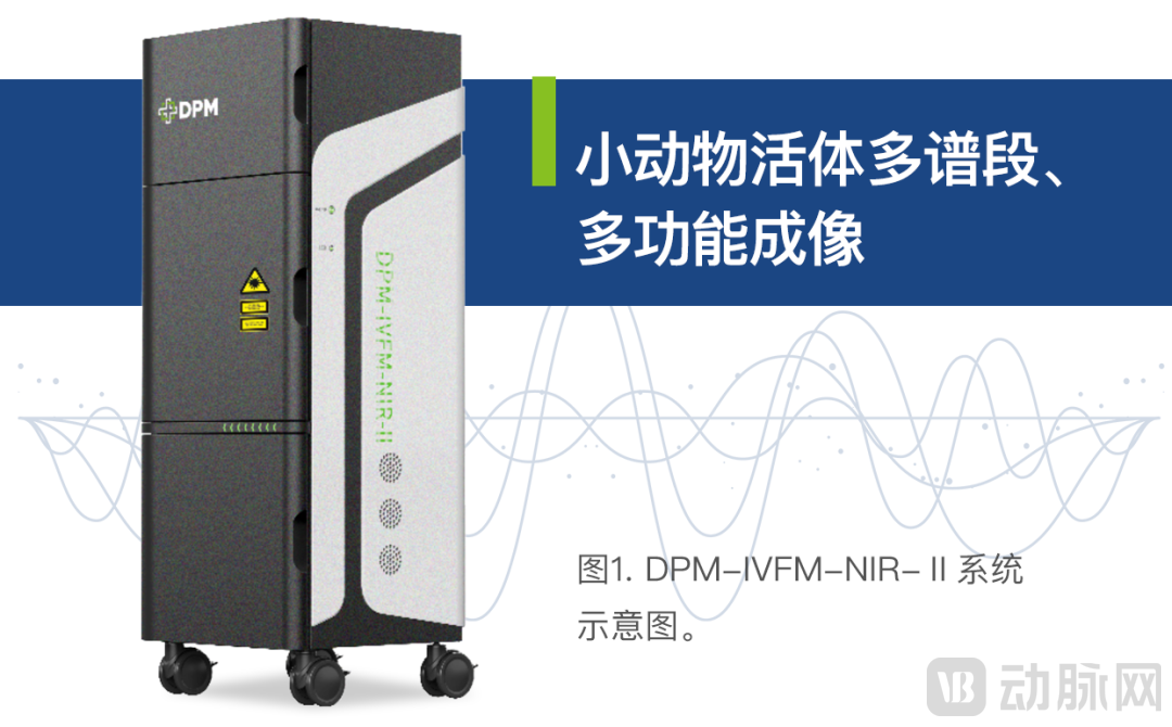
Building on its independently developed imaging system, the DPM team collaborated with Zhujiang Hospital of Southern Medical University and Shenzhen Technology University to design a novel near-infrared II (NIR-II, 1000–1700 nm) rare-earth downconversion fluorescent nanoprobe. Using a thermal decomposition method, they synthesized lanthanide-based fluorescent probes capable of simultaneously emitting fluorescence signals in three distinct spectral bands, and enhanced the biocompatibility of these nanoparticles through liposome encapsulation. Leveraging this probe in conjunction with the DPM team’s DPM-IVFM-NIR-II system (Figure 1), they achieved new applications for multispectral, multifunctional in vivo bioimaging in small animals. The related findings were recently published in Nano Today, a top-tier journal in engineering and technology indexed by the Chinese Academy of Sciences.
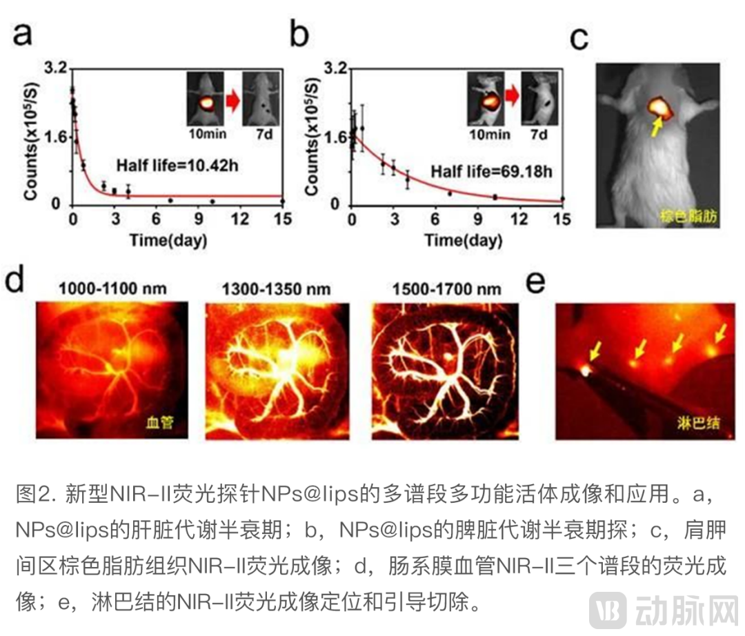
This study synthesized a novel multispectral fluorescence probe for the second near-infrared (NIR-II) window, which can simultaneously emit three distinct spectral fluorescence signals under 808 nm laser excitation, covering 1000–1100 nm (NIR-II), 1300–1350 nm (NIR-IIa), and 1500–1700 nm (NIR-IIb). This enables a series of biomedical applications, including in vivo imaging of brown adipose tissue, microvasculature, lymphatic vessels, and lymph nodes in small animals.
To endow the probe with superior biocompatibility and rapid metabolic clearance, this study modified lanthanide nanoparticles with liposomes to facilitate their rapid excretion from the hepatosplenic system (Fig. 2a,b), thereby reducing the biological toxicity associated with nanoparticle retention in vivo. In in vivo applications, high-sensitivity imaging of brown adipose tissue was achieved by leveraging the NIR-II multispectral fluorescence emitted by this novel probe (Fig. 2c). Brown adipose tissue is a crucial metabolic organ that holds significant value in combating metabolic disorders such as obesity and type 2 diabetes. The new imaging method established in this study provides a novel strategy for promoting in vivo detection of brown adipose tissue and facilitating the exploration of new drugs and therapies. Furthermore, the new probe and system enabled in vivo multispectral imaging of mesenteric blood vessels, demonstrating that both imaging resolution and signal-to-noise ratio could be further enhanced as the imaging wavelength increased (Fig. 2d). Additionally, NIR-II imaging allowed for the in vivo localization of sacral and popliteal lymph nodes in mice, as well as fluorescence image-guided lymph node resection (Fig. 2e).
Building on preclinical studies at the animal level, the DPM team has engaged in in-depth interdisciplinary collaborations with multiple Grade A tertiary hospitals and research institutes in China, thereby advancing novel clinical applications of NIR-II imaging technology.
In response to the persistently high incidence of liver cancer in China, a life-threatening disease, the DPM team successfully developed a clinically applicable NIR-II fluorescence imaging technology and surgical navigation system through nearly three years of interdisciplinary collaboration between medicine and engineering. This achievement provides robust support for the implementation of precision surgery for liver cancer.
DPM Novel NIR-II Fluorescence Imaging Technology
Application in the Clinical Treatment of Liver Cancer
For a long time, surgical intervention has played a pivotal role in the clinical management of liver cancer. Complete resection of hepatic tumor lesions can increase the 5-year survival rate by more than 20%. However, accurately delineating tumor boundaries and identifying micrometastases within the limited visual field obscured by connective tissue and vasculature remains a significant clinical challenge in achieving precise hepatic surgery.
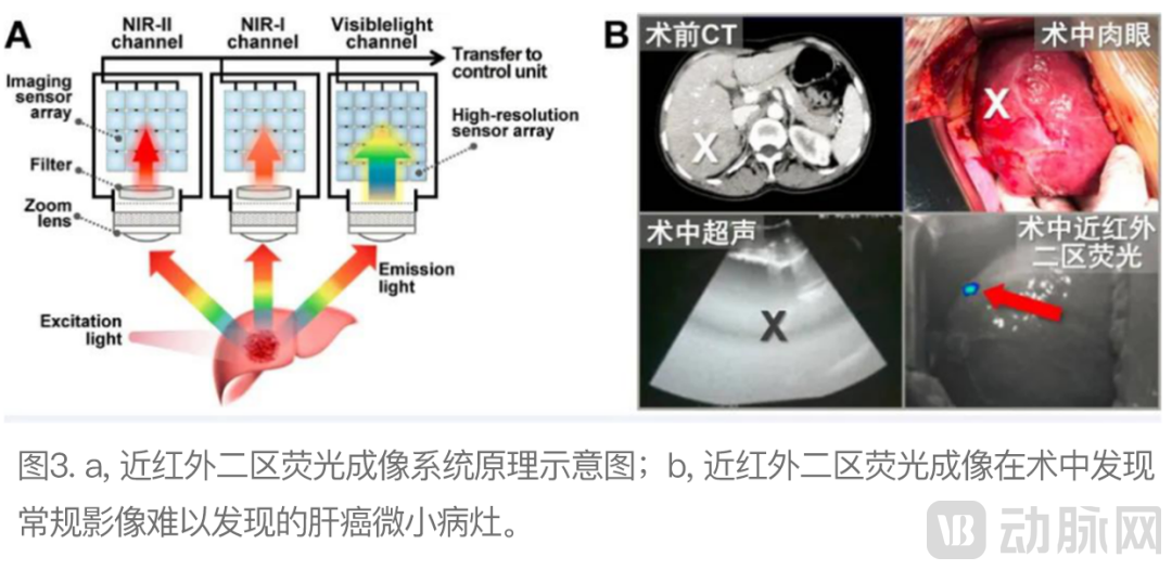
By leveraging novel NIR-II fluorescence imaging technology and intraoperative navigation systems, in conjunction with the fluorescent probe indocyanine green (ICG), it is possible to safely and efficiently achieve "optical labeling" of liver cancer lesions. This approach "illuminates" tumor foci during surgical procedures, accurately revealing the distribution of hepatocellular carcinoma cells and tumor morphology, thereby assisting surgeons in making precise intraoperative decisions and advancing toward the goal of precision resection: "minimizing resection margins to reduce recurrence while preserving maximal liver function."
Clinical trial results demonstrate that NIR-II fluorescence imaging effectively “extends” the surgeon’s visual capabilities, accurately detecting minute hepatocellular carcinoma lesions (2–3 mm in diameter) that are difficult to identify using conventional clinical imaging modalities (such as CT, MRI, and intraoperative ultrasound) or intraoperative visual inspection. Furthermore, after resection of the primary tumor, multispectral fluorescence-guided surgery helps surgeons precisely identify residual hepatocellular carcinoma tissue on the surgical margin. Statistical data from enrolled patients, corroborated by pathological validation, indicate that the application of multispectral fluorescence-guided surgery significantly increases the R0 resection rate for hepatocellular carcinoma and improves the five-year survival rate of patients.
Related work was recently published in Nature Biomedical Engineering, a prestigious journal in the field of biomedical engineering. Upon publication, the study received acclaim from international peers, including Dr. P. Pàmies, Editor-in-Chief of Nature Biomedical Engineering, and Professor H. Choi of Harvard Medical School, who described it as “the first-in-human.” These evaluations highlighted that NIR-II fluorescence imaging technology, “after a decade of fundamental research, has finally been successfully applied in clinical settings for the first time worldwide (the first-in-human), opening the door to its widespread medical application and ultimately benefiting patients.”
DPM Novel NIR-II Fluorescence Imaging Technology
Application in the Clinical Treatment of Gliomas
Furthermore, the DPM team has proposed a novel method for real-time intraoperative diagnosis of gliomas, leveraging NIR-II fluorescence imaging and deep convolutional neural networks, to address the challenges posed by complex tissue architecture and high surgical difficulty. This approach enables rapid and accurate prediction of pathological diagnoses for glioma tissue specimens during surgery. Consequently, it assists surgeons in efficiently achieving "maximal safe resection," thereby protecting patients' functional brain areas while removing tumor cells as comprehensively as possible, ultimately improving postoperative quality of life.
Gliomas account for 75% of primary malignant brain tumors in adults. More than half of these glioma cases are the most lethal type, glioblastoma, with a mean overall survival of only 14.6 months. Neurosurgical resection is the primary treatment modality for gliomas; however, the difficulty in precisely identifying tumor boundaries during surgery often leads to residual tumor tissue and early recurrence. Therefore, rapid and accurate intraoperative diagnosis of tissue specimens is critical. However, the existing hematoxylin and eosin (H&E) staining method is complex and has limited accuracy, making it difficult to meet the clinical needs for rapid intraoperative diagnosis of gliomas.
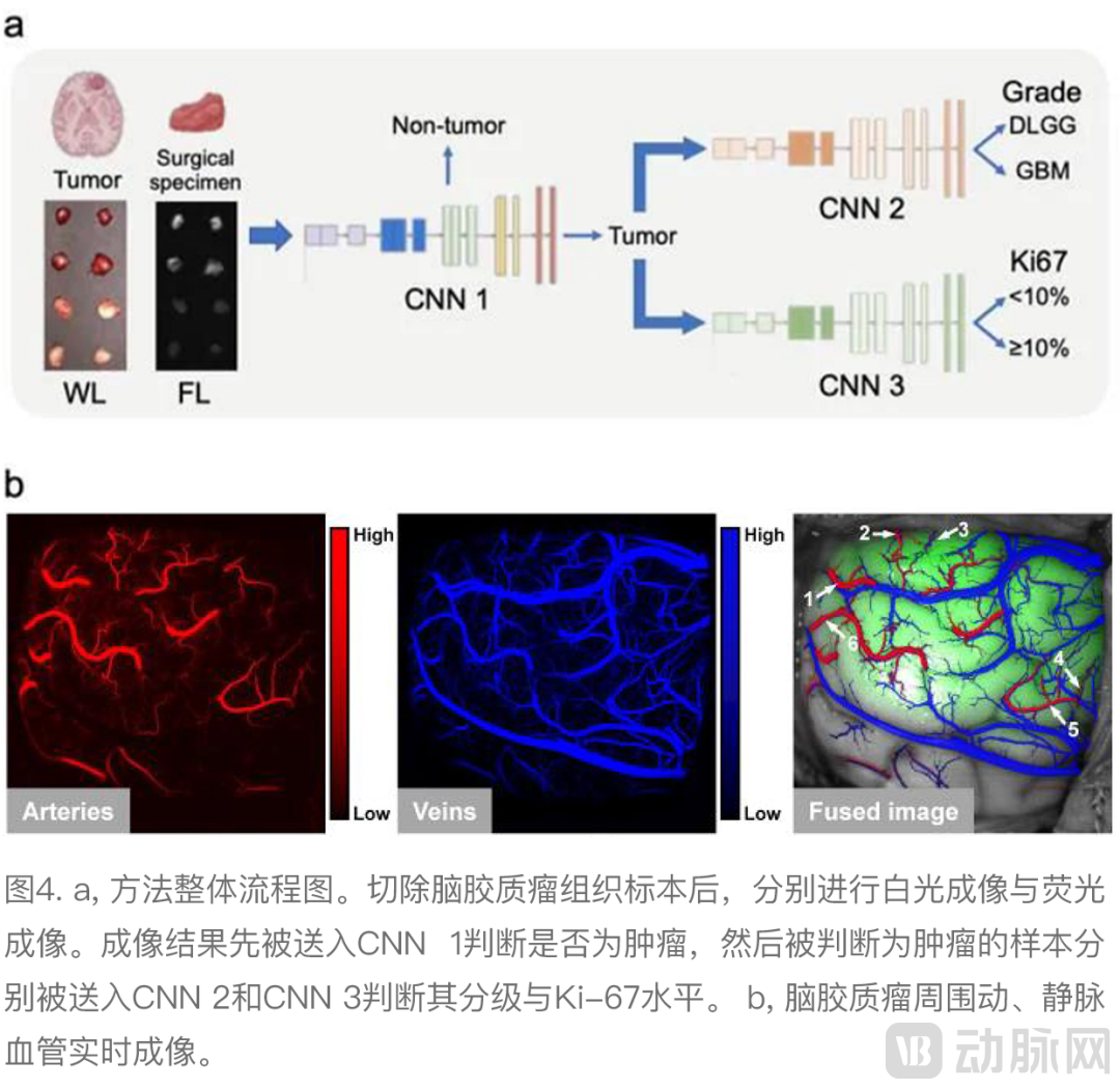
To achieve rapid and accurate intraoperative diagnosis of glioma lesions, the DPM team collaborated with Beijing Tiantan Hospital, Capital Medical University, to conduct a clinical trial on NIR-II intraoperative fluorescence imaging, enrolling 23 patients with malignant gliomas. Each patient received an injection of indocyanine green (ICG) prior to surgery, followed by fluorescence-guided glioma resection. A total of 1,874 tissue samples were collected during surgery, with both white-light and NIR-II fluorescence images acquired for each sample. These images were analyzed using a deep convolutional neural network to predict whether the specimen was tumorous, as well as to determine the tumor grade and Ki-67 index. The postoperative pathological results of each tissue specimen served as the gold standard to validate the performance of the novel NIR-II fluorescence imaging technique. Additionally, gradient-weighted class activation mapping (Grad-CAM) was employed to visualize and interpret the model’s predictions. To further evaluate the diagnostic performance and clinical value of the model, its diagnostic results were compared with the intraoperative diagnoses made by three neurosurgeons.
The results indicate that the integration of NIR-II fluorescence molecular imaging with deep learning models can effectively predict pathological outcomes of glioma tissue specimens intraoperatively. For the task of predicting whether a specimen is tumorous, the NIR-II fluorescence deep learning model achieved an AUC of 0.945 on the test set. Compared with visual assessment by physicians, this novel intelligent NIR-II fluorescence imaging method demonstrated higher sensitivity (93.8% vs. 82.0%, P<0.001) while maintaining comparable specificity (>80%). Analysis of specific diagnostic results further revealed that the proposed novel intelligent NIR-II fluorescence imaging method could correct more than 70% of diagnostic errors made by neurosurgeons. Moreover, it can provide real-time information on lesion grading and Ki-67 expression intraoperatively, with AUCs reaching 0.810 and 0.625, respectively—capabilities unattainable through naked-eye examination by physicians during surgery. These findings were published in the European Journal of Nuclear Medicine and Molecular Imaging, a top-tier medical journal indexed by the Chinese Academy of Sciences.
Leader in the Post-Precision Medicine Era
In summary, while continuously innovating theories and methods in molecular imaging, the DPM team has vigorously advanced the clinical translation of next-generation molecular imaging technologies for the diagnosis and treatment of various malignant diseases, actively promoting the large-scale clinical application of these novel technologies. Guided by the philosophy of “identifying problems from clinical practice, developing solutions that transcend current clinical standards, and evaluating outcomes through clinical application,” the DPM team focuses on the clinical needs for precise diagnosis and treatment of major malignant diseases such as cancer. The team conducts collaborative research and development of novel molecular imaging technologies along two primary directions: First, to achieve real-time in vivo pathological visualization by “clearly visualizing” tumor margins and micrometastases at the molecular and cellular levels, thereby constructing novel tumor information acquisition devices to assist surgeons in improving surgical outcomes. Second, to quantitatively characterize tumor imaging features at the molecular and cellular levels, enabling AI-based image recognition to approximate pathology-based clinical understanding, thus innovating new methods for quantitative tumor information analysis to assist physicians in making precise diagnoses and prognostic predictions. Ultimately, this approach aims to realize individualized precision diagnosis and treatment for patients, providing robust technical support for the rapid development of the new era of precision medicine.