Stanford Bio-X Awards $4 Million in Seed Grants to 10 Interdisciplinary Medical Projects Targeting Regenerative Medicine, Antibiotic Resistance, and Precision Diagnostics
Focusing on original innovation and encouraging free exploration remain the enduring mantra for translating scientific achievements into practical applications. Recently, the New Cornerstone Investigator Program, one of the largest philanthropic initiatives in China funding basic scientific research with private capital, officially announced its first cohort of grantees, among whom 28 scientists were selected from the fields of biology and medical sciences.
On the distant West Coast,Stanford UniversityThe Bio-X Interdisciplinary Initiatives Program (IIP) seed funding scheme was launched in 2000. Reportedly, Stanford Bio-X awards approximately $4 million in two-year seed grants every other year. To date, the program has provided critical early-stage funding for 252 interdisciplinary projects, strengthening interdisciplinary research within the Stanford scientific community in the fields of bioengineering, biological sciences, and biomedical-related areas.
So, which medical seed projects did Stanford Bio-X select in 2022? And what trends do they foreshadow?
Deep Cross-Disciplinary Integration: Ten Major Projects Deployed in Frontier Fields Including Genetics, Structural Biology, and Infectious Diseases
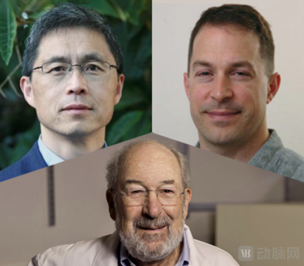
1Deep Learning for the Regulatory Genomics of Whole-Body Regeneration
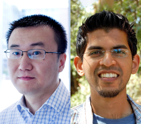
Wang Bo – Assistant Professor of Bioengineering and Assistant Professor of Developmental Biology
Anshul Kundaje—Assistant Professor of Genetics and Computer Science
Thousands of genes must be upregulated or downregulated in space and time to coordinate the responses of various cell types. To understand how this complex molecular choreography is encoded in the genome, the team established a novel laboratory model that combines single-cell multi-omics sequencing with deep learning models to identify regulatory sequences. They then elucidated the genotype–phenotype relationships by computationally predicting the functional effects of mutations in these regulatory sequences, and finally validated these predictions through in vivo experiments using genome editing.
This enables researchers to understand the regulatory genome controlling molecular responses after wounding, thereby informing new strategies to redirect destructive wound responses (such as those commonly observed in humans) toward reparative responses, with the promise of enhancing patients’ regenerative recovery after injury and disease.
2Development of Novel Polyacrylamide Copolymers as Next-Generation Broad-Spectrum Antibiotics Targeting Immune Resistance Mechanisms
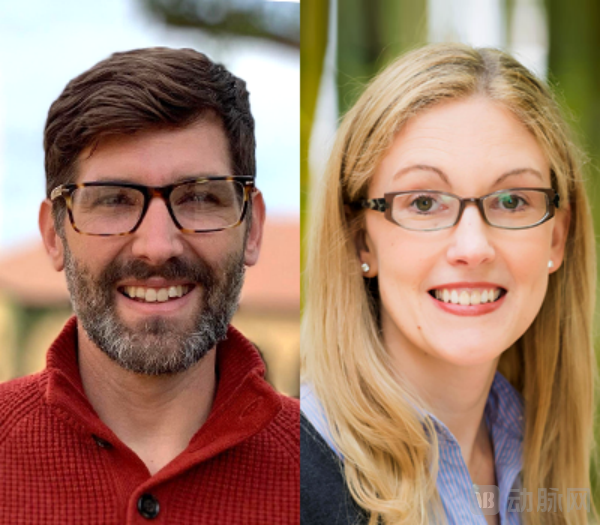
Eric Appel - Assistant Professor of Materials Science and Engineering
Lynette Cegelski – Associate Professor of Chemistry
In recent years, there has been little progress in the development of new antibiotics, while antibiotic-resistant strains continue to emerge worldwide at an alarming rate. To address this unmet clinical need, Eric Appel and Lynette Cegelski have developed a novel class of inexpensive, stable, broad-spectrum polyacrylamide-derived copolymer antibiotics by combining rational chemical design with high-throughput synthesis and screening methods.
It is reported that the project will bring together researchers from diverse backgrounds to collaboratively develop novel broad-spectrum antibiotic copolymers. These copolymers offer advantages such as low cost, scalability, high stability, and resistance to antibiotic resistance mechanisms. Furthermore, due to their selective mechanism of action, they are non-toxic to mammals, thereby addressing the critical global demand for new antibiotics.
3Elucidating the Mechanisms of Wnt Signalosome Formation and Function through Single-Molecule Counting and DNA Origami

William Weis – Professor of Structural Biology, Molecular and Cellular Physiology, and Photon Science at the William M. Hume School of Medicine
Alexander Dunn - Associate Professor of Chemical Engineering
Wnt/β-catenin signaling controls the differentiation and renewal of stem cells during tissue development and maintenance in adult organisms. Wnts are growth factors that bind to cell surface receptors, inducing changes in gene expression that regulate cell growth and differentiation. Mutations in components of this pathway underlie many diseases, including cancer, which results from uncontrolled cell growth.
Although the core molecular components and basic circuitry of this pathway have been established, the molecular mechanisms underlying Wnt/β-catenin signaling remain unclear, hindering the development of therapies targeting this pathway. In a recent study, the team discovered that this pathway employs a novel mechanism for transmembrane signal transduction. They will combine advanced optical microscopy and single-molecule imaging with engineered ligands to elucidate the precise mechanisms controlling Wnt/β-catenin signaling.
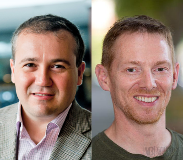
4Bacteriophages as Novel Pathogenic Factors in Lung Transplant Rejection

Paul Bollyky - Associate Professor of Medicine (Infectious Diseases) and Microbiology & Immunology
Markus Covert - Professor of Bioengineering and Professor of Chemical and Systems Biology
This project proposes a breakthrough hypothesis: certain bacteriophages may promote transplant outcomes. This hypothesis stems from a novel epidemiological association between specific bacteriophage families and lung transplant rejection. The team has isolated and cultured bacteriophages from these specific families, discovering that they enter human cells and trigger robust innate immune responses. The proposed model suggests that these bacteriophages bypass the pathways of conventional immunosuppressive drugs used in lung transplantation to activate lymphocytes, thereby driving chronic rejection.
In subsequent work, the team will elucidate the mechanism by which this phage is internalized by human cells, clarify the intracellular signaling events mediating phage-induced immune activation, and finally determine how these phages influence B cell activation and antibody responses in a mouse model of lung transplantation. It is understood that this project will lay the foundation for R01-level research on phages as biomarkers and therapeutic targets for human lung transplant rejection.
5Non-Invasive Virtual Biopsy for Diagnosing Basal Cell Carcinoma Using Machine Learning

Adam de la Zerda – Associate Professor of Structural Biology
Kavita Sarin – Associate Professor of Dermatology
The team’s latest research demonstrates that a novel alignment method combined with OCT2Hist can be used to train neural networks to generate virtual microscopic images of normal skin from OCT scans, enabling clinicians to achieve “virtual biopsy” results without invasive skin excision. As part of this project, the team plans to expand the OCT2Hist framework for basal cell carcinoma (BCC) detection, developing a real-time, non-invasive “virtual skin biopsy” tool for BCC screening.
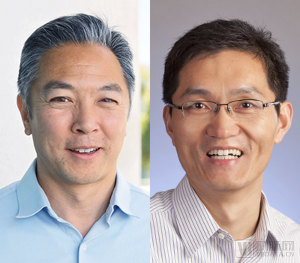
6Extracellular Vesicles and Their Cargo Atlas in Prostate Cancer Cells
Utkan Demirci - Professor of Radiology and Electrical Engineering
Andrew Gentles - Assistant Professor of Medicine
With the emergence of new molecular analysis technologies, liquid biopsy has garnered attention for its role in cancer identification and progression monitoring through biomarkers circulating in the blood. Among these, extracellular vesicles (EVs) are nanosized vesicles shed by cells that transport proteins, nucleic acids, metals, lipids, and metabolites; in other words, circulating EVs in the blood carry cargo that reflects the characteristics of their cellular origin.
In this project, the team will apply state-of-the-art extracellular vesicle (EV) isolation techniques, combined with in vitro models, clinical samples, downstream analyses, and data analytics, to create an EV atlas of prostate cancer. This initiative aims to develop EVs as non-invasive or minimally invasive biomarkers and to establish novel early detection and diagnostic approaches for solid organ tumors.
7Impaired Cellular RNA Editing Is a Cause of Inflammation in Inflammatory Bowel Disease
Calvin Kuo - Professor Maureen Lyles D'Ambrogio
Jin Billy Li - Assistant Professor of Genetics
Currently, little is known about the initial triggers of inflammatory bowel disease (IBD), leading to delays in the development of new therapies.
To address this issue, the team identified a novel genetic risk variant enriched in patients with inflammatory bowel disease (IBD) and other immune-mediated disorders. These variants impair the ability of the protein ADAR1 to edit double-stranded RNA (dsRNA). In the absence of ADAR1-mediated editing, these dsRNAs are mistakenly recognized by cells as viral infections, thereby triggering inflammation.
To verify this possibility, the team first measured the activity of modified ADAR1 in a controllable form within the mouse intestine. Additionally, the team will examine whether DNA from adult patients with inflammatory bowel disease harbors genetic risk variants.
8Multiparametric Imaging for Immune Checkpoint Inhibitor Therapy
Jianghong Rao - Professor of Radiology and Chemistry
Craig Levin - Professor of Radiology
Ronald Levy - Professor of Medicine
Currently, diagnostic imaging based solely on tumor size measurements is insufficient for assessing the initial response to immunotherapy and disease evolution. Therefore, there is an urgent need to develop new non-invasive imaging assays to determine whether a patient’s cancer responds to immunotherapy at an earlier time point than allowed by current methods.
To this end, the research team leveraged expertise in chemistry, physics, engineering, molecular imaging, and cancer immunology to develop a novel non-invasive multiparametric imaging assay capable of sensitively detecting two biomarkers associated with response to immunotherapy in tumors. This new imaging assay will be evaluated in preclinical mouse models of colorectal cancer to facilitate effective patient selection and accurate monitoring of cancer immunotherapy.
9Label-Free and Non-Transgenic Detection of Neurotransmitters in the Enteric Nervous System Using Ultraflexible Intraneural Electronics
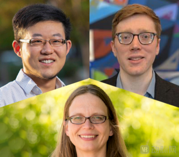
Guosong Hong - Assistant Professor of Materials Science and Engineering
Andrew J. Mannix - Assistant Professor of Materials Science and Engineering
Julia Kaltschmidt - Associate Professor of Neurosurgery
The team has developed an atomically thin MoS2 nanosensor to provide molecular insights into the high spatiotemporal resolution dynamics of neurotransmitters and neuropeptides within the enteric nervous system. Specifically, the MoS2 nanosensor can be injected into the myenteric plexus (MP) layer of the gastrointestinal wall, establishing an intimate interface between the sensing device and enteric neurons. This enables the recording of local nitric oxide (NO) concentrations and real-time monitoring of NO release with high spatiotemporal resolution during colonic motility assays, thereby elucidating the role of NO in regulating intestinal motility.
10Enzymatic Editing of Biofilms for Next-Generation Therapies

Steven Banik - Assistant Professor of Chemistry
Polly Fordyce - Assistant Professor of Genetics and Bioengineering
Many drivers of tumorigenesis and neurodegenerative diseases are proteins, yet most methods for delivering proteins via endosomal membranes rely on protein transduction domains. These domains must be present at very high concentrations to disrupt the lipid bilayer, which can lead to non-specific targeting, thereby limiting the types and sizes of proteins that can be delivered. Consequently, there is an urgent need for alternative strategies.
The team integrates techniques from chemical biology, protein engineering, cell biology, microfluidics, and biochemistry to explore phospholipase A (PLA) editing. By creating gaps in the bilayer membrane to drive the endosomal escape of protein therapeutics, they are developing PLA into a protein transduction reagent for modulating and targeting specific targets.
Conclusion
As a pioneer in translating scientific research into commercial applications, Stanford University has fostered the development of Silicon Valley and the biotechnology industry in Northern California. From the mid-1980s through the 1990s, companies founded by Stanford faculty and students, or those with university affiliations, accounted for more than 70% of Silicon Valley’s enterprises, setting a benchmark for universities and research institutions across the United States and around the world.
Even so, Stanford University has not slowed its pace. In September 2022, the Stanford School of Medicine launched the West Coast’s first Master’s program in Translational Research and enrolled six students. Reportedly, the program accepts students from diverse backgrounds and integrates clinical science and laboratory experience from the Stanford School of Medicine with those of biotechnology or pharmaceutical companies. The curriculum focuses on drug discovery and development, clinical trials and clinical research, as well as research design and analysis. Additionally, there is an applied medicine track emphasizing immunotherapy, gene therapy, vaccines, and biomarkers.
In 2023, with the launch of this course, Stanford University will also make more steady and sustained progress in the translation of scientific research achievements.