Breakthrough in Lung Cancer Organoid Research: Chuangxin International and Guangdong Provincial People's Hospital Publish Landmark Study in Cell Reports Medicine
Recently, a team from Guangdong Provincial People's Hospital, in collaboration with Chuangxin International (Guangzhou) Biotechnology Co., Ltd., published their findings in *Cell*, a journal dedicated to translational and clinical medicine.Cell) sub-journal Cell Reports Medicine (Cell Reports Medicine, IF: 16.988) published the article “Using patient-derived organoids to predict locally advanced or metastatic lung cancer tumor response: A real-world study.” Professor Yang Jinji from Guangdong Provincial People’s Hospital served as the lead contact, with Professor Wu Yilong and Director Chen Huajun as co-corresponding authors. Wang Hanmin, Zhang Chanyuan, Peng Kaicheng, and Chen Zexin, R&D Director at Chuangxin International, were co-first authors. This study was funded by the Guangdong High-Level Hospital Construction Project, as well as the National and Provincial Natural Science Foundations. An abstract of partial results from this study was accepted as a poster presentation by the European Society for Medical Oncology (ESMO) in 2021 and published in the prestigious journal Annals of Oncology (Annals of Oncology) published in the supplement.
Lung cancer ranks first in both incidence and mortality among malignant tumors in China. The prognosis for patients with advanced lung cancer complicated by malignant serous effusion (MSE) is often poorer. With the advent of the era of precision medicine, molecular targeted therapy can prolong overall survival (OS) and improve quality of life in patients with advanced lung cancer. However, some patients exhibit primary resistance to targeted drugs, and even those who initially respond inevitably develop secondary resistance. Currently, the resistance mechanisms in a significant proportion of patients remain unclear. Therefore, there is an urgent need to establish preclinical models that recapitulate the morphological and genomic characteristics of the original tumors to predict the efficacy of targeted therapy and chemotherapy in lung cancer.
To overcome this bottleneck, this study conducted the first large-scale collection of serous cavity effusions or tumor tissues from patients with advanced lung cancer to establish in vitro 3D organoid models. A total of 214 organoid models were successfully cultured, the vast majority of which were derived from malignant serous cavity effusions. The study evaluated the accuracy of predicting clinical efficacy based on Lung Cancer Organoid Drug Sensitivity Testing (LCO-DST) results. This research represents a breakthrough in the field of precision therapy for lung cancer. The results indicate that drug sensitivity testing of tumor organoids can be used to formulate personalized treatment strategies for advanced lung cancer.
Lung cancer is characterized by high tumor heterogeneity, exhibiting phenotypic and genotypic diversity, which poses significant challenges to precision or personalized medicine. Approximately 40% of patients with non-small cell lung cancer (NSCLC) harbor sensitive gene mutations; however, not all of these patients benefit from targeted therapy. Patients with the same histological type and genotype may exhibit vastly different responses to drugs and therapeutic outcomes, and tumors from different sites within the same patient may also vary. Consequently, researchers are eager to establish reliable preclinical models to evaluate tumor treatment efficacy. However, traditional models, such as cell lines, fail to adequately reflect the original characteristics of tumors. Patient-derived xenografts (PDXs) are both expensive and time-consuming, with a modeling success rate of only 20%–30%, falling far short of the demands of personalized medicine. With this objective in mind, the research team led by Professor Yang Jinji from Guangdong Provincial People’s Hospital and the Guangdong Lung Cancer Institute has been dedicated to constructing 3D in vitro lung cancer organoid models since December 2019, conducting globalFirst Large-ScaleReal-World Study Cohort: Exploring the Application Value of Organoids in Advanced Lung Cancer
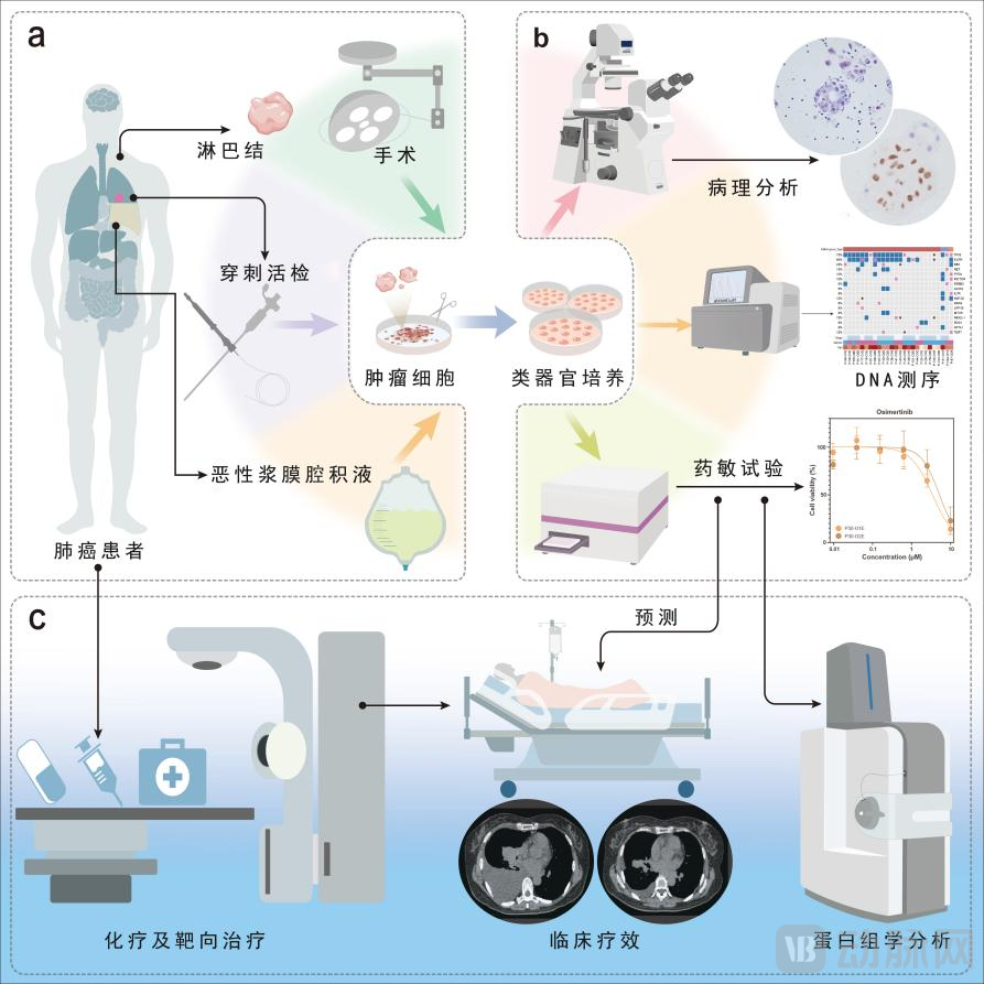
Graphical Abstract of This Study
This study established 214 organoid models from 107 patients, with the majority of samples derived from malignant serous effusions (MSE). The success rate for modeling organoids from MSE was 81.5% (132/162). Using the 214 successfully cultured organoids derived from tissues and MSE, we first performed histopathological or cytological validation, which showed a concordance rate of 77.6% (59/76) between the organoid samples and their matched clinical counterparts (Figure 1). Second, we validated the genomic concordance between organoids and clinical samples; using matched MSE genomes as the reference, the sensitivity for detecting all somatic mutations in organoid samples was 70.1%, with a specificity of 97.7% (Figure 2). These results demonstrate that organoids can adequately reflect the pathological phenotypes, genetic backgrounds, and gene expression profiles of their homologous tissues.
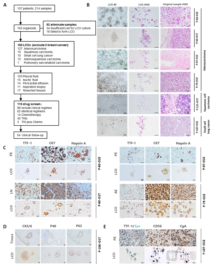
Figure 1. Flowchart of patient screening and pathological validation of LCOs.
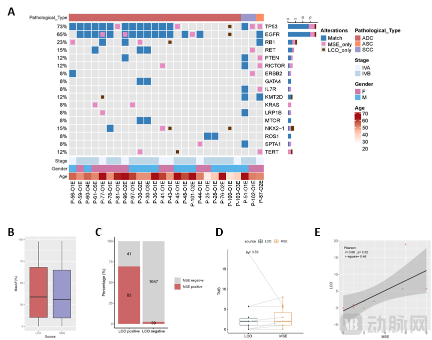
Figure 2. Genomic analysis of lung cancer organoid samples.
The most significant innovation of this study lies in the team’s use of organoid-based models to conduct sensitivity tests for targeted therapies and chemotherapy drugs, thereby predicting clinical tumor treatment efficacy. Clinical treatment regimens were categorized into four major groups: the osimertinib group, the chemotherapy group, the combination targeted therapy group, and other targeted therapy groups. The concordance rates between organoid drug sensitivity results and clinical efficacy were 86.7% (13/15), 83.3% (10/12), 100% (10/10), and 70.6% (12/17), respectively, with an overall accuracy of 83.3% (45/54) (Figure 3). To date,This is the largest real-world study with sample size in the international field of lung cancer organoids for predicting the efficacy of targeted therapy and chemotherapy.。
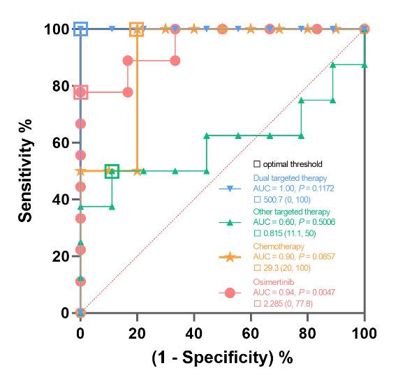

Figure 3. Concordance between drug sensitivity results in lung cancer organoids and clinical tumor response.
Tumor heterogeneity in lung cancer poses a significant challenge to clinical treatment. To comprehensively characterize this heterogeneity, Professor Yang Jinji’s team employed sequential sampling from multiple tumor sites followed by organoid culture. Their results demonstrated that in vitro organoid models recapitulate both the concordance and heterogeneity of the primary tumors, providing deeper insights into phenotypic and genotypic features and offering a more accurate representation of the overall tumor landscape (Figure 4).
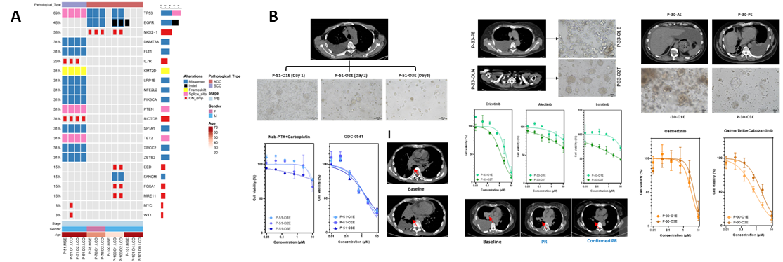 Figure 4. Concordance between drug sensitivity results of lung cancer organoids and clinical tumor efficacy.
Two Typical Cases of Advanced Lung Adenocarcinoma in This Study: One CaseEGFRGene Mutations Combined with PrimaryMETGene Amplification, Another CaseEGFRGenetic Mutations Combined with AcquiredRETGene fusion. Compared with single-agent targeted therapy, drug sensitivity results from two patients demonstrated a higher tumor control rate with combination targeted therapy (osimertinib plus savolitinib/cabozantinib). This was further corroborated in clinical practice, where both patients achieved partial response (PR) with the dual-targeted regimen, suggesting that combination targeted therapy may be a superior option for patients harboring such dual-gene alterations (Figure 5).
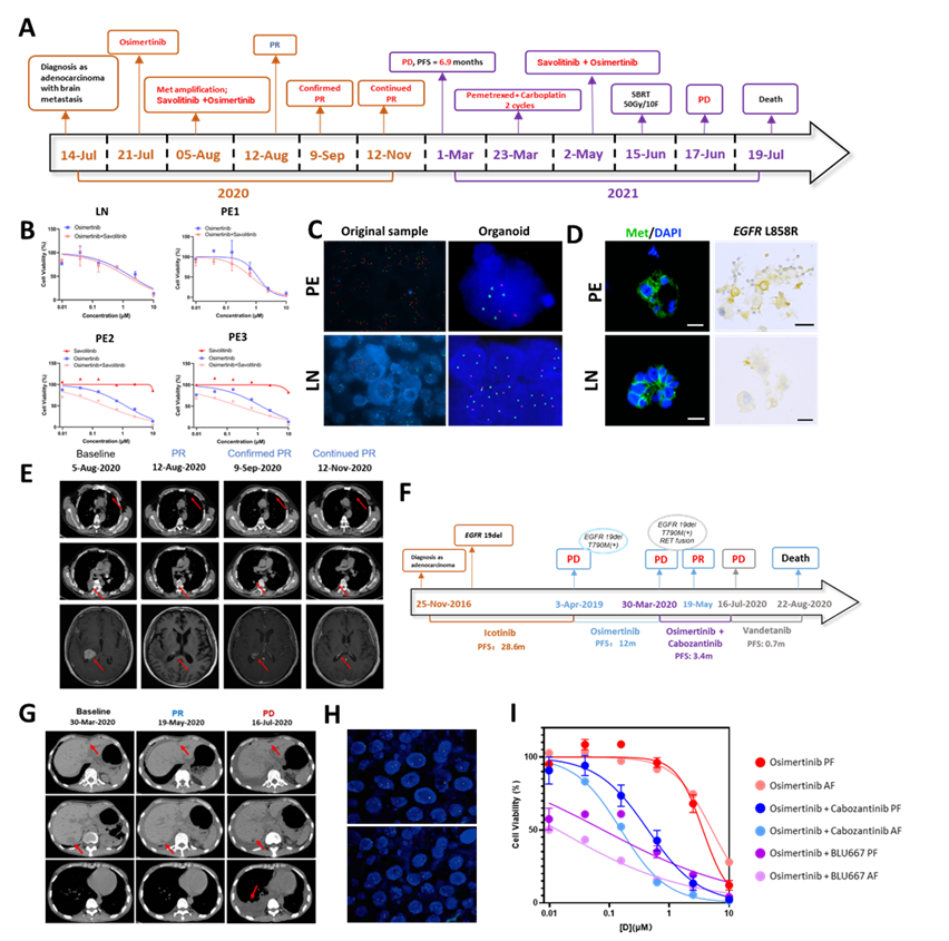 Figure 5. LCO predicts the efficacy of dual-targeted therapy.
Meanwhile, this study conducted proteomic analysis to further explore the molecular mechanisms of dual-targeted therapy, inEGFRMutation CombinationRETIn fusion-positive patients, dual-targeted therapy significantly induced cell death compared with single-agent targeted therapy, providing evidence to elucidate the efficacy of dual-targeted therapy (Figure 6).
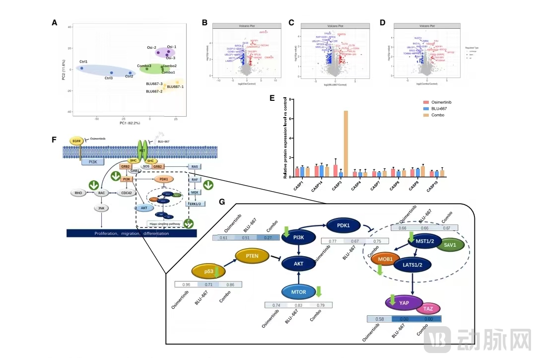
Figure 6. Proteomic analysis of the molecular mechanisms of dual-targeted therapy.
In this study, drug sensitivity testing based on lung cancer organoids accurately predicted the clinical efficacy of antitumor therapy in patients with advanced lung cancer within the cohort. This suggests that drug sensitivity testing using tumor organoids, as an in vitro model for lung cancer, serves as an effective predictive tool for precision or personalized medicine in advanced lung cancer. Furthermore, it holds significant potential for elucidating drug mechanisms of action, thereby helping patients achieve more efficient and precise treatment and improving survival outcomes. In the future, prospective multicenter clinical studies are still needed to further validate its clinical value in the treatment of lung cancer and other solid tumors.