SmartBrain: A 6-kg Wearable Brain PET System Enabling Dynamic Molecular Imaging Beyond the Scanning Room

GE Healthcare
Digital Solution Provider

How far are these scenarios from clinical practice — if epilepsy patients could undergo brain function imaging during seizures, if children didn’t have to remain still inside a confined scanning chamber, and if neuroscientists could observe changes in brain metabolism while a person walks and thinks?
This is not a fantasy. A nameSmartBrain's wearable brain Positron Emission Tomography (PET) system is attempting to liberate brain imaging from fixed examination rooms. Recently, a research team from Shenzhen Bay Laboratory, Harbin Institute of Technology, and the Seventh Affiliated Hospital of Sun Yat-sen University published a paper in the "Journal of Nuclear Medicine," reporting the complete performance evaluation results of this system.The system weighs only about 6 kilograms, has passed the full set of tests according to the National Electrical Manufacturers Association (NEMA) NU 2-2018 standard, and was compared side by side with GE HealthCare's Discovery MI PET/CT (DMI) system.
The "Static Dilemma" of Brain PET
Brain PET is a powerful imaging tool that directly reflects brain metabolic activity.However, traditional brain PET usually requires subjects to remain seated or supine during scanning. This poses a real challenge for children, epilepsy patients, or other populations with neurological disorders, and also limits brain research under natural behavioral states.
The concept of wearable brain PET has been brewing for many years.In 2011, Yamamoto et al. developed the seated PET system PETHat; subsequently, Tashima et al. introduced a similar HelmetChin PET. However, these systems still required a fixed installation platform. In 2017, Majewski et al. developed the semi-mobile AM-PET, while Xu et al. launched the Mind-tracker PET, which has no external support structure and weighs 3.5 kilograms.But the axial fields of view (FOV) of these early systems are only 50 millimeters and 30 millimeters, respectively.
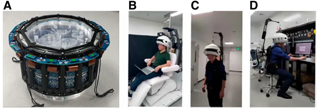
Figure 1: SmartBrain Wearable Brain PET System and Its Application Scenarios.
SmartBrain: Weaving 192 Detectors into a Wearable Helmet
In response to the aforementioned limitations, the research team proposed a 16-sided full-ring structure.SmartBrain has an inner diameter of 207.44 millimeters, an axial FOV of 121 millimeters, and consists of 192 detector modules arranged in 6 rings, with 32 modules per ring.
Each detector module contains a 6×6 array of lutetium-yttrium oxyorthosilicate (LYSO) crystals, with individual crystal dimensions of 3×3×5 mm³, optically coupled to a 3×3 silicon photomultiplier (Silicon Photomultiplier, SiPM) array. The system employs a channel reduction strategy, decreasing the readout channels from 1728 to 432. To reduce weight, the crystal thickness is chosen to be 5 mm, but this comes at the cost of some sensitivity and signal-to-noise ratio.
The total weight of the device is approximately 6 kilograms, supporting list-mode acquisition, with an energy window of 461–611 kiloelectronvolts (keV) and a coincidence time window of 1 nanosecond.The research team also developed two types of mechanical support solutions: a wearable backpack-style and a hanging-style. The temperature is stabilized within 0.5 degrees Celsius through external fans and air-cooling ducts.
NEMA Testing: Measuring Mobile Imaging by Commercial Standards
To verify whether SmartBrain meets the basic standards for commercial PET, the research team conducted a comprehensive physical performance evaluation according to the NEMA NU 2-2018 standard.
In terms of spatial resolution,The average value measured by two-dimensional Filtered Backprojection (FBP) at the center of the axial FOV is 2.29 mm. To further evaluate the intrinsic resolution capability of the system, the central point source data were reconstructed using the Maximum Likelihood Expectation–Maximization (MLEM) algorithm with 1000 iterations, achieving a spatial resolution close to 1.7 mm.
The system sensitivity is 720.2 counts per second per megabecquerel (cps/MBq).。TOF resolution reaches 234 picoseconds, with an energy resolution of 10.8%. The peak Noise-Equivalent Count Rate (NECR) is 4.67 kilocounts per second (kcps), occurring at an activity concentration of 10.1 kilobecquerels per milliliter (kBq/mL), with a scatter fraction of 29.5%.
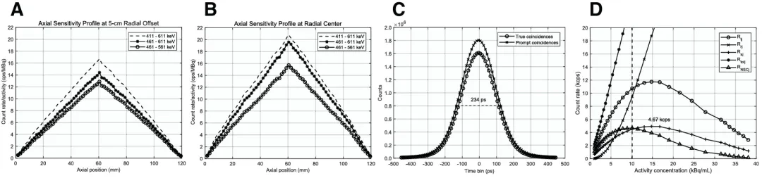
Figure 2: Physical performance test results of the SmartBrain system.
In the image quality test, six hot spheres (diameter 10–37 mm) had a background activity ratio of 9.4:1. The images were reconstructed using the ordered subset expectation maximization algorithm (12 iterations, 4 subsets), with only point spread function modeling (2 mm Gaussian kernel) and attenuation correction applied.
Phantom to Human Brain: 1.7 mm Resolution with Gyrus Restoration
Phantom experiments further validated the reproducibility of the aforementioned values.
As shown in Figure 3, hot rods with a diameter of 1.7 mm can be clearly resolved in the multi-layer Derenzo phantom imaging.At the center of the FOV, the valley-to-peak ratio of the 1.7 mm hot rod is 0.433, which is below the Rayleigh criterion threshold of 0.735, indicating clear inter-rod separation. Even without TOF reconstruction, the 1.7 mm hot rods remain visually distinguishable at both the center and 5 cm off-center positions.
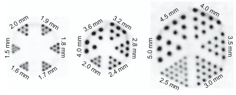
Figure 3: Imaging results of multi-layer Derenzo phantom.
As shown in Figure 4, axial, coronal, and sagittal images in the customized Hoffman brain phantom clearly display the distribution of cortical gray matter activity, forming a striking contrast with the non-radioactive white matter and ventricular regions.The image restores the complex morphology of the cerebral gyri, demonstrating the system's capability to image fine brain anatomical structures.
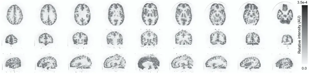
Figure 4: Imaging results of the customized Hoffman brain phantom.
On the Same Stage as DMI: Imaging Quality of 190-Minute Delayed Scans
A 43-year-old male epilepsy patient participated in the comparative study. His fastingBlood glucose was 100.8 mg/dL, and 318.2 megabecquerels (MBq) of 18F-fluorodeoxyglucose (18F-FDG) were injected. First, an 8-minute DMI PET/CT scan was performed approximately 60 minutes after injection; subsequently, a 60-minute SmartBrain scan was conducted at 190 minutes post-injection (residual activity approximately 95.9 MBq).
As shown in Figure 5, the transverse, coronal, and sagittal images of SmartBrain clearly present the cortical uptake pattern, with distinct gray matter distribution and well-preserved gyral anatomy. The white matter and ventricular areas show low or no activity, consistent with the normal distribution pattern of 18F-FDG.
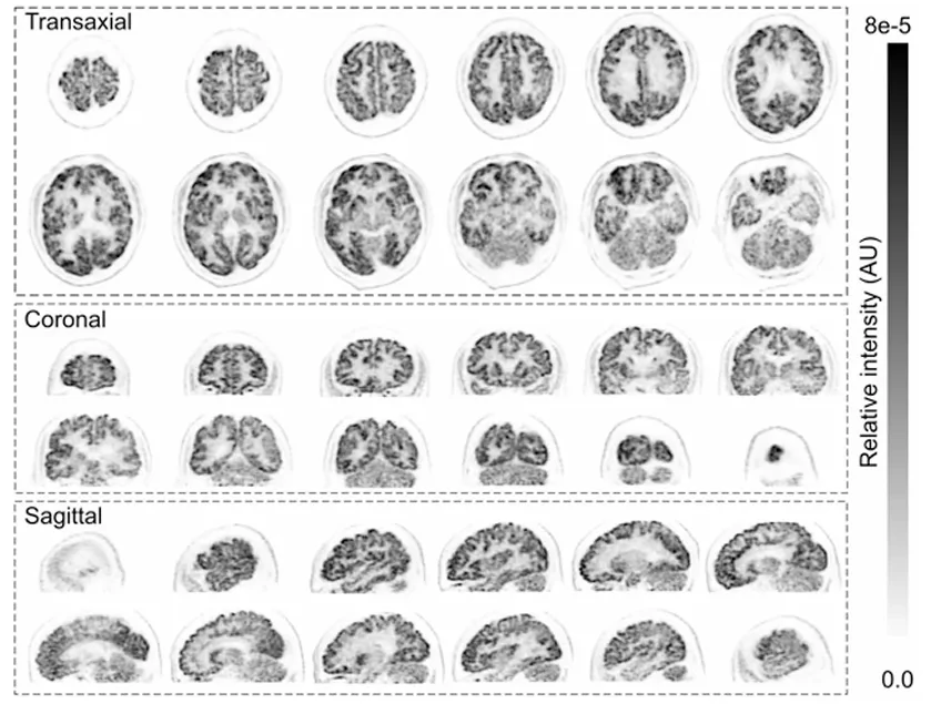
Figure 5: Human brain 18F-FDG images acquired by SmartBrain.
As shown in Figure 6, compared with the DMI image, SmartBrain still exhibits a similar cortical uptake pattern under delayed scanning and low activity conditions, with clearer delineation of sulcal details (red arrow in Figure 6). This suggests that,Although the sensitivity of wearable systems is lower than that of DMI, sufficient diagnostic-grade images can still be obtained under low-dose conditions with the help of TOF reconstruction and long acquisition times.
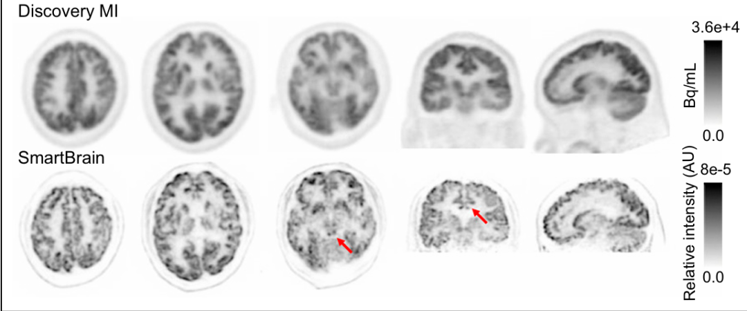
Figure 6: Comparison of DMI and SmartBrain Human Brain 18F-FDG Images.
Future: Brain PET Moving from "Fixed Gantry" to "Portable Diagnosis"
SmartBrain represents one of the few truly wearable brain PET systems capable of imaging during free movement.Its full-ring configuration ensures complete FOV coverage, the 5mm short crystal reduces depth of interaction (DOI) uncertainty and Compton scattering within the crystal, the 3mm crystal spacing limits inter-crystal scattering, and the 20cm small aperture reduces the impact of non-collinearity effects.
However, the current limitations are also clear.Thinner crystals result in relatively lower sensitivity; the system is not yet equipped with pile-up correction, limiting performance under high count rate conditions; fast dynamic studies remain challenging.Moreover, the detector coverage and geometry still need to be optimized to support clinical needs for higher sensitivity.
The research team pointed out that future improvements include enhancing detector geometry design, developing advanced correction algorithms, and introducing AI-assisted image reconstruction. These optimizations are expected to further reduce acquisition time, improve image quality, and expand its applications in clinical and neuroscience fields.Scope.
When brain PET can be worn like a helmet, the application scenarios of neuroimaging will be rewritten. From the seizure period of epilepsy.From real-time monitoring, to the assessment of the natural state of children's neurodevelopment, and further to brain science research on motor-cognitive coupling, wearable PET may be standing at a turning point.
Reference: Han Liu, Wenkang Qu, Da Liang, Xin Yu, Yuejie Lin, Haoyu Zou, Siyuan Han, Zhijun Zhao, Ying Lin, Xiaoyin Zhang, Jinyong Tao, Wenbin Li, Huiping Zhao, Yibin Zhang, Gongning Luo, Ningyi Jiang and Qiyu Peng. Journal of Nuclear Medicine April 2026, jnumed.125.271350.

Brain-computer interface community is in ChinaThe First Brain-Computer Interface (BCI) Industry Service Platform。Mainly provide the following services for enterprises, research teams, investment institutions, and practitioners:
Publicity Report:Report on corporate dynamics, technology interpretation, and product introductions in the form of images, short videos, and live broadcasts to increase exposure and industry influence.。
Resource Docking:Match resources such as capital, supply chain, clinical institutions, and channel partners based on demand, complete real对接s, and promote cooperation.
Achievement Transformation:Assist the technical team in finding industry partners, investors, and implementation scenarios to promote the transformation from technology to product.
Event Planning and Execution:Undertake the planning and execution of online and offline roadshows, salons, forums, and other events.
Other Customization Requirements:Including personalized services such as report customization, market research, and talent recruitment support.
Business Cooperation Discussion,Please contact WeChat: ZuoLeiLeiya
(Note: Name - Organization - Collaboration)
Submission | Become a Creator,Please contact WeChat: RoseBCI

?Starred and Top-pinned?
Don't Miss Any Frontier Advances in Brain-Computer Interface

One-click triple action: "Share," "Like," and "View"
Welcome to chat in the comment section