Zhongke Weiyi Medical Technologies Files IPO Prospectus: Pioneering Compact, Specialized, and Mobile MRI Solutions Starting with Head and Neck Imaging
Although as early asIn 1973, American scientist Paul Lauterbur published the MRI imaging technology. But 50 years later, approximately two-thirds of the world’s population still lack access to MRI.
According to data from the Huajing Industry Research Institute, Japan’s per capita MRI equipment ownership reached as high as 57.4 units per million people in 2022. The United States, Greece, South Korea, Germany, Italy, Finland, and Norway all reported MRI equipment ownership rates exceeding 30 units per million people. In the same year, China’s per capita MRI equipment ownership stood at only 9.38 units per million people, indicating a substantial gap compared with developed countries.
“There is an urgent clinical demand for magnetic resonance imaging (MRI); nearly all medical departments rely on MRI for disease diagnosis.“Unless it’s an emergency patient who cannot undergo MRI due to time constraints,” the director of the neurosurgery department at a Grade 3A hospital in Shanghai told VCBeat.
Currently, the price of a single MRI unit on the market typically ranges from RMB 6 million to RMB 20 million, with substantial subsequent maintenance costs, making it largely unaffordable for primary care hospitals. Large tertiary Grade-A hospitals are the primary adopters of MRI systems. For hospitals that have already deployed MRI, the pain points associated with its use are clearly defined.
First, MRI equipment is immobile and cannot meet the needs of patients with high transfer risks.MRI scanners are bulky and heavy, typically installed on the first floor of a hospital’s radiology department. Being immobile, they require patients to be transported to the equipment for each examination. This poses significant challenges for patients with high transport risks, such as comatose ICU patients whose conditions cannot be adequately assessed by CT scans but who cannot be safely moved for MRI examinations, ultimately hindering accurate diagnosis. Furthermore, transporting neonates carries substantial risks, and large-scale MRI systems are not well-suited for neonatal imaging.
Second, it fails to meet the personalized needs of specialized departments.There are significant differences in MRI usage requirements across various departments. For instance, the emergency department emphasizes the timeliness of examinations; inpatient wards require high-frequency use; dentistry needs to account for oral-specific characteristics; orthopedics requires patients to be scanned in a sitting position with higher demands on image clarity; neonates need to be examined in the delivery room or operating theater; and ICU patients cannot undergo long-distance transfers.
Third, long waiting times for MRI examinations and limited patient capacity.MRI scans at most hospitals require appointment scheduling and queuing. Acute patients who cannot undergo magnetic resonance imaging (MRI) in a timely manner may miss the critical window for diagnosis and treatment. For instance, in cases of acute cerebral infarction, lesions are not visible on CT scans within the first 24 hours after onset. Diagnosis, subsequent treatment decisions, and confirmation of infarction all rely on MRI examinations. However, due to limited capacity in MRI and radiology departments, frequent queue-jumping would disrupt normal scheduled appointments.
The director further added that although the state is vigorously developing and constructing diagnostic and treatment systems for chest pain centers, such as those for myocardial infarction and cerebral infarction, magnetic resonance imaging (MRI) has not been included in the clinical pathways. This omission is not due to a lack of necessity, but rather takes into account the actual medical conditions in China. “If MRI equipment could be deployed in emergency departments, it would certainly be prioritized over CT.”
At that time, Jiang Weiping, who was in charge of the transfer and commercialization of scientific and technological achievements at a research institute under the Chinese Academy of Sciences (CAS), noticed a compact nuclear magnetic resonance (NMR) project developed by a bioelectromagnetic technology research team during the project screening process. “I found that this project closely aligned with our selection criteria—absolute global technological leadership, high barriers to entry, and proximity to industrialization. Furthermore, its applications were well-suited to the national direction of tiered diagnosis and treatment as well as DRG-based payment reform.”
To further validate the clinical demand for this technical product, he spent more than four months traveling to cities such as Beijing and Shanghai. After visiting over forty department directors, he gradually gained clarity in his mind—“Miniaturization, specialization, and visualization are the trends in the future development of MRI equipment.”
So, he rolled up his sleeves and got to work, determined to fully drive the project forward.
MRI systems can be classified into low-field, mid-field, high-field, and ultra-high-field categories based on magnetic field strength. MRI systems with a field strength of less than 0.5 T are referred to as low-field MRI, which typically utilize permanent magnets. MRI systems with a field strength between 0.5 T and 1.0 T are termed mid-field MRI. MRI systems with a field strength greater than 1.0 T but less than 2.0 T are classified as high-field MRI.
Generally, an increase in magnetic field strength can lead to improvements in imaging and image quality. However, as the field strength increases, the magnet becomes larger in volume and heavier in weight, resulting in higher manufacturing costs. Meanwhile, higher field strengths amplify certain field-dependent effects (such as chemical shift artifacts and BOLD effects, which can generate artifacts and interfere with diagnostic interpretation) and increase the specific absorption rate (SAR). Furthermore, higher field strengths require larger MRI facilities and installation rooms.
Therefore, although higher field strength yields higher image signal-to-noise ratio, it is not always the case that higher field strength is better. In clinical applications, each level of field strength has its own advantages and disadvantages.
In terms of magnetic field strength, although low-field MRI systems have lower field strengths, they can utilize permanent magnets. Their advantages include no requirement for liquid nitrogen or liquid helium, no need for shielded rooms, stable field strength, lower costs, and a smaller footprint. It is worth noting that the annual maintenance cost for high-field MRI systems is typically in the hundreds of thousands of yuan range. Furthermore, due to constraints related to size, weight, and environmental requirements, MRI equipment is generally limited to installation on the first floor.
“Mobility is at the core of compact MRI systems,” Jiang Weiping, founder of Zhongke Microlight, told VCBeat. Currently, MRI equipment follows two technological pathways: superconducting and permanent magnet. Among them,Superconducting MagnetTo maintain the superconducting performance of the superconducting coils, a cryogenic environment must be sustained, requiring a dedicated refrigeration system capable of maintaining such low temperatures. Except in cases of accidents or maintenance, the system must remain powered on continuously once activated; power loss and shutdown are strictly prohibited.Permanent MagnetGenerally made from rare-earth materials, such as neodymium-iron-boron (NdFeB) and iron-cobalt-nickel alloys. Permanent magnets can retain their magnetism indefinitely, feature a compact size, and do not require an external power source.From the perspective of current technology, small permanent magnet MRI systems are more suitable for mobile applications.
“Small-scale MRI is not a miniaturized version of large-scale MRI; it follows a unique technical pathway and presents significant technical challenges.”Jiang Weiping told VCBeat that although relevant approved products already exist both domestically and internationally, constraints such as excessively low magnetic field strength, limited clinical problem-solving capabilities, and the need to outsource core components make it difficult to balance product performance with cost, leaving these solutions still some distance away from true specialization.
Miniaturized, Portable, Departmentalized, Lightweight, Self-Shieldedis a distinctive feature of Zhongke Microlight’s MRI products.
“All of our products are custom-built to meet the specific MRI requirements of clinical departments; they are distributed exclusively to specialized clinical departments, not to radiology departments.”Jiang Weiping explained that MRI requirements vary across different departments. For instance, joint MRI examinations require patients to be scanned in a seated position, while neonatal MRI units need to be located within obstetrics and gynecology departments, necessitating comprehensive consideration of factors such as image clarity, compatibility with other medical equipment, and spatial constraints. Zhongke Microlight customizes the design, magnetic field strength, imaging sequences, and software of its MRI systems according to the specific requirements of each specialty department regarding image clarity, scanning speed, and imaging protocols.
According to Jiang Weiping, since the advent of MRI, 50% of MRI scans have been performed on the head and neck, and the queue for MRI examinations is primarily caused by the high volume of head and neck scans. Zhongke Microlight Medical Research Center (Xi'an) Co., Ltd. took this as a breakthrough point and pioneered the development of0.2T Mobile Head and Neck MRI, enabling MRI systems to break free from the constraints of radiology and imaging departments and become intelligent point-of-care diagnostic devices, thereby achieving a leap from “patients seeking equipment” to “equipment seeking patients.”
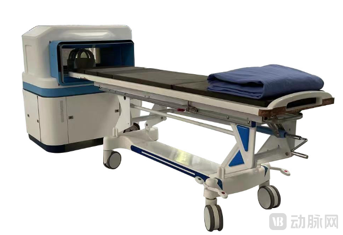
0.2T Mobile Head and Neck MRI
The magnet is the “heart” of the magnetic resonance system. To achieve device miniaturization while maintaining product performance, Zhongke Microlight adoptsLightweight magnet design, unique imaging sequence technology, miniaturized spectrometer design...encompassing comprehensive innovations from hardware to software. The device features a gradient magnetic field strength of 16 mT/m and a maximum slew rate of 40 mT/m/s. Weighing only 750 kg, it is equipped with omnidirectional casters at the base for effortless mobility and operates at room temperature.
This device is easy to operate,Diagnostic results can be synchronized and accessed in real time. By integrating remote diagnosis with AI technology, it facilitates physicians’ workflow and enables the provision of optimal treatment plans for patients.
Furthermore, the device features self-shielding and is compatible with other medical equipment, eliminating the need for additional shielded rooms. This reduces manufacturing and operational costs while significantly enhancing the convenience and accessibility of MRI examinations, addressing limitations that prevent large-scale MRI systems from reaching deeper clinical applications.Emergency Department, Intraoperative, ICU, Pediatrics, Orthopedics, Neurology, Neurosurgery, Stomatology, Primary Healthcare Institutions, Field Training, Mobile Healthcare, Physical Examination Centers, Veterinary Hospitals, and other scenarios.
“The sole rationale for the existence of medical devices is their ability to address clinical problems. Our design philosophy prioritizes solving clinical issues first, followed by considerations of miniaturization, specialization, and portability. For clinical diagnostic needs in head and neck applications, a precision of 0.2T/1mm fully meets physicians’ requirements,” summarized Jiang Weiping.
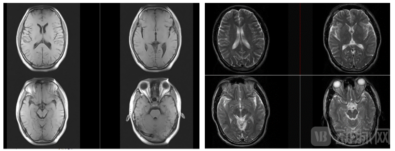
Imaging Performance of Zhongke Microlight’s 0.2T Mobile Head and Neck MRI
Leveraging the product technology of its 0.2T mobile head-and-neck MRI, Zhongke Microlight has also expanded its applications into another blue-ocean market with immense potential—veterinary healthcare—and launched0.2T Dedicated Veterinary MRI Scanner.
The core technical specifications of this imager are essentially consistent with those of human medical devices, while its hardware and software have been optimized specifically for small animals. For instance, a series of imaging sequences have been developed based on the characteristics of small animals; the MRI bore is utilized as an animal bed to save space; and the device can be operated by simply plugging it into a standard 220V household power supply. Currently, the device has completed research, development, and production, and is poised for deployment in veterinary hospitals.
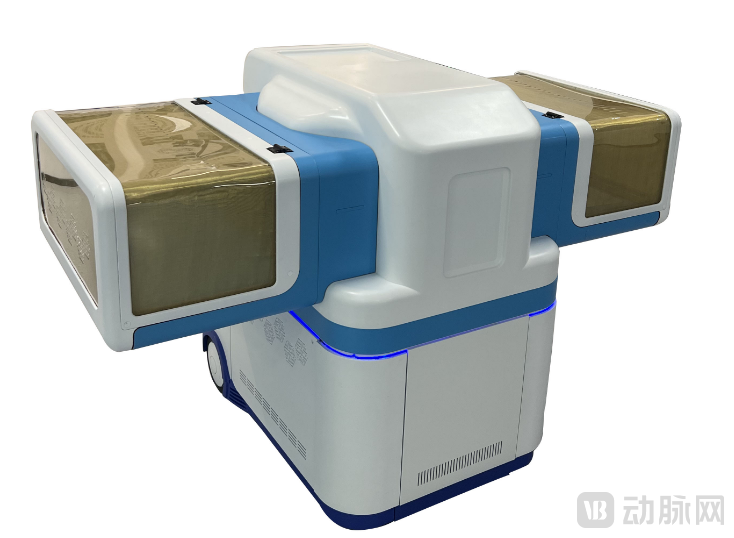
0.2T Dedicated Veterinary MRI Scanner
In the process of rapidly advancing R&D, Zhongke Microlight is also continuously strengthening its patent moat. It is reported that the company has filed a total of 82 patents, with more than 55 published; two are utility model patents, and the rest are all invention patents. Additionally, there are two software copyrights.
With the widespread adoption of precision medicine, the miniaturization, portability, and digitization of medical imaging have become an inevitable trend. Miniaturized devices not only enhance the efficiency of imaging diagnosis but also help hospitals increase revenue.
In the future, Zhongke Microlight will continue to deepen its R&D efforts in specialized MRI fields while simultaneously expanding into the small-scale MRI market for pets. Leveraging the “Small MRI, Broad Applications” intelligent point-of-care diagnostic solution, we aim to fully empower MRI technology and eliminate waiting times for MRI examinations.