Case Report: Prof. Xie Feng’s Team at Liaoning JinQiu Hospital Successfully Treats Hepatocellular Carcinoma with Vispearl® Radiopaque Drug-Eluting Embolic Microspheres

H&H Healthcare
R&D and Producer of Interventional Medical Devices for Heart Disease
Basic Information:56-Year-Old Male
History of Present Illness:Primary liver cancer, recurrence after right hepatectomy, new intrahepatic lesions discovered in March 2026.
Past Medical History:In 2022, primary liver cancer was diagnosed, BCLC-B stage. A right hepatectomy was performed at the First Hospital of China Medical University, and postoperative pathology confirmed hepatocellular carcinoma. In February 2024, lung metastasis was found during a follow-up examination, and treatment with lenvatinib was initiated, later switching to regorafenib. In March 2025, brain metastasis was discovered, and surgical resection was again performed at the First Hospital of China Medical University. In October 2025, metastasis to the right thigh was found, and the patient was admitted to our department for O+Y (nivolumab + ipilimumab) treatment. After only one treatment session, further treatment was discontinued due to personal reasons. In March 2026, new intrahepatic lesions were detected, and the patient was admitted to our department for TACE combined with O+Y treatment. The thigh lesion showed slight shrinkage, followed by biopsy.
Laboratory Examination:AFP normal, ALT 81 UI/L, AST 107 UI/L, Total Bilirubin 31.74 umol/L
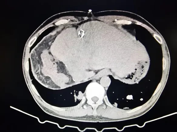
Imaging Examination:Preoperative enhanced CT showed a 3.0 cm lesion in the left lateral lobe of the liver, with significant enhancement in the arterial phase and washout in the delayed phase.
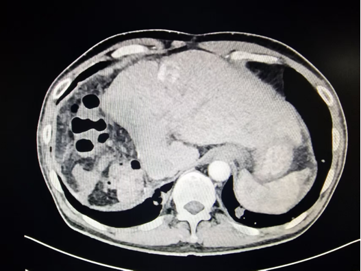
Imaging shows: a 3cm newly developed lesion in the left lateral lobe of the liver.
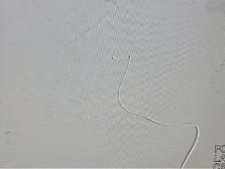
Percutaneous Right Femoral ArteryPuncture, A 5F arterial short sheath was inserted, and an RH catheter and guidewire were introduced through the guidewire to perform a common hepatic artery angiography.
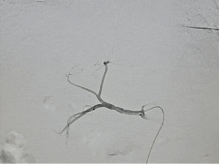
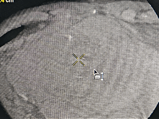
Contrast imaging showed: A round-like tumor staining was observed in the left lobe of the liver, supplied by branches of the left hepatic artery.
The microcatheter was inserted into the tumor's blood-supplying artery. After confirmation by CBCT, Vispearl was injected through the microcatheter.®️Contrast-enhanced drug-loaded embolic microspheres 70-150μm 1g, loaded with 40mg of epirubicin.
After the contrast agent clearance, CBCT angiography was performed again, showing complete tumor embolization with good embolic agent filling.
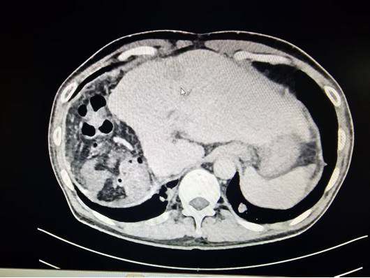
One and a half months after the surgery, the lesion decreased from 3.0cm preoperatively to 1.7cm. Both lung metastases and right thigh metastases were significantly reduced. According to the mRECIST criteria, the treatment efficacy was evaluated asCR. The deposition of visible microspheres within the lesion was clearly demonstrated, with no reflux observed in the surrounding liver tissue, highlighting the advantages of precise embolization.
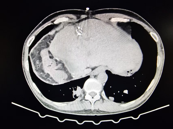
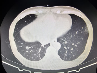
CT scan shows: Tumor activity has basically disappeared.
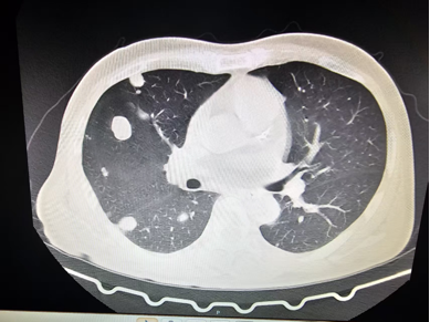
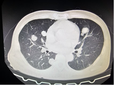
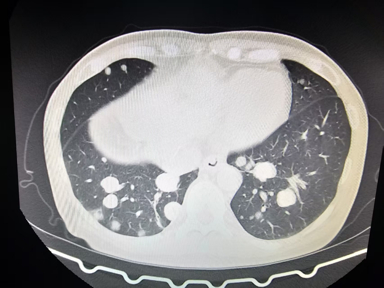
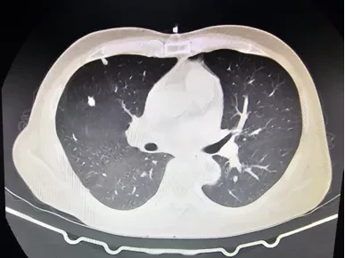
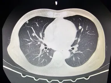
Significant reduction in lung lesions
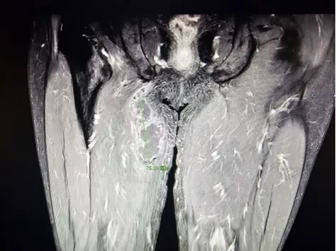
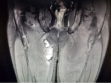
The lesion in the thigh has slightly shrunk, and the pathological result of the biopsy is necrotic tissue.
Professor Xie Feng stated that the use of visible microspheres + CBCT has important guiding significance for TACE treatment, allowing immediate assessment of whether the lesion embolization is complete during the procedure.The visible microspheres are not only suitable for super-selective fine embolization, but their visibility can also be used forBaseline embolization before ablation provides guidance for subsequent combination therapy.
Expert Profile

Doctor of Imaging and Nuclear Medicine,Chief Physician
Director of the Interventional Department, Golden Autumn Hospital, Liaoning Province
Doctoral Supervisor at Liaoning University of Traditional Chinese Medicine
Master's Graduate Supervisor at China Medical University and Dalian Medical University
Vice Chairman of the Nuclear Medicine Branch of Liaoning Medical Association
Vice Chairman of the Tumor Minimally Invasive Treatment Committee, Liaoning Province Primary Health Association
Member of the Radiology Branch of Liaoning Provincial Medical Association
Member of the Comprehensive Interventional Radiology Group, Interventional Medicine Branch, Liaoning Medical Association
Standing Committee Member of the Tumor Minimally Invasive Special Committee of Liaoning Province Anti-Cancer Association
Member of the Liver Cancer Special Committee of Liaoning Province Anti-Cancer Association
Standing Committee Member of the Radiation Intervention and Immunology Branch of Liaoning Province Immunology Society
Standing Committee Member of the Multidisciplinary Team (MDT) Diagnosis and Treatment Professional Committee of the Liaoning Province Cell Biology Society Tumor
1. Comprehensive interventional treatment for liver cancer and other benign and malignant liver tumors
2. Interventional Treatment for Knee Arthritis (GAE)
3. Interventional Treatment for Benign Prostatic Hyperplasia
4. Interventional Treatment of Obstructive Jaundice
5. Comprehensive Interventional Treatment of Lung Cancer and Pulmonary Metastases
6. Interventional Treatment of Uterine Fibroids and Adenomyosis
7. Interventional treatment for liver cysts, liver abscesses, renal cysts, etc.
8. Interventional Treatment of Acute Cholecystitis
9. Percutaneous biopsy of tumors in various parts of the body
Interventional Department of Liaoning Jinqiu Hospital: Specialties include comprehensive interventional treatment for liver cancer and other benign and malignant liver tumors, interventional treatment for knee arthritis (GAE), interventional treatment for benign prostatic hyperplasia, interventional treatment for obstructive jaundice, comprehensive interventional treatment for lung cancer and lung metastases, interventional treatment for uterine fibroids and adenomyosis, interventional treatment for liver cysts, liver abscesses, renal cysts, etc., interventional treatment for acute cholecystitis, and percutaneous biopsy for tumors in various parts of the body. In the past five years, the department has led two provincial natural science foundation projects and published more than 10 academic papers in domestic and international journals (including 7 SCI-indexed articles with a total impact factor of 22.951, and 8 core journal articles).