Xi'an Jiaotong University Second Affiliated Hospital Team Pioneers Intravital Placental Imaging Technology to 'Visualize' Pregnancy-Related Diseases
The placenta is crucial for promoting fetal development.A clear understanding of the placenta can help researchers address clinical issues such as miscarriage, stillbirth, and preeclampsia. To further investigate placental function and its role in maternal-fetal diseases, there is an urgent need for a method that enables in vivo placental research.
Recently,Professor Huang Qiang, Department of Pediatric Surgery, The Second Affiliated Hospital of Xi’an Jiaotong UniversitywithDuke University Department of Biomedical EngineeringIn collaboration, we developed a technique for in vivo placental imaging in mice. In models of alcohol exposure, maternal cardiac arrest, and pregnancy-induced hypertension, the team meticulously observed changes in placental blood oxygen saturation and vascular circumference associated with these conditions.For the first time, high-resolution imaging of the placenta in maternal-fetal diseases has been achieved, laying the foundation for studying the role and mechanisms of the placenta in these conditions.
Published as a cover article in Science Advances under the title “Longitudinal intravital imaging of mouse placenta,” this achievement...
 Science Advances Cover (Source: Science Advances)
I. Further Extension of the “Abdominal Window” Technique
As early as 2020, Huang Qiang’s team developed a live imaging technique for mouse embryonic development, which, through different developmental stages,"Abdominal Window", achieving for the first time high-resolution imaging observation of the continuous developmental process of mouse embryos from embryonic day 9.5 to birth.
This achievement enables researchers to continuously and clearly observe the developmental process of mouse embryos, while also laying the foundation for today’s “in vivo placental imaging technology in mice.”
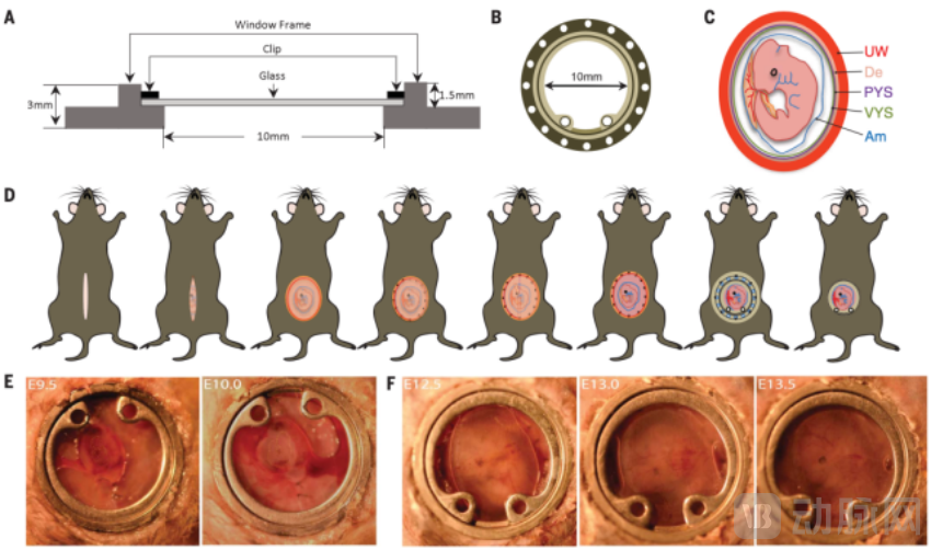 “Abdominal Window” Developed by Huang Qiang’s Team (Image source: Science)
In 2024,Huang Qiang’s team combined the “abdominal window” technique with the latest photoacoustic microscopy and two-photon microscopy technologies to successfully observe the developmental process of the mouse placenta from its early formation stage until just before delivery.Although this technology is currently limited to mouse models, it lays the foundation for future clinical applications.
In the future,This technology can help physicians better understand placental function during pregnancy and its role in pregnancy-related complications, thereby improving prenatal management and intervention strategies.. Furthermore, this technology can be leveraged to develop novel diagnostic tools that support the early identification of pregnancy risks and facilitate the development of new therapies for pregnancy complications.
The team stated that it will leverage artificial intelligence technologies to further enhance the resolution and accuracy of imaging, applying this technology to a broader range of biomedical fields.
II. In Vivo Imaging Technology: “Multimodal” as the New Trend
In life sciences and medical research, imaging technology is a core driver of technological advancement. In vivo imaging techniques enable researchers to observe cellular and molecular activities, thereby facilitating the identification of disease pathogenesis and the development of therapeutic strategies.
Currently, in vivo imaging technologies are primarily categorized into five major types: visible light imaging, radionuclide imaging, magnetic resonance imaging (MRI), computed tomography (CT), and ultrasound imaging. These modalities differ in their application domains and exhibit distinct advantages and disadvantages.
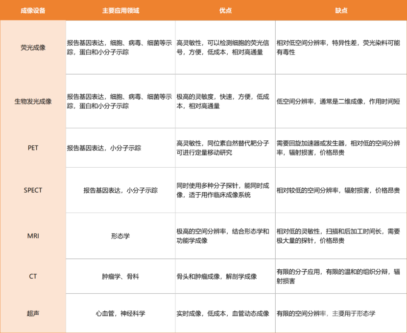 (Source:(Kaiji Biotechnology; Table compiled by VCBeat Orange Bureau)
To enhance the efficiency of in vivo imaging technology, multiple imaging modalities have been integrated in recent yearsMultimodal Fusion Imaging Systemhas become the new direction pursued by the market.
In 2023,Ruishi MedicalLaunched the independently developed IMAGING 1000, a three-dimensional multimodal precision imaging system. This device integrates multiple imaging modalities, including Micro-CT, 3D bioluminescence imaging, and 3D fluorescence molecular imaging, achieving a combination of functional and structural imaging. Based on this technology, users can accurately detect true 3D signals within living organisms, enabling precise localization and diagnosis in three-dimensional space.
andBoluotengShortly thereafter, in early 2024, the SkyView multimodal fusion imaging system for small-animal in vivo CT was developed. Reportedly, this imaging system integrates micrometer-scale 3D imaging with CT and optical imaging, addressing the limitations of conventional optical imaging, such as insufficient penetration depth and the ability to only two-dimensionally observe signal intensity. It not only enables non-invasive, high-precision visualization of internal structures in laboratory animals but also enhances imaging speed, efficiency, and operational convenience.
Furthermore, multimodal fusion imaging systems have also secured a place in the construction of national major scientific and technological infrastructure. In 2022Multimodal Cross-Scale Biomedical Imaging FacilityCompleted in Beijing. Its primary purpose is to develop more advanced in vivo imaging technologies, enabling traditional studies of anatomy and physiological functions to delve into the cellular and molecular levels.
Whether it is the multimodal in vivo imaging system or the “in vivo placental imaging technology for mice” developed by Huang Qiang’s team, both represent comprehensive achievements resulting from interdisciplinary integration. Interdisciplinary collaboration is not only a significant trend in current technological development but also a key driving force for future scientific and technological progress. In the future, this interdisciplinary research approach will not only effectively address challenges that are difficult to solve within a single discipline but also foster more innovative and practical scientific outcomes.