Ultra-High-End Static CT Clinical Data Unveiled: Nanovision on the Verge of Mass Production

Nanovision
X-ray Detector and Static CT Product R&D Provider
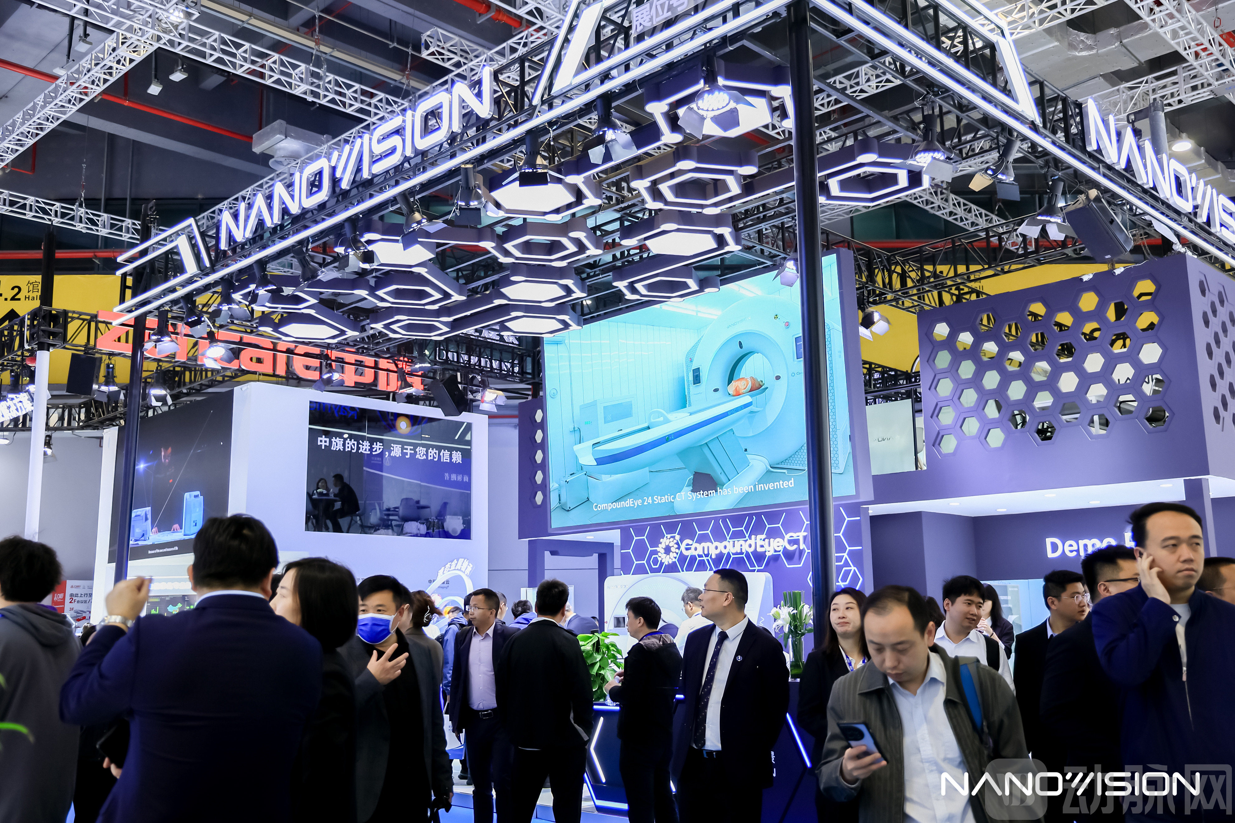
On the first day of CMEF, Nanovision, a domestic manufacturer of ultra-high-end CT scanners, announced the latest clinical progress of its static CT system, “Compound Eye 24,” at its booth. The fiercely competitive ultra-high-end CT market now faces further uncertainty.
Relevant information indicates that Nanovision has designed a clinical trial protocol for its static CT scanner in accordance with the "Technical Review Guidelines for the Registration of X-ray Computed Tomography Equipment," and has fully entered the clinical testing phase.
At this juncture, Nanovision is only one step away from achieving large-scale independent R&D and manufacturing of this domestically produced ultra-high-end CT scanner.
The so-called "static CT" refers to a "static" dual-ring structure composed of an "X-ray source ring" formed by multiple X-ray tube–high voltage generator sets and a "detector ring" formed by a full-ring detector. It achieves 360° data acquisition through sequential pulsed exposure of the X-ray sources controlled by timed electronic switching, thereby transforming the dynamic "mechanical rotation" in spiral CT into static "optical rotation."
Through this specialized imaging approach, static CT avoids the substantial centrifugal forces generated by the high-speed rotation of spiral CT during scanning and eliminates trailing artifacts, thereby surpassing the rotational speed limits of spiral CT and significantly enhancing the spatial resolution of CT images.
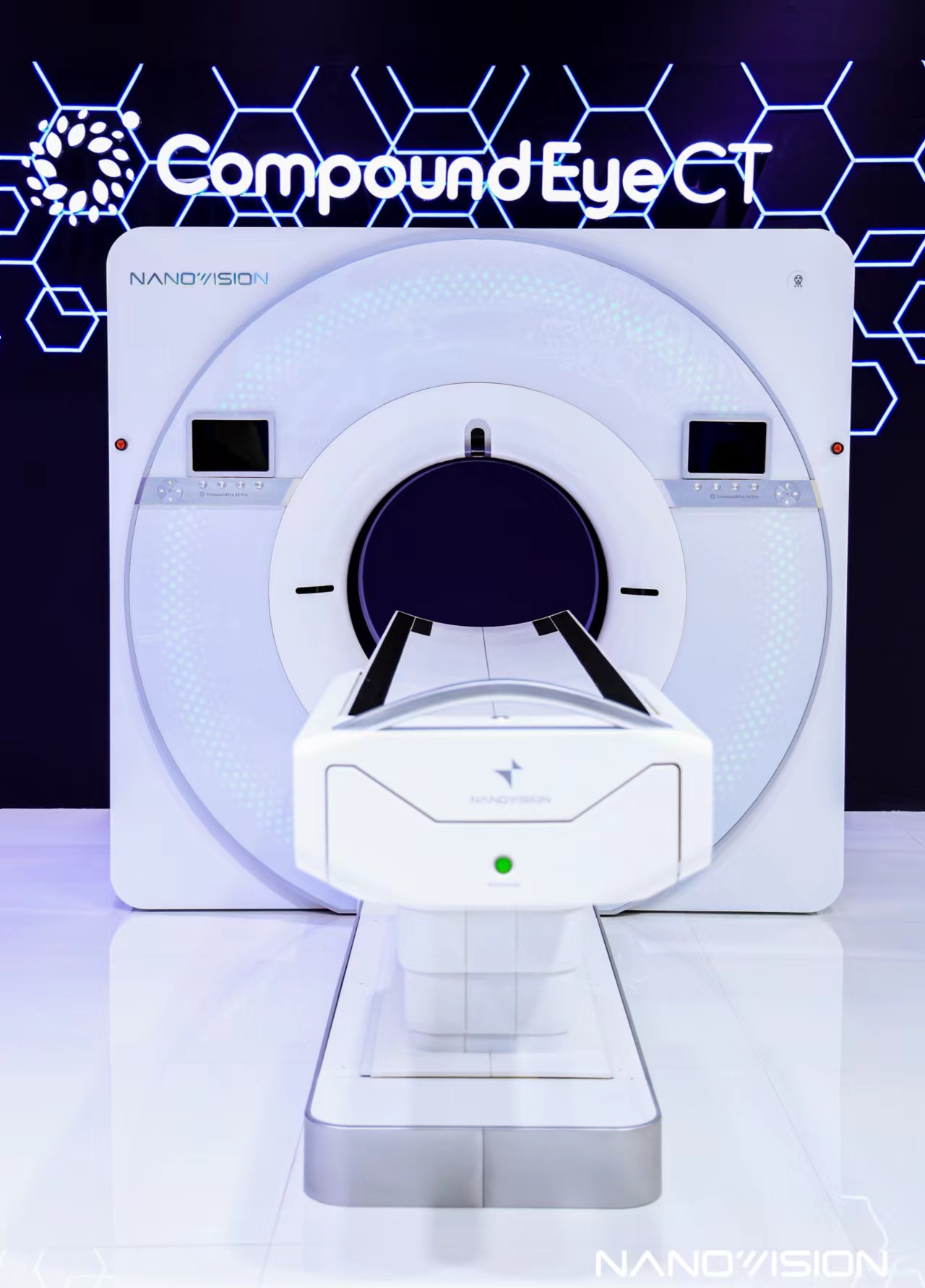
According to Nanovision, the thinnest scan slice thickness of its first-generation static CT system, “Compound Eye 24,” has reached 0.165 mm and features isotropic resolution, significantly enhancing spatial resolution capability.
In the face of Nanovision’s relentless pursuit of ultimate spatial resolution, many may question its significance. After all, current ultra-high-end CT systems have already conquered the most challenging cardiac imaging, and with AI empowerment, they are capable of addressing the vast majority of clinical issues.
However, humanity’s exploration of the microscopic world is unending. As we capture increasingly rich information, our conclusions will become ever more precise. Therefore, the pursuit of CT systems with higher resolution always holds practical significance.
Applying high-resolution features to clinical practice, the technology's promotional effect on pulmonary nodule diagnosis is evident. Currently, images of nodules smaller than 6 mm generated by conventional CT usually lack sufficient precision and have blurred edges. Therefore, international guidelines use 6 mm as a threshold, recommending clinical evaluation for pulmonary nodules with a diameter greater than 6 millimeters, waiting until the nodules grow to a size that meets the capabilities of imaging equipment for further diagnosis.
In contrast, static CT imaging produces virtually no artifacts, enabling comprehensive visualization of the details of 4-mm nodules and even clear delineation of the surrounding vasculature. This allows physicians to determine the benign or malignant nature of nodules at an earlier stage, thereby further reducing the difficulty and risks associated with surgical resection of malignant nodules. If this technology achieves large-scale clinical adoption, it could potentially reshape the clinical guidelines for lung cancer diagnosis.

Imaging Comparison of Ultra-High-End Spiral CT and “Compound Eye 24” Static CT for a 4-mm Pulmonary Nodule in the Right Upper Lobe of a Volunteer
Another piece of evidence supporting the clinical value of static CT stems from the treatment of liver cancer.Currently, most physicians opt for surgical resection to remove hepatocellular carcinoma tissue; however, 70%–80% of patients experience recurrence within five years postoperatively. This is primarily because it is difficult to achieve complete tumor removal under conventional CT-guided navigation.
Nanovision told VCBeat, “Liver cancer tissues are typically rich in nutrient-supplying blood vessels that feed the tumor. However, these vessels are too small to be visualized by conventional contrast-enhanced CT. Consequently, during preoperative assessment, the extent of liver resection is often underestimated, leaving behind many infiltrating and micro-infiltrating vessels, which ultimately leads to hepatocellular carcinoma recurrence.”
Theoretically, the high-precision images from static CT can reveal subtle clues overlooked by conventional CT. According to Nanovision, the company is collaborating with a leading Grade A tertiary hospital on a research project funded by the National Natural Science Foundation of China. This innovative technology is expected to revolutionize surgical planning for liver cancer in the future, significantly improving patients’ five-year postoperative survival rates.
Beyond breakthroughs in scientific research and clinical practice, the successful commercialization of static CT will also facilitate further development and optimization of CT equipment.
“Now, there are fewer and fewer chief specialists dedicated to CT research, and most academic papers focus on magnetic resonance imaging. This is because the development of spiral CT has stagnated for many years, with most research topics already exhausted, and it is difficult to provide adequate support for particularly complex equipment,” Nanovision told VCBeat. “The emergence of static CT can break this status quo. We hope to facilitate more in-depth scientific research and uncover new value from a more microscopic perspective.”
Although the various parameters and performance metrics of static CT far exceed those of traditional spiral CT, only a handful of companies are engaged in tackling the challenges of static CT in the market. Fundamentally, core components are the key constraint limiting the development of this type of CT.
The first obstacle comes from the detector's segmentation. On spiral CT, scintillators are typically segmented via physical patterning to achieve pixelation. Since the pixel size of static CT detectors is 1/16 that of spiral CT detectors, new technologies must be employed to attain such fine pixelation precision.
The Second Obstacle Comes from the Detector. In actual R&D, Nanovision found that existing photon-counting detectors were inadequate: “The count rate is insufficient, and they saturate under high doses,” failing to meet the requirements of static CT. To address this critical issue, it is necessary to develop a new generation of detectors.
The third obstacle comes from the narrow-pulse high-voltage generator.In spiral CT, the X-ray tube emits continuous X-rays. In static CT, there is no rotation; instead, electronic pulses control each X-ray tube to emit X-rays sequentially in brief instants. Therefore, rapid switching must be achieved through high-speed narrow-pulse technology. There are two challenges in this process: first, the emitted X-rays must reach a high voltage of 140 kV; second, fast switching with microsecond-level control must be realized.
Currently, only a handful of companies in the global market have independently developed even one of the aforementioned components, with Nanovision being the sole entity to have overcome all associated challenges.
Specifically, Nanovision has not only developed the world’s first photon-flow detector, achieving a maximum spatial resolution of 25 LP/cm at an MTF of 10%, but also pioneered a novel material growth technology that enables ultra-fine pixelation for static CT detectors. In terms of narrow-pulse high-voltage generators, its self-developed ultra-high-speed high-voltage generator can generate or cut off the stable high voltage required for the high-speed directional movement of activated electrons within 10 μs, thereby meeting the X-ray generation requirements during static CT scanning.
Furthermore, to achieve complete control over its supply chain, Nanovision has independently developed key components such as static CT-specific chips and small-array X-ray tubes, thereby realizing truly end-to-end in-house research, development, and manufacturing for this ultra-high-end imaging equipment.
Having conquered the core technologies, Nanovision has long since laid out its mass production strategy. Previous reports indicate that Phase I construction of Nanovision’s Chengdu Production Center was completed in 2022. At full capacity, the facility can produce approximately 500 units of the “Compound Eye 24” static CT scanner annually.
With the support of multiple policies, domestically produced ultra-high-end CT scanners are seizing an unprecedented opportunity to break through market barriers. In a few months, when Nanovision successfully completes the regulatory review and approval process, it will swiftly enter the commercialization phase, having completed all necessary preparations. At that point, Nanovision may overtake its competitors and reshape the current market landscape for ultra-high-end imaging equipment.