Harbin Institute of Technology's Cold-Responsive Team Files Patent for Electrospun Occlusion Membrane in Cardiac Devices
In August 2018, the team led by Professor Leng Jinsong of Harbin Institute of Technology, an academician of the Chinese Academy of Sciences, submittedA Patent on the Preparation of Occluder Flow-Blocking Membranes for Cardiac Applications via Electrospinning. This patent addresses the issue of slow endothelialization caused by the use of dense flow-blocking membranes in existing occluders, and entered the patent publication stage in the same November.
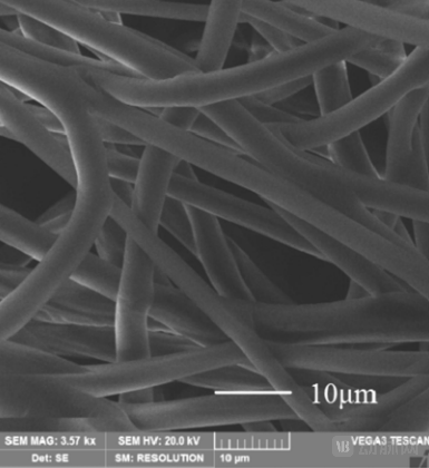
High-magnification microscopic morphology of fibers in the prepared flow-diverting membrane, sourced from the patent specification
It is understood that the flow-obstructing membranes in existing occluders are mostly made of dense polyester material. The dense structure of this material slows down the endothelialization rate, and the material remains in the body for a long time even after endothelialization.The electrospun porous material employed by Professor Leng Jinsong’s team facilitates cell adhesion and proliferation, thereby not only enhancing the utilization rate of drugs within the barrier membrane but also reducing the drug release rate to achieve sustained drug release.
Electrospinning Holds Great Promise in the Cardiovascular Field
Electrospinning offers significant advantages and broad application prospects in cardiovascular research, owing to its high porosity, large specific surface area, favorable mechanical properties, and structural similarity to the natural extracellular matrix.
In the field of vascular tissue engineering, electrospun artificial blood vessels are composed of stacked fiber membranes of a certain thickness. These fiber membranes are formed by numerous fibers arranged either randomly or in an oriented manner, which significantly influence cellular behaviors in vivo, such as inducing cell adhesion, migration, and differentiation.
In 2023, the research group led by Li Long at Guizhou University employed a stepwise electrospinning technique to sequentially fabricate one randomly oriented fiber layer and two aligned fiber layers, which were then subjected to folding and rolling operations to obtain a tri-layered vascular scaffold. This tri-layered vascular scaffold holds significant potential to biomimic the native three-layer structure of blood vessels and guide the spatial arrangement of cells akin to that in natural vessels, thereby further promoting comprehensive vascular reconstruction and regeneration.
In 2024, a team led by Li Jiashen at the University of Manchester developed a novel strategy for fabricating polylactic acid artificial blood vessels with controllable macroscopic structures by combining electrospinning with mold-assisted acetone treatment. Furthermore, these artificial blood vessels exhibit excellent structural stability, tunable mechanical strength, superior biocompatibility, and a hierarchical porous structure.
In the field of drug development, in 2021, Professor Zhao Qiang’s team from the College of Life Sciences and the State Key Laboratory of Medicinal Chemical Biology at Nankai University utilized electrospinning technology to fabricate functional cardiac patches for the treatment of myocardial infarction.
Multiple Companies Have Already Entered the Market
Due to its structural similarity, high porosity and specific surface area, excellent biocompatibility, tunable mechanical properties, ease of processing and modification, and superior drug delivery capabilities, electrospinning has attracted significant interest from numerous companies. Its applications extend beyond the treatment of cardiovascular diseases to encompass orthopedics, medical dressings, and other diverse scenarios.
In the cardiovascular field, Abbott developed the first everolimus-eluting poly(lactic acid) stent for human use in 2006. The stent struts had a thickness of 150 μm, and the scaffold material was primarily PLLA. In March 2009, research by Patrick et al. demonstrated favorable absorption of the stent two years after implantation, with no luminal restenosis or in-stent thrombosis, leading to its subsequent clinical application.
In addition, Roumai Medical has also designed an artificial vascular stent. This stent employs electrospinning technology to separately spin poly(lactic-co-glycolic acid) (PLGA) and polycaprolactone (PCL) materials, enabling the composite material to self-roll into a three-layered, biomimetic tissue-engineered blood vessel with a diameter of 3 mm.
In the future, electrospinning technology will also be combined with other methods, such as solution bath spinning, electrohydrodynamic direct writing, and melt electrospinning direct writing (MEDW), to enhance its application in the cardiovascular field. The research group led by Dietmar Hutmacher at Queensland University of Technology has utilized emerging MEDW 3D printing technology in combination with polycaprolactone/reduced graphene oxide nanocomposite inks to produce customizable scaffold-like structures with fine struts, thereby improving mechanical properties. It is reported that graphene-based materials are easy to print and exhibit excellent endothelialization and anti-platelet aggregation effects, holding promise for development into future biodegradable cardiac stents.