Shanghai Ninth People's Hospital Transfers Two Auriculocephalic Angle Scaffold Patents for RMB 1 Million
Recently, Shanghai Ninth People’s Hospital, Shanghai Jiao Tong University School of Medicine, announced that it would transfer the patent rights for “A Cranioauricular Angle Stent” (ZL2020208104303.3) and “Cranioauricular Angle Stent” (ZL202030223106.4) to Taizhou Laser Medical Apparatus and Instruments Co., Ltd. for RMB 1 million.
The patent originates from the team led by Zhang Ruhong in the Department of Plastic and Reconstructive Surgery at Shanghai Ninth People’s Hospital, Shanghai Jiao Tong University School of Medicine. The team has produced substantial academic achievements in facial plastic and aesthetic surgery, as well as in the repair and reconstruction of auricular deformities.
The transferee in this transaction, Taizhou Laser Medical Apparatus and Instruments Co., Ltd., was established in 2011, according to Tianyancha data. Its business scope includes the production and sales of Class I, II, and III medical devices.
The core patent involved in this transfer—the cranioauricular angle stent—features a compact footprint and excellent strength. It can replace cartilage in cranioauricular angle reconstruction, providing robust support while maintaining optimal angle and depth, thereby shaping a stable cranioauricular angle and an aesthetically pleasing cranioauricular sulcus.
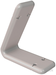
Cranioauricular Angle Bracket, Image Source: Patent Specification
The external ear is a three-dimensional superficial organ comprising 15 anatomical structures, making the reconstruction of a realistic three-dimensional auricle highly challenging. Furthermore, reconstructing the cranioauricular angle—a subunit structure that combines aesthetic form with functional integrity—represents a critical step in total ear reconstruction.
Cranioauricular Angle ReconstructionIn layman's terms, cranioauricular angle reconstruction refers to "propping up" the ears. Many surgeons in China often perform this procedure during a second-stage operation.
The primary objective of cranioauricular angle plasty is to achieve an upright, stable, and symmetric appearance of the reconstructed ear. The procedure addresses two key issues: the placement and fixation of the postauricular support framework, and the coverage of this framework with fascial tissue.
The most commonly used support materials are autologous costal cartilage or bone cement. However, as scaffolds, these materials occupy the limited volume of the cranio-auricular sulcus, resulting in a shallow contour of the sulcus, or cause reduction of the cranio-auricular angle due to cartilage resorption, thereby compromising the final aesthetic outcome and functional recovery.
The cranio-auricular angle bracket has been optimized to address the aforementioned issues. It comprises an upper support plate and a lower support plate, designed to support the auricle and the postauricular cranium, respectively. These two components combine to form a bent structure with an included angle of 55–65°, conforming to the natural physiological morphology of the cranio-auricular angle.
Specifically, the upper support plate (dimensions: length 0.8–1.2 cm, width 0.4–0.6 cm, thickness 0.5–0.7 mm) is responsible for stably supporting the auricle, while the lower support plate (dimensions: length 0.7–1.0 cm, width 0.4–0.6 cm, thickness 0.5–0.7 mm) fits closely against the postauricular skull to provide a stable foundation. Each support plate features two perforations at its proximal end, specifically designed for wire fixation, with grooves added on the outer side of the perforations to ensure secure and non-slipping wire fixation.
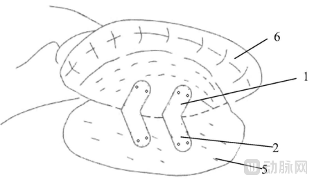
Schematic Diagram of the Usage Status of Cranioauricular Angle Stents. Image Source: Patent Specification
In actual surgical practice, physicians may flexibly select two or more cranioauricular angle scaffolds to support the auricular sulcus based on the patient’s specific conditions. The inferior supporting plate is placed against the postauricular skull, while the superior supporting plate elevates the auricle. Fixation is achieved by threading steel wires through perforations, and the surface is covered with fascial tissue and skin grafts.
Notably, the edges of the opposing inner surfaces and the two corners at each end of both the upper and lower support plates are meticulously rounded to prevent sharp edges or corners from puncturing the overlying fascia and skin grafts, thereby ensuring smooth postoperative healing. In contrast, the edges contacting the skull and auricular cartilage remain unchamfered; their flat, square corners provide more stable support, as the tissue thickness in these areas is sufficient to withstand the impact of such edges.
The core advantages of the cranio-auricular angle scaffold lie in its compact design, minimal space occupation, excellent strength, and high adaptability. It serves as an ideal substitute for cartilage, providing stable support in cranio-auricular angle reconstruction, precisely maintaining the ideal angle and depth of the cranio-auricular angle, thereby shaping a stable cranio-auricular angle and an aesthetically pleasing cranio-auricular sulcus. Furthermore, physicians can flexibly modify or replace the scaffold according to individual patient differences to deliver superior therapeutic outcomes.
Microtia is a common congenital craniofacial anomaly, second only to cleft lip and palate. According to data from the Plastic Surgery Hospital of the Chinese Academy of Medical Sciences (also known as Ba Da Chu), there are approximately 600,000 patients with microtia in China. In addition, the rising aesthetic trend of “elf ears” has further stimulated the development of auricular reconstruction. Taking this opportunity, VCBeat’s Orange Fruit Bureau has compiled an overview of the ear reconstruction methods and products currently available on the market.
l Autologous costal cartilage: Widely used in clinical practice, but requires consideration of patient age and the surgeon’s carving expertise
Costal cartilage, as the most commonly used autologous material for ear framework construction, offers unique advantages by avoiding rejection reactions and resulting in fewer postoperative complications. Ear reconstruction requires harvesting the 6th, 7th, and 8th costal cartilages from the patient. However, there are certain age requirements; since younger children have insufficient cartilage volume, surgery is generally performed after the age of six.
Autologous costal cartilage auricular reconstruction is widely used in clinical practice due to its significant advantage of excellent biocompatibility; however, it demands high carving proficiency from the surgeon, and complications such as thoracic deformity and pneumothorax occasionally occur.
Biomaterials: From Medpor to Su-por, no rib cartilage harvesting is required; suitable for both adults and children, but carries risks of rejection and exposure.
Both Medpor and Su-Por are made of porous high-density polyethylene, an inert material with excellent malleability that can be cut and welded after heating. Its porosity facilitates cell proliferation and microvascular ingrowth, offering exceptional biocompatibility and good in vivo stability. The advantage of this material is that ear reconstruction surgery can be completed in a single stage, reducing surgical steps, shortening the recovery period, and avoiding the pain associated with harvesting rib cartilage, making it a viable option for both children and adults.
However, Medpor is relatively rigid, and the reconstructed auricle lacks the flexible, bendable physiological elasticity of natural tissue. Once necrosis of the overlying soft tissue occurs and the framework becomes exposed, spontaneous healing is difficult, necessitating surgical revision. Furthermore, auricular reconstruction using synthetic materials is technically demanding, requiring surgeons to possess extensive experience, advanced technical skills, and refined aesthetic judgment.
Su-Por material features an optimized manufacturing process that enhances its toughness and elasticity, resulting in a softer texture. This reduces the clinical risks of stent exposure and fracture. The material has received dual certification from the U.S. FDA and China’s NMPA. Currently, Su-Por custom-made products enable precise reconstruction through implantation based on patients’ 3D CT scan data. The one-piece ear scaffold provides greater intraoperative convenience for clinicians by eliminating the need for trimming and shaping, significantly shortening surgical time and thereby reducing patients’ potential anesthesia-related risks.
The emergence of Su-Por has gradually replaced the use of Medpor. However, both materials remain foreign bodies to the human body, which may lead to postoperative rejection reactions. Over time, there is also a risk of exposure of the ear framework.
Research on Auriculocephalic Angle Reconstruction Remains Highly Active; Tissue Engineering and Other Technologies May Become the Breakthrough for Localized Solutions
Through the above analysis, it is evident that while there is significant demand for ear reconstruction, available treatment options remain limited. In particular, biomaterials such as Medpor and Su-Por rely heavily on imports, and domestically developed alternatives are still insufficient. As mentioned at the beginning of this article, the cranioauricular angle scaffold technology represents one of the innovative efforts in China within this field.
It is particularly important to emphasize that auricular reconstruction, as one of the most challenging procedures in plastic surgery, not only requires the reconstructed ear to have a realistic appearance and strong three-dimensionality, but also to exhibit good symmetry with the contralateral healthy ear. As a key structure for maintaining the three-dimensional contour of the auricle, the construction of the cranioauricular angle has long been a focal point of research among plastic surgeons.
Furthermore, we have observed that tissue engineering technologies have made significant advances in recent years across multiple fields, including vascular, neural, and skeletal reconstruction. Their application in auricular reconstruction is also likely to demonstrate substantial potential. With the continuous advancement of tissue engineering techniques, cartilage tissue engineering is poised to become a key approach for further enhancing auricular reconstruction technologies.
References:
[1] “Research Progress on Cranioauricular Angle Plasty in Ear Reconstruction,” Chinese Journal of Plastic Surgery
[2] “Efficacy of Digital 3D Silicone Costal Cartilage Models in Ear Reconstruction,” Chinese Journal of Medical Aesthetics
[3] “Building Ears” for Children, Ba Da Chu Plastic Surgery Hospital