Cision Vision Announces IPO Filing: Pioneering SWIR Imaging Platform InVision Set to Transform Cancer Staging
Lymph nodes are part of the body’s immune system, interconnected via lymphatic vessels and distributed throughout the body, with visible lymph node clusters in regions such as the neck, abdomen, axillae, and groin. However, lymph nodes also serve as pathways for cancer metastasis. Determining whether cancer cells have spread from the primary tumor site to regional lymph nodes is a critical factor in guiding subsequent treatment decisions and predicting prognosis, representing one of the most essential tasks in TNM staging of tumors.
Decades of clinical publications have demonstrated that the number of lymph nodes retrieved is closely associated with survival rates in cancer patients. [1]According to the 8th edition of the American Joint Committee on Cancer (AJCC) Cancer Staging Manual, “the number of lymph nodes removed and retrieved from surgical specimens is associated with improved survival, likely due to enhanced staging accuracy.”
Given the significant correlation between lymph node count and patient survival rates, all examination protocols require pathologists' assistants (PAs), residents, and pathologists to identify “all lymph nodes” or “as many lymph nodes as possible.” Lymph node count is also used as a quality indicator: “At least 12 lymph nodes must be examined for each colorectal cancer case.””, which includes AJCC, CAP, and NCCN, among othersMany medical institutionsPrescribed standards.[1]
On August 13, local time, the White House announced a major initiative, allocating $150 million to eight institutions to advance precision oncology. The recipients include seven top-tier universities and a Silicon Valley-based medical imaging startup, Cision Vision, which utilizes short-wave infrared technology to visualize hidden lymph nodes in surgical biopsy samples, thereby enhancing the efficiency and precision of cancer treatment.
On August 15, Cision Vision received a $22 million grant from the U.S. Advanced Research Projects Agency for Health (ARPA-H) for its Precision Surgical Intervention (PSI) program.
“The true number of lymph nodes in the specimen has always been a mystery.”
Cision Vision’s official website features the following statement: “The true number of lymph nodes in specimens has been a mystery.”
In the United States, pathologists' assistants (PAs) and pathology residents are typically responsible for examining lymph nodes, with the latter also usually undertaking substantial academic duties within hospitals.
The two most critical factors influencing the difficulty of lymph node sampling are the size of the tissue specimen and whether the patient received preoperative treatment. The former is significant because pathological examination is limited to the volume of tissue surgically resected. The latter is important because preoperative chemotherapy can shrink both the tumor and lymph nodes; lymph nodes measuring 1 mm or smaller are neither visible nor palpable, making it extremely challenging even for the most skilled pathologists’ assistants (PAs) to identify all such nodes.
In addition to the aforementioned issues, traditional diagnostic pathways are often cumbersome and complex, involving multiple steps such as needle biopsy sampling and staining, followed by meticulous examination by pathologists under a microscope. Furthermore, the number of lymph nodes in specimens of varying sizes is inconsistent, which undoubtedly increases the risk of missed diagnoses.
MIT Team Cleverly Uses “Short-Wave Infrared + AI”"ColorDifferentiation“Presenting Lymph Nodes”
So, is there a method that can help physicians intuitively, rapidly, and accurately detect lymph nodes?
Cision Vision was created precisely for this purpose. Founded in 2020 as a spin-off from the Massachusetts Institute of Technology (MIT), the company leverages breakthrough imaging technology co-developed by MIT postdoctoral researcher Jeremy Li and Professor Angela Belcher, Chair of the Department of Biological Engineering at MIT. This technology utilizes short-wave infrared (SWIR) light to reveal natural differences in chemical composition between lymph nodes and adipose tissue.

The two founders of Cision Vision, image from the company's official website
The principle behind this technology is not complex. Short-wave infrared (SWIR) light has a wavelength range of approximately 1,000–2,000 nanometers. Since major components of the human body, such as water, fat, and collagen, exhibit natural absorption peaks within the SWIR spectrum, these constituents display distinct “colors” under SWIR illumination, thereby enabling their differentiation.
The water content contrast among lymph nodes, lymphatic vessels, and surrounding adipose tissue generates distinct “colors.” Cision Vision leverages this principle by employing structured light for hyperspectral imaging, assisting pathologists in locating lymph nodes within surgical specimens, thereby enhancing the efficiency and accuracy of lymph node identification.
Certainly, the refinement of technology cannot be separated from practical validation and clinical feedback. To this end, the Cision Vision team interviewed over 1,000 clinicians and underwent nine rounds of optimization and iteration before finally developing InVision.

More than ten hospitals in the United States are now using InVision. Image source: The Pathologist
InVision is a phase-contrast microscope consisting of two components: a display screen and an observation area. According to founder Jeremy Li, the device is only “the size of a women’s shoebox,” allowing it to fit easily into any limited space.
InVision’s imaging system, based on short-wave infrared technology and integrated with AI, assists pathologists in performing lymph node examinations more efficiently and accurately by providing contrast images of lymph nodes and surrounding adipose tissue (with lymph nodes appearing dark and surrounding adipose tissue appearing bright), thereby facilitating cancer detection and staging. Furthermore, the system can be seamlessly integrated into existing anatomical pathology workflows without requiring changes to established procedures, making it more convenient, user-friendly, and easier to adopt.
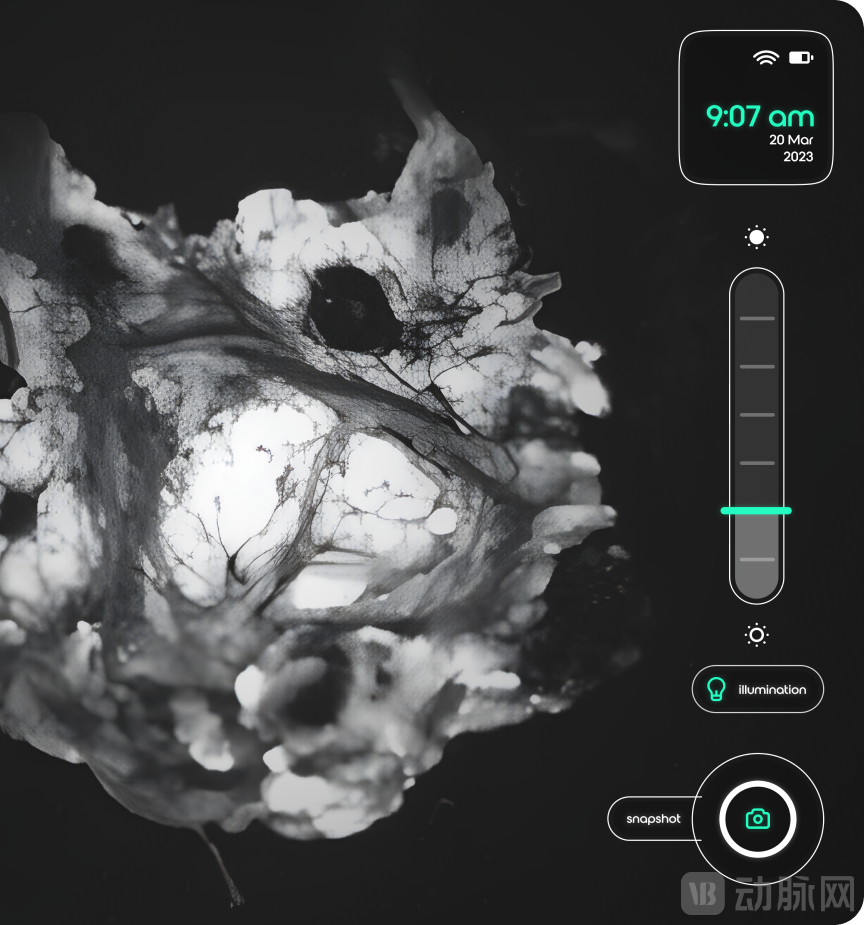
InVision Imaging System Operation Page Display
Its adjustable tilt screen ensures an optimal viewing angle and delivers real-time, high-resolution imaging (70 μm) with a latency of less than 0.01 seconds. The touchscreen surface remains highly responsive even when the user is wearing gloves or has wet hands. Below the display is a spacious observation area that allows direct visualization of tissue samples up to 2–3 mm in thickness and measuring up to 8 cm × 6 cm. The system also features a specimen snapshot function, enabling one-click capture and saving at any time. Furthermore, InVision supports both plug-and-play operation and battery power, offering approximately three hours of battery life.After use, simply wipe the observation area clean with an alcohol swab.
andSentinel Lymph Node Biopsy, etc.Compared with conventional methods, the InVision system eliminates the need for complex pre-processing steps such as tissue puncture biopsy and staining. It is radiation-free and does not require injections, thereby reducing patient discomfort and streamlining the diagnostic workflow. More importantly, it enables physicians to simplify complex procedures whileAvoid Omissions,Achieve more accurate cancer staging.
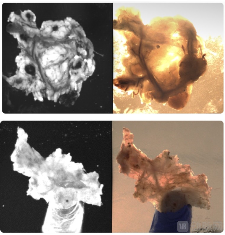
From top to bottom: mesenteric lymph nodes, submillimeter head and neck lymph nodes; the left image shows InVision imaging.
Notably, the InVision imaging system is the first to utilize short-wave infrared technology to differentiate lymph nodes from surrounding adipose tissue. It has not only received FDA Class I medical device clearance but also been honored with TIME Magazine’s Best Inventions of 2023 award and the Red Dot Design Award.
Currently,InVisionCompleted atDeployed in more than ten U.S. hospitals. With the support of ARPA-H, its adoption will further increase.
ChinaDomestically, the focus is more on contrast agents and surgical navigation systems.
Looking at the domestic market, there are also many tumor-related products based on infrared technology, but they focus more on contrast agents and surgical navigation systems. Currently, there is noSimilarInVision’s devices. Here, VCBeat has compiled a brief overview based on publicly available information from the past two years. The following list is presented in no particular order. If any items have been omitted, please feel free to contact the author via the details provided at the end of the article.
NC527-X, a Near-Infrared Hypoxia-Targeted Contrast Agent Independently Developed by Zhejiang Haibo Biotechnology Co., Ltd.By integrating tumor-targeting technology with near-infrared (NIR) fluorescence imaging, this agent uniquely targets and binds to tumor tissues for precise identification. Meanwhile, NIR fluorescence imaging enables high-definition, high-sensitivity visualization of tumor lesions without damaging normal tissues. In July 2024, it received implicit approval for an Investigational New Drug (IND) application from the U.S. Food and Drug Administration (FDA), becoming the world’s first-in-class NIR fluorescent agent for precise tumor imaging and the first such drug from China to obtain FDA IND clearance.(Recommended reading:[Exclusive] Haibo Biologics Completes Tens of Millions in Pre-A Financing, Exclusively Invested by Puhua Capital)
Xindou Biotechnology (Suzhou) Co., Ltd. independently developed the near-infrared tumor-targeted fluorescent contrast agent product, DGPR1008 Injection,It is indicated for use in patients undergoing prostate cancer surgery as an adjunctive tool for intraoperative identification of malignant lesions, representing the first product approved for clinical use in this field in China. DGPR1008 injection demonstrates high sensitivity, strong specificity, and a favorable tumor-to-background ratio (TBR), enabling surgeons to clearly delineate tumor location and boundaries, minimize damage to normal tissues, and identify microscopic tumor foci and metastatic lymph nodes. In May 2024, it was announced that its investigational new drug application had received implicit approval from the Center for Drug Evaluation (CDE) of the National Medical Products Administration of China (Acceptance Number: CXHL2400243).(Recommended Reading: First Near-Infrared Tumor-Targeted Fluorescent Contrast Agent Clinical Trial Approved, Filling the Domestic Gap in This Field!)
Furthermore,Beihang University, in collaboration with the Institute of Automation, Chinese Academy of Sciences, and other institutions,Developed a near-infrared fluorescence surgical navigation system for tumors. This system enables non-invasive, real-time visualization of biochemical events at the molecular and cellular levels of tumors during surgery, providing surgeons with objective, real-time evidence at the molecular pathological level to support intraoperative decision-making. Related products have been promoted and applied in more than 1,000 Grade A tertiary hospitals across China, with cumulative sales exceeding RMB 130 million, thereby breaking the long-standing monopoly held by foreign competitors in this field.[2]
Professor Wang Dong from the AIE Research Center at Shenzhen University and Professor Zhu Shoujun from Jilin University, both members of Academician Tang Benzhong’s teamIn July 2022, a research paper titled "Molecular engineering of AIE luminogens for NIR-II/IIb bioimaging and surgical navigation of lymph nodes" was published in the journal *Matter*. This study employed molecular engineering strategies to construct near-infrared II (NIR-II) aggregation-induced emission (AIE) molecules with the longest absorption wavelength reported to date. Compared with indocyanine green (ICG), an FDA-approved contrast agent widely used in clinical practice, this material enables sentinel lymph node imaging and surgical navigation with prolonged retention, deep tissue penetration, and a high signal-to-noise ratio.
Professor Xi Rimo’s and Associate Professor Meng Meng’s research groups, School of Pharmaceutical Sciences, Nankai University,20In December 2021, a research paper titled “A Tumor-Targeting Near-Infrared Heptamethine Cyanine Photosensitizer with Twisted Molecular Structure for Enhanced Imaging-Guided Cancer Phototherapy” was published in the prestigious international academic journal J. Am. Chem. Soc. The research team designed and synthesized a tumor-targeting near-infrared photosensitizer, T780T, by introducing a twisted tetraphenylethylene (TPE) structure between two molecules of the cyanine photosensitizer IR780 to enhance its photothermal conversion efficiency and photothermal stability. The constructed T780T molecules can self-assemble into uniform nano-aggregates in aqueous phase. After intravenous injection via the tail vein in tumor-bearing mice, the T780T aggregates demonstrated significant tumor accumulation. A treatment regimen involving a single administration followed by two laser irradiations achieved markedly enhanced tumor inhibition.
References:
[1] <What is the Most Common Lymph Node Yield for Colorectal Cancer Cases? The results may scare you.>,Cision Vision;
[2] https://www.cma.org.cn/attach/0/9ab917ad252449d8bd6cc1c4ee44ded1.pdf
[3] <MIT’s tiny technologies go to Washington>
[4] <CisionVision Receives Prestigious ARPA-H Funding>
[5] <Invision: A Clear View>