Canon Medical Unveils Groundbreaking Innovations at CIIE with Multiple China Debuts Targeting Global Healthcare Challenges
Highlights:
AI Super Brain Iterates Again, Empowering the Entire Equipment Lineup
PIQE CT Makes Its China Debut, Ushering in the Era of “4K” Ultra-High-Definition Imaging
Ultimate 3T MRI: Breaking the “Trade-off Triangle” to Achieve High-Resolution Spinal Imaging in 59 Seconds
The Industry’s Highest-Resolution Angiography System Makes Its China Debut, Redefining Confidence in Interventional Procedures
……
On November 5, 2024, Canon Medical Systems made its appearance at the 7th China International Import Expo (CIIE). Marking its seventh consecutive year at the CIIE, Canon Medical Systems participated under the theme “A Century of Chasing Light, Innovating for a Far-Reaching Future.” Showcasing numerous innovative achievements that integrate global resources with Chinese expertise, the company highlighted advancements in precision medicine, digital transformation, and AI empowerment. Several products were exhibited in the Chinese market for the first time. Leveraging the CIIE platform, Canon Medical Systems is committed to ensuring that patients and healthcare professionals in China have simultaneous access to the latest medical technological advancements worldwide, thereby fulfilling its long-term commitment to serving Chinese users through concrete actions.
Tadashi Taguchi, Chief Representative of Canon Medical Systems in China, stated: “The China International Import Expo (CIIE) serves as a vital platform for bridging resources between China and the world and sharing development opportunities, as well as a key stage for foreign-invested enterprises to showcase new products and technologies. This year marks Canon Medical’s seventh consecutive participation in the CIIE, with the number of new product exhibits reaching an all-time high. We have brought our latest global AI-enabled products and solutions in computed tomography (CT), magnetic resonance imaging (MRI), and angiography. Notably, the Supreme 3.0T MRI made its Chinese debut ahead of RSNA 2024. As an active participant in China’s healthcare sector, we are approaching our 50th year of development in the country. Moving forward, we will continue to deepen our presence in China, deeply integrating global innovation resources with the local market. Leveraging our century-honed expertise in imaging technology, our forward-looking innovations in AI and digitalization, and our comprehensive value-based collaborations with Chinese partners, we aim to accelerate our integration into the new landscape of new quality productive forces in China’s healthcare sector, thereby contributing to the high-quality development of Healthy China.”
World-Class Intelligent New Products Directly Address Core Medical Needs
4K Ultra-HD Cardiac Imaging with 95% Dose Reduction
Aquilion ONE with PIQE CT
- China Premiere -
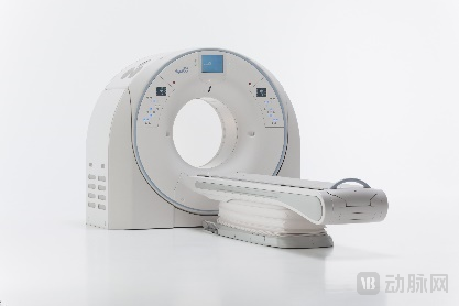
Cardiovascular and cerebrovascular diseases have become the leading cause of death threatening human health. Data from the "Report on Cardiovascular Health and Diseases in China 2023" shows that the number of patients with cardiovascular and cerebrovascular diseases in China has reached as high as 330 million, with a trend toward affecting younger populations. In actual clinical diagnosis, due to the limitations of traditional CT in scanning speed, temporal/spatial resolution, and contrast imaging technology, accurate diagnosis in challenging scenarios such as coronary artery calcification, coronary stents, and prosthetic heart valves has remained difficult to achieve.AIThe rapid development of imaging technology has brought about new imaging solutions. Canon Medical is showcasing its next-generation high-definition CT for the first time in China, equipped with the newly developed HD AIEnginePIQE(Precise IQ Engine), breaking through traditional CTImaging Contraindication, CTCardiac imaging has entered the “4K“Era.
Traditionally,To obtain images with higher image quality,RegardingMore NeededRadiation DoseQuantity,And Canon Medical's high signal-to-noise AIAiCE Engine2.0Revolutionarily Breaks Through"Low Radiation" and "High Image Quality"betweenCompromise relationship. The dose for a routine non-contrast chest CT scan is approximately 1 millisievert.(Approximately equal to 1000microsievert), AiCE 2.0 can reduce the doseSignificantly reduced by 95%,Reduced to 50 microsieverts—Approximately equivalent to the natural background radiation exposure received during an international flight from Shanghai to New York—this is achieving CTA Major Leap in Ultra-Low-Dose Imaging。
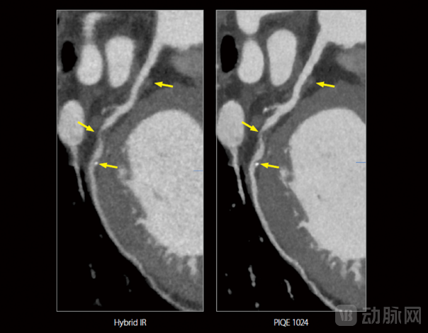
In the diagnosis of coronary heart disease (CHD), positive remodeling is one of the markers indicating a high risk of coronary plaque rupture. Failure to accurately identify positive vascular remodeling may lead physicians to underestimate the patient’s risk of acute cardiovascular events (such as myocardial infarction), resulting in missed or misdiagnosis. The image above illustrates an extremely small lesion captured with the assistance of PIQE imaging during follow-up diagnosis of a CHD patient. As shown, compared with the conventional hybrid iterative reconstruction image (left), the PIQE image (right) more clearly demonstrates severe stenosis (>70%) in the left circumflex artery (LCX, a major branch of the coronary arteries) caused by soft plaque, along with outward/positive remodeling of the mid-segment vessel. It also reveals that the stenosis is located approximately 10 mm proximal to the first obtuse marginal branch, with a tiny calcified plaque present distal to the stenotic site. In response to this newly developed lesion, the physician promptly adjusted the treatment plan.
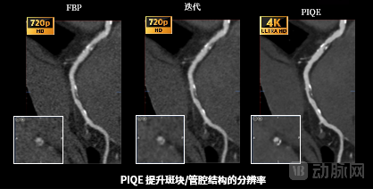
The image above presents a comparative imaging analysis of intravascular soft plaques in patients with coronary heart disease. For the visualization of microplaques, neither Filtered Back Projection (FBP) nor iterative reconstruction images can clearly delineate the edges, which hinders the assessment of the intravascular conditions. However, with the aid of PIQE, the plaque margins appear sharper and the luminal borders are more distinct, thereby facilitating more accurate clinical judgments regarding changes in the lumen structure.
Breaking Through the “Balance-of-Power Triangle”
Supreme Ultra-HD 3.0T MRI
- Premiered in China Prior to RSNA -
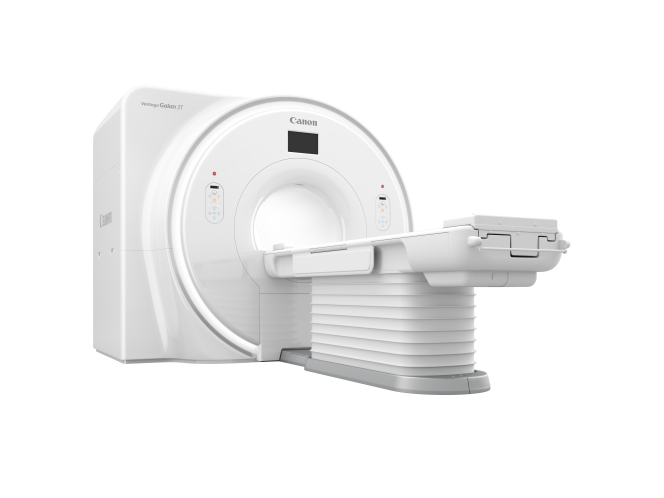
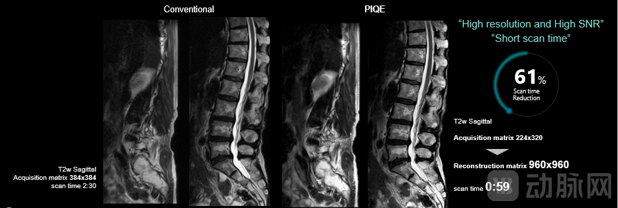
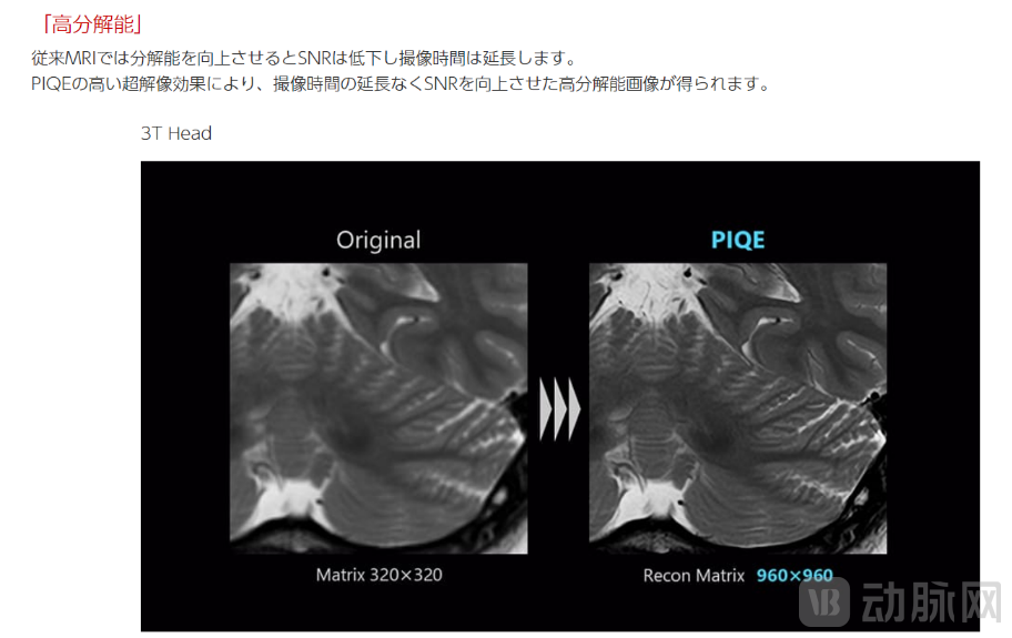
In magnetic resonance imaging (MRI), the signal-to-noise ratio (SNR), spatial resolution, and scan time are known as the “trade-off triangle.” These three factors interact with and constrain one another, jointly influencing image quality and diagnostic accuracy. Historically, acquiring high-resolution images often required prolonged scan times, which inevitably hindered imaging efficiency. The high-definition AI engine PIQE enables a perfect “trio” of benefits: increasing the image matrix, reducing image noise, and shortening scan time. Furthermore, when integrated with the Real-time platform, it significantly accelerates data exchange and processing, thereby enhancing clinical diagnostic performance.
59-Second High-Resolution Spinal Imaging
In spinal imaging, the Supreme system leverages PIQE technology to acquire high-resolution images with a 960×960 matrix in just 59 seconds of ultra-fast scanning, creating new value for the rapid diagnosis and treatment of trauma patients.
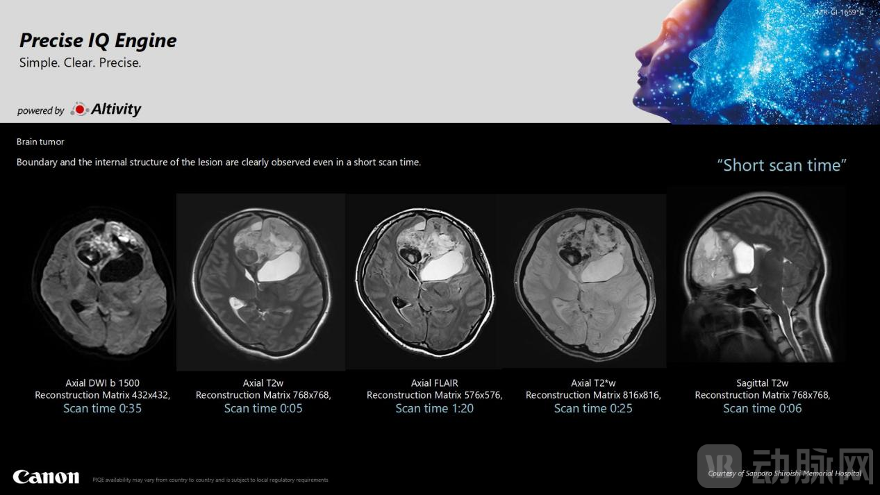
2-Minute Brain Imaging
Conventional non-contrast cranial CT scanning, which typically involves five sequences, takes approximately 7–10 minutes. In contrast, under the same conditions, PIQE can reduce the scan time to just 2.5 minutes. Even with this significant reduction in scanning duration, lesion boundaries and internal structures remain clearly visualized, substantially improving diagnostic efficiency and accuracy. For acute cerebral conditions such as acute cerebral infarction and intracerebral hemorrhage, this technology helps secure valuable time for treatment, thereby contributing to improved patient prognosis.
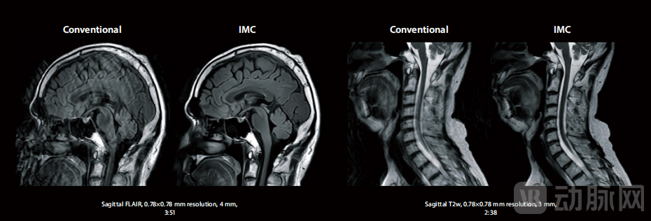
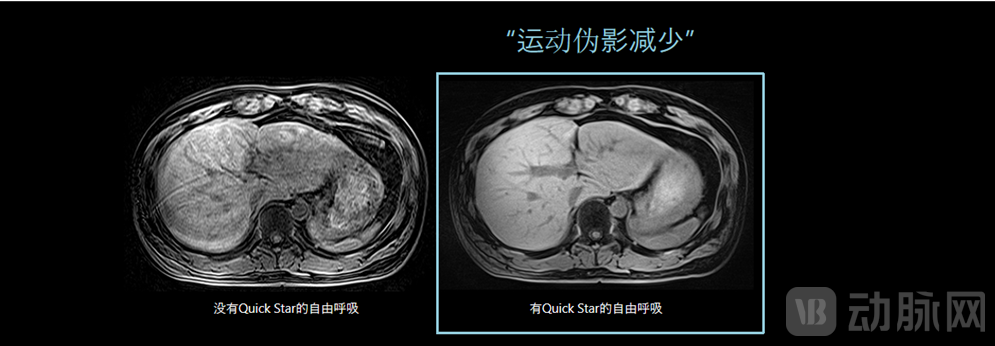
Comfortable and Unobtrusive, Patient-Friendly
Patients with liver disease, individuals who have difficulty holding their breath, and pediatric patients often exhibit insufficient cooperation during magnetic resonance imaging (MRI) scans, leading to motion artifacts in the images that compromise diagnostic accuracy. IMC employs a deep learning-based approach for motion correction, effectively mitigating artifacts caused by involuntary patient movement. Furthermore, the Quick Star free-breathing technique significantly reduces artifacts induced by respiratory motion, markedly improving the success rate of MRI examinations in pediatric patients. Meanwhile, the Real-time platform, which integrates "exchange, correction, and reconstruction" functionalities, can identify and suppress useless RF waveforms that generate artifacts, thereby reducing interference and ensuring the accuracy and reliability of the data.
The Industry’s Only Ultra-Micro HD Flat Panel Detector: Redefining Confidence in Interventional Procedures
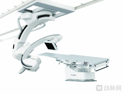
Alphenix Sky+ Microscopic HD Dual Slip-Ring Interventional System
- China's Debut Exhibition -
Unlike CT and MRI angiography, angiography systems provide real-time, dynamic vascular imaging, enabling rapid and precise localization and accurate depiction of lesion details. They can also detect concealed intravascular lesions at an earlier stage, playing an irreplaceable role in the precise diagnosis and interventional treatment of cardiovascular and cerebrovascular diseases.
76-Micron High-Definition Microscopy: Breaking the Limits of Imaging
Alphenix Sky+ features the world’s first microscopic high-definition detector, with a breakthrough pixel size of 76 microns—just one-quarter the area of traditional detectors. Its spatial resolution has doubled compared to previous models, reaching 6.6 lp/mm, thereby surpassing the magnification limits of conventional imaging technologies. This enables operators to perform interventional procedures as if under a microscope, allowing for precise visualization of minute lesions and accurate navigation. Combined with hardware and software innovations such as the full 16-bit high-definition imaging chain and Alphenix 2.0 intelligent applications, Alphenix Sky+ delivers clinical-grade microscopic high-definition imaging for refined and complex interventional surgeries in neurology, cardiology, oncology, and peripheral large vessels, thereby enhancing physicians’ confidence during procedures.
Breaking Through Spatiotemporal Limits: High-Speed Whole-Body 3D Imaging
In interventional therapy, rapid angle switching and position adjustment are key to improving surgical efficiency. The Alphenix Sky+ adopts a dual-slip-ring C-arm 9-axis flexible gantry design, breaking through the "spatiotemporal" limitations of traditional DSA equipment. It enables high-speed 3D acquisition at any position within a 201 cm range across the entire body during free breathing, with second-level synchronization for single acquisitions. This provides more efficient and precise intraoperative 3D angiography for interventional treatment of pelvic tumors and complex lower limb diseases, effectively overcoming respiratory motion artifacts, enhancing image quality, and improving surgical efficiency.
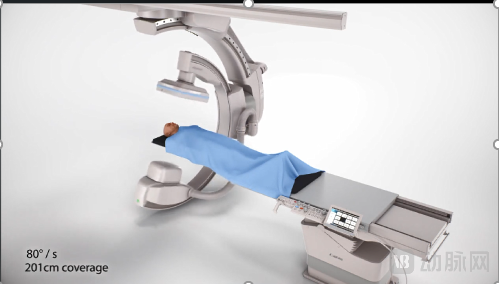
(Dual Slip-Ring C-Arm with 9-Axis Agile Gantry Design)
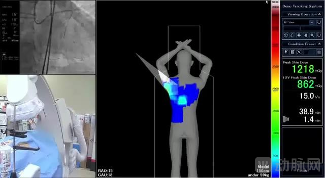
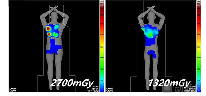
Intelligent Dose Control: Safeguarding the Safety of Patients and Medical Staff
Unlike other radiological diagnostic procedures, interventional radiology results in significantly higher radiation doses for both patients and medical staff. Therefore, strengthening dose protection for both parties has always been a critical focus in the development of interventional radiology. Equipped with DoseRite, the industry’s only intelligent dose visualization technology, the system enables real-time tracking and control of radiation dose, ensuring uniform dose distribution and consistent image quality, thereby safeguarding the safety of both patients and healthcare providers.
Multimodal Fusion Significantly Enhances Diagnostic and Therapeutic Efficacy in Critical Care
Collaborative Imaging Ultra-HD Comprehensive Solution
- China's Debut Exhibition -
Currently, the global healthcare landscape is facing challenges such as high medical costs, heavy burdens on healthcare professionals, and limited accessibility to high-quality medical resources. Committed to helping clinicians achieve more with fewer resources, Canon Medical has launchedCollaborative Imaging Ultra-High-Definition Integrated Solution (hereinafter referred to as “CI”), and was exhibited for the first time in the Chinese market。CIthe birth, enabling coverage of CT、MREmpowered by artificial intelligence, the full portfolio of products in angiography and ultrasound synergizes through multimodal fusion to highly integrate ultra-high-definition diagnostic data across various fields. This not only provides a more comprehensive analytical perspective for disease diagnosis and enhances diagnostic precision, but also improves interdisciplinary collaboration efficiency. It delivers comprehensive solutions for oncology, cardiovascular, neurological, and sports medicine, thereby empowering and enhancing the diagnostic and therapeutic efficacy for critical and severe conditions.

Taking sports medicine as an example, CI enables ultra-high-definition collaborative imaging of CT, MRI, and ultrasound for sports injuries. It integrates data from different diagnostic stages and various diagnostic devices. Based on extensive multi-dimensional diagnostic data, it enhances the efficiency of diagnosis and treatment and the scientific rigor of clinical decision-making, thereby supporting early diagnosis and treatment of sports injuries, as well as injury prevention and rehabilitation.
Canon Medical collaborates with renowned sports clubs such as Manchester United, FC Barcelona, and Real Madrid, as well as leading experts, to continuously drive innovation. It has published more than 40 studies on musculoskeletal (MSK) and cardiac imaging, contributing valuable academic expertise to the sports medicine community and supporting athletes in managing sports-related injuries and making optimal return-to-play (RTP) decisions.
In addition to new products making their debut launches and exhibitions, Canon Medical also showcased several star products at this year's CIIE.
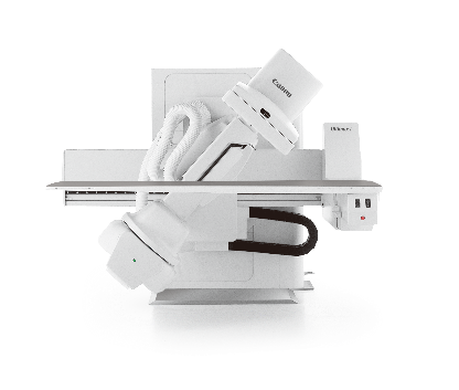
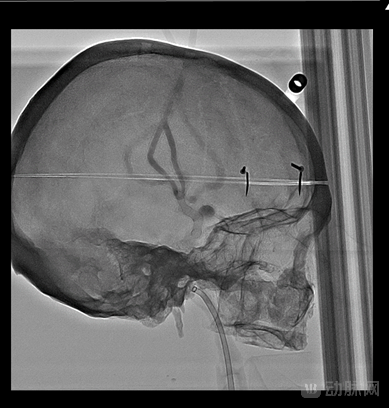
Ultimax-i is a rare X-ray imaging system in the industry that ingeniously integrates a C-arm angiography unit with a tiltable gastrointestinal table. Equipped with multiple functional modules, it is widely applicable across various fields, including radiology, gastroenterology, pulmonology, interventional medicine, urology, and obstetrics/gynecology. It supports multi-departmental collaborative applications as well as complex diagnostic and therapeutic tasks within individual departments. Furthermore, the upgraded Ultimax-i features Accent, an intelligent high-definition algorithm jointly developed by Canon Medical and Olympus. Combined with high-definition flat-panel detectors and a new digital system, it significantly reduces radiation dose while enhancing image processing precision and the ability to capture subtle lesions. This achieves high-resolution dynamic imaging, making diagnostic and therapeutic procedures easier and more reliable, with precise and efficient diagnostic results. To meet the cutting-edge demands of the Chinese market, Canon Medical will soon launch the China-produced Ultimax-i and introduce new products in China.
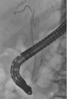
Standard Conditions
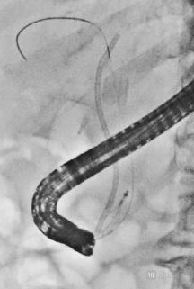
After Applying Accent
In the patient shown in the image, a plastic stent was implanted due to residual gallstones after lithotripsy. The stent had migrated into the biliary tract, necessitating retrieval of both the stent and the residual stones using a stone extraction basket. After applying Accent, the distal opening of the stent, as well as the guidewire and guide wire of the stone extraction basket, were clearly visualized, enabling the physician to successfully complete the stent retrieval procedure.
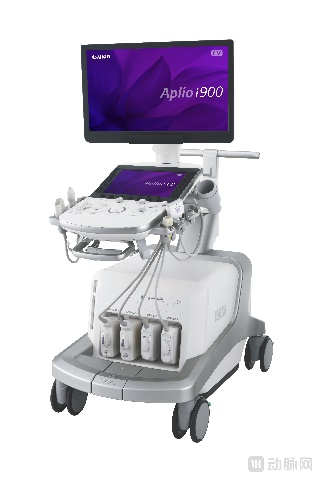
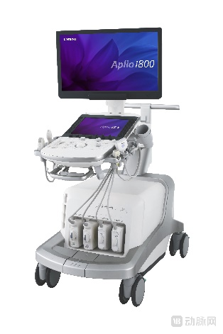
As Canon’s flagship ultrasound product line, the Aplio i series incorporates numerous advanced technologies, such as high-frequency transducer arrays reaching up to 33 MHz and 24 MHz, innovative i-series transducers, and iDMS (intelligent Dynamic Micro Slicing) technology, which delivers ultra-thin beams to enhance imaging resolution.as well as the highly sensitive iSMI (iSuperb Microvascular Imaging) technology. Additionally, the Liver Package offers a non-invasive, quantitative solution for the entire course of liver disease management. By integrating advanced technologies such as ATI (Attenuation Imaging) and SWE (Shear Wave Elastography), it enables comprehensive, multi-angle assessment of diffuse liver diseases, covering various pathological states including hepatocellular inflammation, necrosis, fibrosis, and steatosis, thereby supporting whole-course disease management. In 2024, leveraging the Aplio i series’ liver disease solutions, global and Chinese multicenter projects supported by Canon Medical Systems published their latest research findings in the RSNA academic journal Radiology, providing forward-looking insights into the exploration of liver diseases.
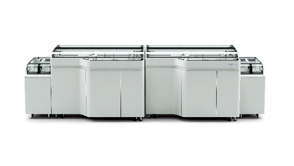
The TBA-FX8 Fully Automated Biochemistry Analyzer is equipped with a variety of innovative intelligent applications that significantly enhance testing efficiency. Featuring a 3D intelligent robotic arm for sample aspiration, it offers rapid processing speed, large reagent storage capacity, and high testing throughput, thereby empowering the optimization of laboratory workflows. It also supports expandable connectivity to laboratory automation track systems. Equipped with an advanced optical system with high spectrophotometric precision, it enables laboratories to perform a broader range of test items. Additionally, it features a dedicated STAT loading port and a bidirectional track sampling mode to meet the demands for rapid emergency testing. Currently, the TBA-FX8, powered by V2.0 software, has been launched in the Chinese market, supporting hospitals in building smart laboratories.
Looking ahead, leveraging the platform of the China International Import Expo (CIIE), Canon Medical will continue to introduce advanced medical technologies and products, actively promote the implementation of various achievements, and work together with partners from all sectors to continuously support the high-quality development of China’s healthcare industry.