Zhike Lishang Revolutionizes Endoscopy with China's First 500x Cellular Endoscope Enabling Biopsy-Free Precision Diagnosis of Gastrointestinal Cancers
In March 2025, at the endoscopy center of a leading Grade-A tertiary hospital in Shanghai, a microscopic revolution was upending the standards for early gastrointestinal cancer screening. Under the real-time imaging capabilities of a domestically produced 500x cellular endoscope, the esophageal mucosal surface—previously indistinct under conventional endoscopy—was rendered with exquisite clarity: normal mucosal cells were sharply demarcated from abnormally enlarged clusters of cancerous cells, and the distorted morphology of capillaries was clearly visible.
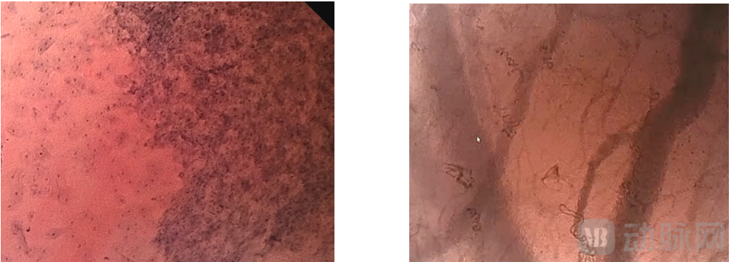
Conventional endoscopy requires the removal of lesion tissue for biopsy to confirm tumors, whereas cell endoscopy enables precise optical biopsy diagnosis directly, allowing physicians to achieve accurate diagnoses without relying on histological biopsy. The cell endoscope currently being operated by the physician is the 500x magnification cell endoscope (Zhejiang Medical Device Registration No. 20232062041), independently developed by Zhejiang Zhike Lishang Medical Technology Co., Ltd. (hereinafter referred to as “Zhike Lishang”).
Cellular endoscopy is an electronic endoscope with ultra-high magnification, representing one of the most advanced endoscopic examination technologies currently available. It enables cellular-level observation, allowing physicians to clearly visualize detailed morphological features of cells, glandular structures, and microvasculature. Previously, such high-precision, cellular-level observation could only be achieved through ex vivo pathological examination; however, innovations by Zhikeli have made in vivo diagnosis possible, providing a novel solution particularly for patients who are unable to undergo biopsy due to anticoagulant therapy.
Previously, Olympus was the only international supplier capable of providing electronic endoscopes with ultra-high magnification. By independently developing core technologies such as optical continuous zoom and optical magnification, Zhikelishang has not only broken this technological monopoly but also holds promise for transforming the traditional biopsy-dependent diagnostic model into a real-time microscopic observation model, thereby revolutionizing early screening and detailed examination of gastrointestinal tumors.
1. The field of view is nearly three times that of imported products.
Achieving the Transition from "Multiple Examinations" to "One-Stop Diagnosis"
The 500x cellular endoscope developed by Zhikeli Shang, leveraging its 140° wide-angle standard field of view and ultra-high resolution of 2–3 μm under microscopic magnification, clearly visualizes the entire gastrointestinal mucosa as well as local glandular structures, cell nuclei, and microvascular networks. The microscopic field of view measures 1,200 μm × 670 μm, which is 2.8 times larger than that of imported products (570 μm × 500 μm). This means that a single scan can cover the area that would require multiple lens adjustments with imported endoscopes, thereby significantly improving examination efficiency. Furthermore, this optical in vivo histopathology capability enables physicians to diagnose early-stage cancer without the need for biopsy, upgrading gastrointestinal endoscopy from “single-mode observation” to “full-scale diagnosis.”
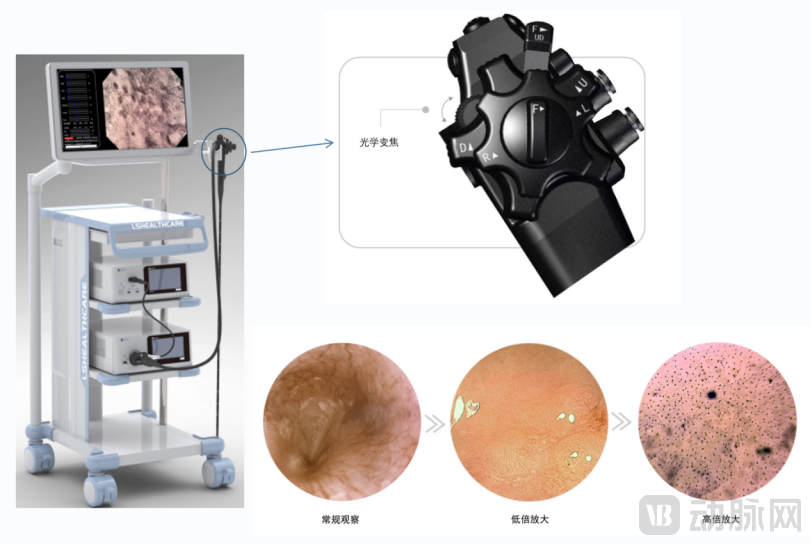
Furthermore, the device integrates a “three-in-one” zoom system, meaning that a single model of zoom gastroenteroscope replaces the functions of three conventional models of electronic gastroenteroscopes: standard, low-magnification (80–135×), and high-magnification (up to 500×). Physicians can switch between these modes simply by operating a zoom lever. This simplifies diagnostic and therapeutic procedures that previously required switching between different device models into a single operation, significantly improving examination efficiency while enhancing diagnostic accuracy.
In addition to capturing static images of cellular structures, this cellular endoscope can clearly visualize blood flow information at lesion sites. It enables intuitive, real-time monitoring of in vivo drug responses during pharmacotherapy without the need for sampling or biopsy, thereby providing continuous dynamic evidence to guide the formulation of personalized treatment plans.
In the field of endoscopy, there exists a technical paradox involving “wide field of view, high resolution, and low cost.” Especially when achieving clear 500x magnification, any minor operational tremor or organ movement can cause image blurring. Zhikeli Shang has successfully overcome this challenge through ingenious preliminary design and innovations in both hardware and software. For instance, by cleverly designing and simplifying aberration-compensating lens groups, they achieved a cross-scale continuous zoom lens. Additionally, intelligent image stabilization algorithms eliminate motion blur caused by breathing or operational tremors by optimizing image processing algorithms and enabling rapid imaging at 60 frames per second.
The 500x magnification cell endoscope not only helps determine the extent of lesions but also assists physicians in making precise differential diagnoses for uncertain suspected lesions. Through in vivo observation with the cell endoscope, doctors can clearly distinguish the characteristic differences between normal cells and early-stage cancer cells, enabling examination, cellular observation, and lesion diagnosis to be completed in a single procedure. This significantly improves clinical workflow efficiency and benefits patients.
Multiple relevant clinical studies have confirmed that confocal laser endomicroscopy offers significant advantages in diagnosing Barrett’s esophagus and differentiating gastric diseases, holding promise for advancing a “diagnosis-and-treatment-in-one” closed-loop care model.
In addition to the remarkable breakthroughs in flexible endoscopes, Zhikeli Shang has also delivered equally impressive performance in rigid endoscopy.
In minimally invasive surgery, accurately marking the lesion area is merely a basic requirement; furthermore, surgeons must identify critical structures such as neural and vascular tissues to avoid inadvertent injury to surrounding tissues and reduce the incidence of postoperative complications.
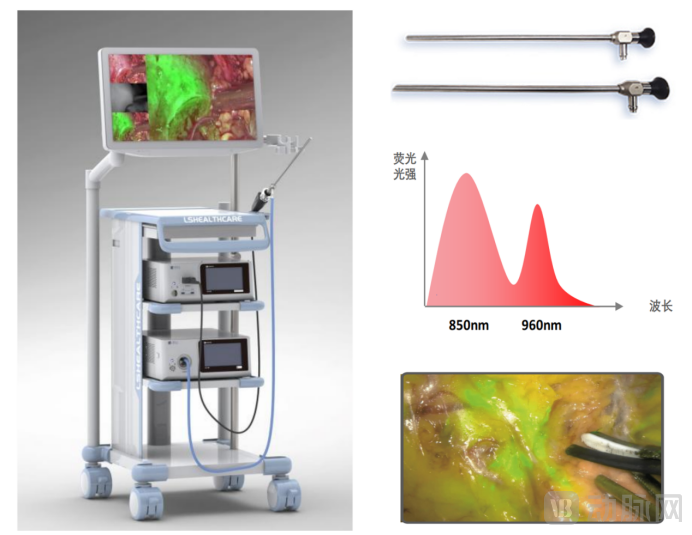
Zhike Lishang’s independently developed 4K dual-fluorescence laparoscope (Zhejiang Medical Device Registration No. 20242061905) was designed precisely for this purpose. By simultaneously capturing dual fluorescence bands at 800–850 nm and 900–1000 nm, it enables specific labeling of different tissues. The short-wave band (800–850 nm) can identify tumor margins or blood vessels, while the long-wave band (900–1000 nm), leveraging its deep tissue penetration advantage, can visualize critical structures such as the ureters and nerve plexuses, thereby enhancing surgical safety and efficacy.
Compared to traditional single-fluorescence systems that focus solely on the lesion area, dual-fluorescence technology enables simultaneous high-definition visualization of the surgical field and protection of critical tissues. This effectively addresses iatrogenic injuries caused by poor tissue identification in minimally invasive surgeries, making it particularly suitable for complex surgical scenarios requiring precise manipulation.
In terms of optical performance, Zhikeli employs aspheric optical design and broadband chromatic aberration correction to ensure large-field-of-view imaging with minimal distortion, enabling physicians to accurately assess dimensions at the field edges. Meanwhile, its triple-band parfocal technology ensures perfect alignment between the visible light and dual fluorescence channels, eliminating the need for refocusing when switching endoscope bodies during clinical procedures, thereby maintaining surgical continuity.
In terms of cost and compatibility, the 4K dual-fluorescence laparoscope from Zhikeli is priced comparably to traditional single-fluorescence laparoscopes, yet offers superior performance. It can operate independently or be used in conjunction with endocytoscopes, thereby fulfilling the need for collaborative “internal–external” diagnosis and treatment of gastrointestinal diseases, and providing a more comprehensive and precise solution for complex surgeries such as those involving the intestine.
The launch of the 4K dual-fluorescence laparoscope not only completes Zhikeli Shang’s product portfolio in high-end flexible and rigid endoscopes, mitigating the dependency risks associated with a single technical approach, but also provides clinicians with a cost-effective, multifunctional, integrated solution.
The source of this endoscopic technology revolution is inseparable from the two decades of technological accumulation by the National Engineering Research Center for Optical Instruments at the College of Optical Science and Engineering, Zhejiang University.
Founder Wang Liqiang enrolled in the integrated bachelor’s, master’s, and doctoral program at the College of Optical Science and Engineering, Zhejiang University, in 1994. He remained at the university as a faculty member after completing his postdoctoral research and has undertaken nearly ten national-level scientific research projects. With over two decades of accumulated expertise, he has achieved outstanding results in optical design, CMOS imaging, embedded systems, and digital image processing. He holds independent intellectual property rights for core technologies, including wide-angle objective lenses for high-definition endoscopes, dynamic multispectral image analysis, 3D dual-optical-path Hopkins systems, and assembly processes for miniaturized CMOS detectors.
The R&D team’s professional expertise comprehensively covers disciplines essential to endoscope development, including optics, precision mechanics, electronics, and software. With over a decade of experience in collaborative industry-academia-research-clinical partnerships, core team members have accumulated profound foundational and applied technical capabilities in endoscopy. These include technologies such as micro optical zoom lenses, wide-angle high-magnification optical amplification, high-definition electronic imaging, multispectral image enhancement, and ultra-miniature CMOS imaging. The team addresses all critical technological milestones in endoscope R&D, possessing full-chain research and development capabilities.
Built on over two decades of expertise in optoelectronics at Zhejiang University, and leveraging the “scientist + engineer + clinical expert” golden triangle model, Zhike Lishang has obtained seven medical device registration certificates within less than four years of its establishment. The company has constructed a product portfolio featuring synergistic and complementary flexible and rigid endoscopes, which are applied in multiple clinical specialties, including gastroenterology, gastrointestinal surgery, hepatobiliary surgery, and urology.
Zhike Lishang is currently driven by a dual-engine strategy of direct supply to medical institutions and ODM partnerships with manufacturers. It has already completed the sales of several cell endoscopy systems and will accelerate its penetration into the domestic market this year.
Wang Liqiang believes that cross-scale imaging, multispectral specific imaging, multimodal imaging, and AI technology will become the future development directions for endoscopes. Based on this, Zhikeli has also carried out related technological research and product reserves.
Furthermore, the company will optimize precision endoscopy techniques for the digestive tract to enable “diagnosis and treatment in a single session,” allowing patients to complete both diagnostic and therapeutic procedures during a single hospital admission. This approach avoids the need for readmission and redundant preparations, thereby reducing medical costs and improving the efficiency of diagnosis and treatment.
From the “microscopic eye” of cell endoscopy to the “multi-dimensional navigation” of dual-fluorescence laparoscopy, Zhikeli Shang is reshaping the discourse power of Chinese-made endoscopes with hardcore optical technology. Its value lies not only in breaking the import monopoly but also in enabling precision medicine to truly permeate the entire chain of minimally invasive diagnosis and treatment through an innovative path of “software-hardware synergy and multi-modal fusion.”