China's Domestic X-ray Source Innovators Reach Global Forefront in High-End Imaging
In the furthest upstream corner of high-end medical equipment, the little-noticed “X-ray source” is brewing a cross-era imaging revolution.
In 1895, Wilhelm Röntgen generated free electrons by heating a tungsten filament, accelerated them under high voltage, and forced them to strike a metal target, thereby successfully generating X-rays and laying the theoretical foundation for modern medical imaging.
Over the past century, medical imaging equipment has undergone dramatic changes, yet the evolution of hot-cathode X-ray sources has remained firmly grounded in this core principle. It was not until the 21st century, with the continuous emergence of innovative imaging devices, that this century-old technology finally faced new competitive challenges.
Taking static CT systems equipped with a large number of X-ray sources and detectors as an example, these systems replace spiral imaging with pulsed exposure of the detector ring, requiring the arrangement of numerous X-ray sources and detectors within the limited space of the gantry. Therefore, the X-ray sources in such CT systems must meet two key criteria: compact size and high intensity. However, since the volume of thermionic anode X-ray sources is directly proportional to their output, they fail to meet these requirements.
To overcome this physical bottleneck, some innovative companies have chosen an alternative path by leveraging cold-cathode X-ray sources to generate high-energy electron beams. In breaking through this impasse, they are also writing a new epic in the field of radiological imaging.
In simple terms, cold cathode X-ray sources utilize carbon nanotube-based cathodes to emit electrons, which are accelerated by an electric field to strike the anode target, thereby generating X-rays. As no heating is required, cold cathode X-ray sources offer advantages over traditional thermionic cathodes, including faster response times, lower power consumption, and easier integration.
When evaluating the performance of cold-cathode X-ray sources, carbon nanotubes can readily serve as an indicator of quality. Carbon nanotubes are one-dimensional quantum materials composed of single or multiple layers of graphene sheets rolled around a central axis at a specific angle to form coaxial, hollow, seamless tubular structures; they emit electrons via field emission. In any field emission process, the electric field strength can be enhanced by increasing the aspect ratio of the emitter surface. Therefore, theoretically, the sharper the tip of the carbon nanotube, the stronger the generated electric field intensity.
In the early 21st century, cold-cathode X-ray sources with beam currents at the 10 mA level were already being mass-produced for industrial and security inspection applications, such as power grid and railway track inspections. Driven by higher power output and longer operational lifespans, cold-cathode X-ray sources rapidly achieved large-scale commercialization.
Witnessing the success of cold-cathode X-ray sources in the aforementioned fields, X-ray tube manufacturers worldwide, including GPS, have rapidly embarked on exploratory efforts. After all, cold-cathode X-ray sources can generate high-energy electron beams at room temperature with stable performance, while effectively reducing the footprint of imaging equipment. In an era where medical imaging devices are becoming increasingly bulky, this technology offers substantial potential for innovation and development.
However, challenges such as the high difficulty of material preparation, unstable field emission performance, complex process integration, and high costs have become prominent during the research and development and commercialization phases.
To ensure clear imaging even with slight patient movement, cold-cathode X-ray sources must be capable of emitting currents exceeding 50 mA, thereby completing exposure within one second. The ability to deliver high-current beams, achieve high-precision control, and maintain stable long-term operation constitutes the critical benchmark underlying the significant advantages that carbon nanotube cold-cathode X-ray sources bring to medical imaging—a feat that few companies could fully accomplish at the time.
It was not until around 2016 that domestic and international enterprises achieved breakthroughs in the preparation of high-performance carbon nanotubes and gradually promoted their application in medical imaging.
Currently, overseas companies such as Varex Imaging, Micro-X, and NanoX, as well as domestic enterprises including XinHong Electronics, Haozhi Imaging, and Aolei Technology, have all achieved scaled mass production through their respective technical pathways. This has validated the advantages of carbon nanotube cold-cathode X-ray sources in terms of high definition, rapid imaging, low radiation dose, compact size, and energy efficiency.
Among these, the two most representative technical approaches are Micro-X’s single-focus integration and the multi-focus array configurations developed by Varex Imaging and XinHong Electronics, with the latter presenting greater technical difficulty and challenges. Currently, Varex Imaging has achieved breakthroughs in mammography systems, while the Chinese enterprise XinHong Electronics has successively launched a series of cold-cathode multi-focus X-ray source products, which are gradually being applied in medical imaging.
In terms of lifespan, manufacturers of medical cold-cathode X-ray sources have also achieved continuous breakthroughs, reaching a level comparable to that of hot-cathode X-ray sources.
In the accelerated lifetime testing of detectors conducted by Micro-X, three self-developed carbon nanotube emitters of identical specifications were subjected to high-speed repetitive exposure. Carbon nanotube A was tested under an exposure condition of 20 mAs (clinically used for abdominal imaging), while carbon nanotubes B and C were tested at 2 mAs and 3.2 mAs (clinically used for chest imaging), respectively. The results showed that when subjected to a maximum exposure of 20 mAs, the emitter maintained operational functionality for approximately 10 years; under chest exposure conditions of 3.2 mAs, the emitter’s lifespan exceeded 20 years.
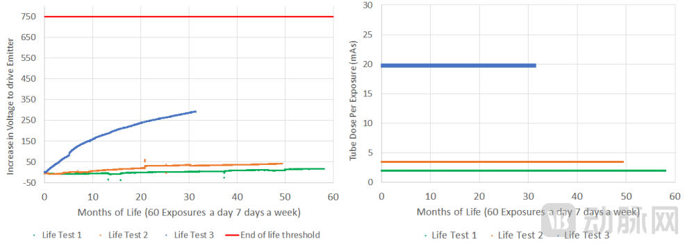 Micro-X Carbon Nanotube Accelerated Life Test (blue line: 20 mAs, orange line: 3 mAs, green line: 2 mAs)
Under the alluring prospect of epoch-making radiological imaging, a large number of investment institutions rapidly initiated their investment layouts as soon as breakthroughs were made in the manufacturing process of medical carbon nanotubes. According to statistics from VCBeat, enterprises in China capable of mass-producing medical cold-cathode X-ray sources have, to varying degrees, received capital support.
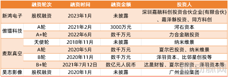 Financing Status of Domestic Companies with Production Lines for Medical Cold-Cathode X-Ray Sources (Incomplete Statistics)
Meanwhile, proprietary products developed by startups have demonstrated substantial potential, having identified unique pathways to empower next-generation imaging equipment such as CT, DR, and dental X-ray systems.
CT
Applications of cold cathode X-ray sources in the field of CT primarily revolve around their "ease of integration," which involves reducing the size of the X-ray source to free up internal space within the CT gantry. This enables either a reduction in the overall size of the CT system or the incorporation of additional precision instruments within the CT gantry. Specific applications include static CT and portable CT.
Static CT is one of the most mature and commercially promising application scenarios for cold-cathode X-ray sources. To simulate the rotational imaging of spiral CT, static CT must deploy a large number of X-ray tubes in a ring configuration within the gantry, with electronic pulses controlling each tube to emit X-rays instantaneously in sequence; this therefore requires substantial support from cold-cathode X-ray sources.
CT scanners are essential imaging equipment that all medical institutions must procure, and the ring-shaped radiation source structure of static CT systems has further expanded demand. As static CT technology continues to advance, the demand for cold-cathode X-ray sources will rise even further.
In the field of portable CT, Australian X-ray manufacturer Micro-X launched a head CT scanner last year, featuring 21 mini X-ray carbon nanotube sources for mobile stroke care.
The device can be stored on the side of an ambulance and assembled by rotation when in use. Weighing approximately 70 kilograms, it not only reduces the need for additional reinforcement of the ambulance but also effectively lowers configuration costs, enabling real-time CT diagnosis in the vast majority of outdoor settings.
Some companies are continuously “streamlining” CT innovation. For instance, the Nanox.ARC, which received FDA approval in 2023, is a multi-source digital 3D tomosynthesis system that reconstructs a series of 2D projection images into a stack of tomographic images (or slices) of the imaged object, thereby enabling 3D visualization.
By boldly eliminating the hot-cathode X-ray source, the previously enclosed, integrated gantry has been replaced by a visibly exposed metal framework. Although this design may appear somewhat rudimentary, Nanox.ARC effectively reduces the cost of 3D imaging while maintaining image quality, making it particularly suitable for resource-limited regions that cannot afford conventional CT equipment.
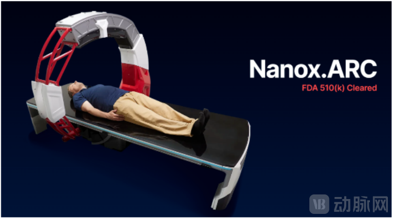 Nanox.ARC Diagnostic Schematic
Periapical X-ray Unit
Current oral imaging is predominantly dominated by two-dimensional (2D) dental radiography and three-dimensional (3D) cone-beam computed tomography (CBCT). In clinical practice, while 2D dental radiography is cost-effective, it frequently suffers from image superimposition and structural distortion. Conversely, although 3D CBCT provides volumetric imaging, it is expensive, characterized by low spatial resolution, prolonged scan times, high radiation dose, and susceptibility to motion artifacts such as those caused by patient respiration.
Under this initiative, XinHong Electronics has introduced cold-cathode X-ray sources into oral imaging, thereby creating a novel intraoral three-dimensional (3D) imaging system that can be regarded as a dental X-ray unit capable of 3D imaging. With radiation doses and operational procedures comparable to those of conventional dental X-ray units, this intraoral imaging system delivers richer 3D imaging details, providing clearer and more detailed visualization of patients’ oral conditions. It enables better identification of subtle dental pathologies, such as occlusal and proximal caries, root fractures, surface injuries and cracks, and anomalous narrow root canals, thus achieving an optimal balance between image quality and radiation dose.
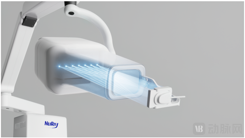 Xinhong Electronics “Intraoral 3D Imaging System”
However, the introduction of cold-cathode X-ray sources has also increased the system’s overall cost. Consequently, after obtaining FDA approval and being covered by U.S. Medicare, XinHong Electronics’ intraoral 3D imaging system is primarily marketed to mid-to-high-end clinics in the United States, with gradual expansion into developed markets such as Europe, Japan, and South Korea.
DR
In 2018, Varex Imaging partnered with H+P Advanced Technology to establish VEC Imaging, making a significant bet on the development of carbon nanotubes. One year later at the RSNA, VEC Imaging unveiled the NT-2518C, a novel nanoscale multi-beam curved-array X-ray tube for 3D breast diagnostics, marking the application of cold cathode X-ray sources in digital radiography (DR).
According to publicly available information, the NT-2518C features a multi-X-ray-beam nanotube with 25 X-ray sources and employs Multibeam Field Emission Cold Cathode Nanotube Emitter technology, making it highly suitable for 3D breast imaging. Compared with conventional 3D mammography systems, it eliminates the need for gantry and X-ray tube rotation; instead, scanning is completed by sequentially activating each nanofilament to emit electron beams, thereby significantly reducing the time required for 3D breast scans.
In China, XinHong Electronics has currently completed the development of relevant solutions for tissues such as the breast, chest, head, and joints, forming a mobile 3D imaging solution. This solution is similar in size to mobile DR systems, with radiation doses far lower than those of large CT scanners. It can be easily deployed in emergency rooms, ICUs, and operating rooms, providing precise and safe diagnostic and treatment options for patients with limited mobility or highly contagious diseases.
From a technical perspective, domestic and international enterprises have largely mastered the preparation technology for high-performance carbon nanotubes and developed complete equipment products with distinct features. However, in the market, there is still a considerable gap before new technologies can be applied on a large scale in complete systems and achieve commercialization at scale.
First, there is still room for further reduction in the price of cold-cathode ray tubes. Currently, mature nanotube fabrication techniques can be broadly categorized into three types: arc discharge, laser ablation, and chemical vapor deposition. Each method has its own advantages and disadvantages in terms of fabrication difficulty, growth rate, and carbon purity; however, all struggle to simultaneously achieve low cost and high yield.
Secondly, it takes time to gradually cultivate physicians’ usage habits. Taking dental X-ray machines as an example, dentists were previously accustomed to making diagnoses based on single-layer images. In contrast, modern intraoral 3D imaging systems generate over 30 layers of data. While these systems provide images with greater detail, lower the diagnostic threshold, and enable more precise diagnoses, they also alter physicians’ cognitive approaches and image-reading habits. Therefore, developers must invest sufficient resources in physician training to gradually build consensus among the medical community regarding new equipment, thereby facilitating large-scale product adoption.
However, from another perspective, these technical and promotional challenges have opened up a new frontier in medical imaging, with significant opportunities hidden within the challenges.
After all, for the past century, China’s core components for medical imaging have consistently lagged behind those developed overseas. Now that domestic and international players are starting from the same baseline, many innovative Chinese enterprises are well-positioned to stand out and seize opportunities in the new era of medical imaging.