GE Healthcare China's 'Integrated Brain-Heart and Oncology Solution' Selected for National High-End Medical Equipment Promotion Program, Powered by AI-Enabled Full-Chain MRI Imaging
Recently, GE Healthcare China announced at the 2025 China Association of Medical Equipment Conference and Exhibition (CAME) that its “Integrated Brain-Heart Management and Precision Oncology Diagnosis and Treatment Solution,” developed based on deep learning-enabled 3.0T magnetic resonance imaging, has been successfully selected for the “2024 Promotion Project for High-End Medical Equipment” (hereinafter referred to as the “Application Project”), jointly released by the Ministry of Industry and Information Technology and the National Health Commission.
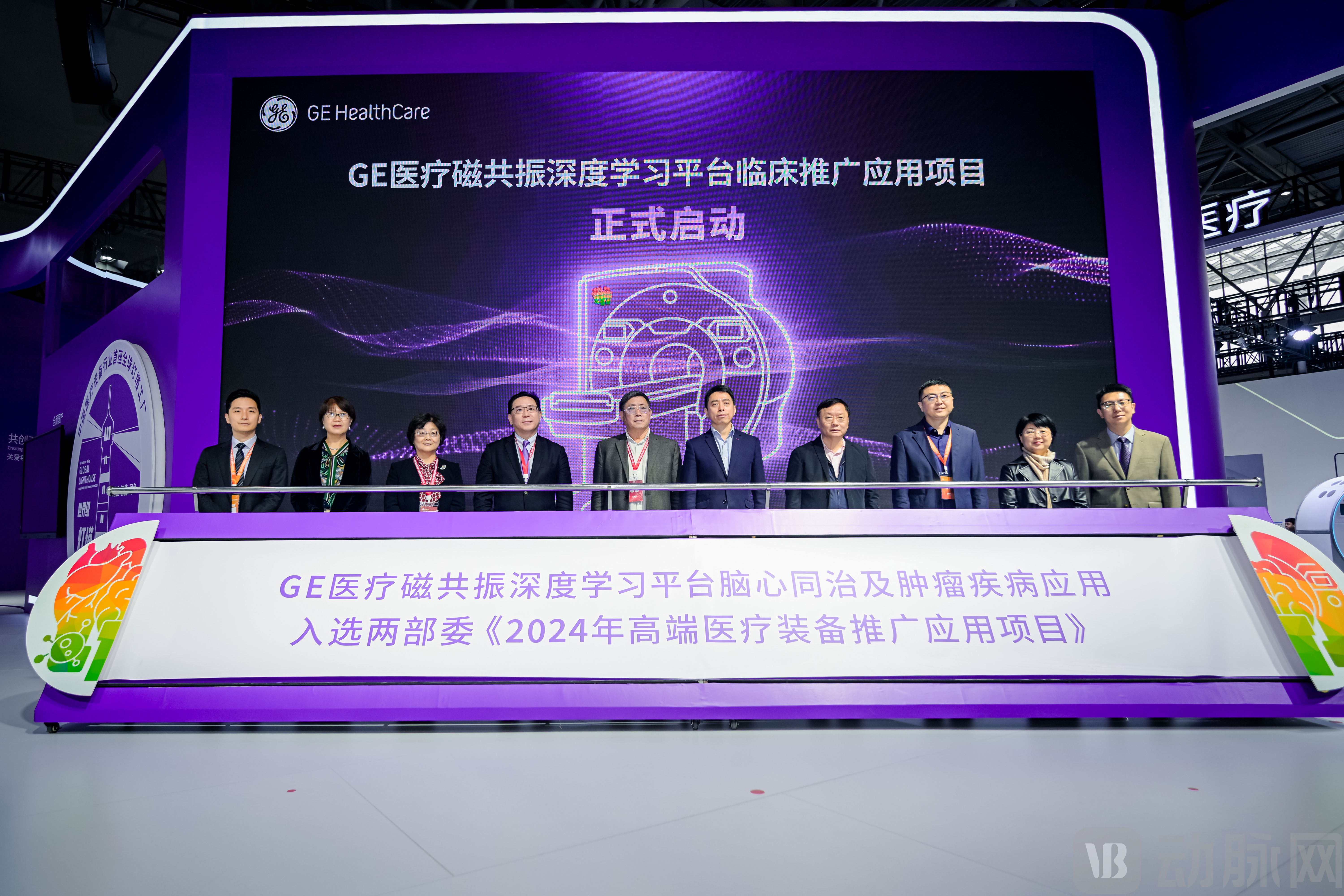
Among the multiple projects selected for promotion by the two ministries in this batch, medical imaging accounted for 11 slots.GE Healthcare China’s “Integrated Brain and Heart Treatment and Precision Oncology Diagnosis and Therapy Application Solution” is the only demonstration case that achieves AI empowerment across the entire imaging chain.
It is reported that this application solution primarily leverages GE Healthcare’s third-generation deep learning Sonic AI platform to construct an imaging chain, thereby enhancing the efficiency and precision of magnetic resonance imaging (MRI) while elucidating more detailed aspects of cardiovascular and cerebrovascular function. This holds significant importance for advancing interdisciplinary precision diagnosis and treatment of cardiovascular, cerebrovascular, and oncological diseases.
Chen Jinlei, Vice President of GE Healthcare China and General Manager of the Medical Imaging Business, stated“This inclusion not only recognizes GE Healthcare China’s ‘Borderless Innovation’ practices and affirms the achievements of its ‘Full Localization’ strategy, but also demonstrates national support for the practical application of technological innovations in high-end medical equipment. AI-driven imaging innovations have already been implemented across GE Healthcare’s entire product portfolio. We will continue to deepen the integration of AI throughout the entire imaging chain, collaborate with multiple stakeholders to tackle innovations in the diagnosis and treatment of prevalent diseases, and enhance diagnostic efficiency and accuracy through intelligent imaging technologies. This will enable a value leap for domestic innovations, transitioning from laboratory breakthroughs to widespread clinical accessibility.”
Cardiovascular and cerebrovascular diseases have long been the leading cause of death among Chinese residents, accounting for 40% of all deaths. The advancement of diagnostic and therapeutic approaches is crucial to improving patient survival rates.
In the past, limited medical resources often confined clinical practice to a symptomatic approach—treating headaches when the head ached and foot pain when the feet hurt. With breakthroughs in medical understanding and technology, the profound connection between heart and brain diseases has become increasingly clear: both are disorders of the circulatory system, sharing numerous risk factors and similar pathogenic mechanisms. In this context, the theory of “simultaneous treatment of brain and heart” has been proposed and validated. This approach shifts the focus from treatment to prevention, effectively reducing the incidence, disability, and mortality rates of cerebrovascular and cardiovascular diseases.
Zhang Jing, Director of the Department of Magnetic Resonance Imaging at the Second Affiliated Hospital of Lanzhou University, believes that this protocol holds significant clinical value for early disease screening and intervention.
In conventional examinations, electrocardiography (ECG) is limited in its ability to depict detailed cardiac structures, while echocardiography cannot simultaneously assess cerebral status; whereasMagnetic resonance imaging, leveraging its multimodal imaging advantages, has become the only diagnostic modality currently capable of simultaneously and precisely assessing both the nervous and cardiovascular systems.Notably, it innovatively integrates high-resolution brain vessel wall imaging with cardiac examination into a “one-stop” workflow, significantly reducing the time required for traditional separate examinations and improving efficiency. Meanwhile, combined cardio-cerebral assessment can be completed with a single contrast agent injection, which also lowers the risk of contrast-induced nephropathy compared to the traditional two-injection protocol.“Simultaneous Treatment of Brain and Heart” technological breakthroughs have simultaneously addressed the two major pain points of patient safety and healthcare costs.Furthermore, the “Integrated Brain-Heart Treatment” protocol provides a feasible pathway for promoting cost-effective, precision diagnosis and treatment at the primary care level through process reengineering and algorithm optimization.
However, translating this theoretical breakthrough into practical application urgently requires technological innovation as its foundation.
GE Healthcare China is addressing this challenge through technological reengineering, developing a precision diagnosis and treatment application solution based on the theory of “simultaneous management of brain and heart conditions.” This solution primarilyAddressing issues such as fragmented imaging assessment dimensions and low diagnostic efficiency in the diagnosis and treatment of cardio-cerebrovascular diseases. Leveraging three core technologies: the latest generation of dual-layer deep learning algorithm models, novel AIR physiological data acquisition, and a high-throughput ultra-computing architecture., enabling intelligent fusion, rapid analysis, and precise diagnosis of multimodal imaging data, thereby advancing the development of precision diagnosis and treatment for related diseases.
In combined brain and heart imaging, it not only evaluates patients' cerebrovascular and cardiovascular status, assists physicians in the early identification and assessment of cardiac injury, and provides comprehensive diagnostic information for the integrated management of brain and heart conditions, but also overcomes the limitations of conventional magnetic resonance imaging. By administering a single contrast injection, it enables a one-stop examination of the nervous system, carotid arteries, and heart, reducing examination time from over one hour to under 30 minutes, thereby providing rapid and precise diagnostic support for complex conditions such as acute chest pain accompanied by cerebral ischemia symptoms.
In Oncology Diagnosis and Treatment, its sub-millimeter lesion detection capability, combined with perfusion quantitative analysis technology, significantly enhances diagnostic accuracy across the entire cycle from early screening to treatment efficacy assessment. Meanwhile, by delivering higher-quality images with greater precision and faster scanning speeds, it effectively alleviates the challenge of prolonged patient appointment wait times, providing robust support for tumor screening, detection, staging, and treatment response evaluation.
Zhao Shihua, Director of the Department of Magnetic Resonance Imaging at Fuwai Hospital, Chinese Academy of Medical Sciences, stated“The integrated management of brain and heart conditions relies on breakthroughs in imaging technology and pathological research, a endeavor fraught with challenges that necessitates a genuine shift in diagnostic workflows from ‘fragmented, stepwise processes’ to ‘one-stop integration.’ Therefore, through the widespread adoption of innovative equipment, AI assistance, standardized training, and multi-center collaboration, we can enhance diagnostic efficiency while establishing a technological foundation for the systematic assessment of cerebrocardiovascular disease risk, thereby translating the medical concept of ‘integrated brain-heart management’ into clinical practice.”
The value is already significant, but to truly achieve technological implementation, clinical validation and standardized promotion remain indispensable.
During the press conference, Lu Hong, Director of the Department of Breast Imaging Diagnosis at Tianjin Medical University Cancer Institute and Hospital, shared the interim results of a multicenter study.: Through deep learning technology, the scanning speed for breast examinations has significantly improved, with the daily patient volume per machine increasing by approximately15%. In addition to shortening scan times, deep learning technology has also improved image quality, enabling hospitals to more accurately capture and analyze the details of lesions. During the scanning process, the incidence of artifacts has also been significantly reduced, with a decrease of approximately28%。
Currently, a multicenter clinical study of this protocol is being conducted under the leadership of Tianjin Medical University General Hospital, in collaboration with multiple hospitals across the Beijing-Tianjin-Hebei region, including Tianjin Medical University Cancer Institute and Hospital, Tianjin Third Central Hospital, TEDA International Cardiovascular Hospital, Tianjin Dongli Hospital, Peking University Sixth Hospital, and the Affiliated Hospital of Hebei University of Engineering.
Going forward, GE Healthcare China will accelerate its expansion into more regions across the country under the joint promotion of two ministries.
At this year’s CAME exhibition, GE Healthcare, under the theme “Pursuing Quality Through Innovation to Create the Future,” focused on domestically produced high-end innovations, primary healthcare support, integrated diagnosis and treatment, digital-physical integration, and medical-engineering collaboration, comprehensively showcasing more than 40 innovative products, technologies, and clinical solutions. In addition to the 3.0T Hero Series MRI, which was selected for the “Application Projects” list, the exhibition also featured a range of cutting-edge technological achievements, including the ultra-high-end quantum CT manufactured through innovation in China, and the PET/CT MAX Apollo, a powerful tool for integrated diagnosis and treatment of critical diseases.
A Review of GE Healthcare China’s Exhibited Products Shows the Company Has Achieved AI-Enabled Full Imaging Chain Integration.
In fact, since introducing deep learning technology to the field of medical imaging, GE Healthcare has continuously driven innovation in this area.
The integrated research and development of its deep learning methods with magnetic resonance systems has evolved to the third generation, achieving comprehensive support for imaging across all mainstream scanning sequences and human anatomical regions. Technical indicators show that the signal-to-noise ratio (SNR) performance of the current solution approaches that of a 6.5T superconducting magnet. During the technology validation period from 2021 to 2025, this technology also underwent extensive validation by the market and clinical experts.
Building on its technological breakthroughs, GE Healthcare has further targeted clinical pain points by applying deep learning technology to achieve dual-dimensional advancements in precision oncology diagnosis and treatment as well as combined cardiac-cerebral screening, significantly enhancing scan speed while improving image quality.
The maturity of deep learning technology represents a paradigm shift in the field of medical imaging. GE Healthcare’s achievement of AI-enabled breakthroughs across the entire imaging chain has reshaped clinical diagnostic and therapeutic efficiency, providing a practical model for innovation in high-end medical equipment.