Phased Array CT 'CompoundEye 24' Debuts at CMEF: Redefining Medical Imaging with 0.1mm Slice Thickness
As the rotation speed of the X-ray tube–detector assembly and the detector width in spiral CT approach their physical limits, it has become increasingly difficult to achieve a qualitative leap in image quality by iteratively improving core components, as was traditionally done.
Therefore, in recent years, manufacturers of high-end imaging equipment have either innovated detector materials or restructured imaging logic to seek a second path that breaks through the existing physical limits of CT.
NanoVision, a Chinese manufacturer of ultra-high-end CT systems, has achieved new breakthroughs by innovating along both aforementioned pathways: it integrates 24 sets of integrated X-ray tube arrays arranged in a ring configuration as radiation sources and 64 detector modules distributed in a ring array within a single CT gantry. By precisely controlling the sequential pulsed exposure of the arrayed X-ray sources through timed electronics for data acquisition, the system is thus named “Phased-Array CT.”
This innovative concept and technical architecture transform the dynamic “mechanical rotation” of spiral CT scanning into static “optical rotation,” while achieving a leapfrog improvement in both temporal and spatial resolution of CT.
Compared with top-tier spiral CT, the first-generation phased-array CT "Compound Eye 24" has been able to increase the temporal resolution by 3 times and the spatial resolution by 64 times, with the thinnest scan layer thickness being only 0.1mm.
Of course, “Compound Eye 24” is merely the beginning of “Phased Array CT.” With continuous iteration of phased array technology and ongoing innovation in product hardware, NanoVision Imaging has the capability to uncover hidden biological information within human medical images at faster and more precise scales.
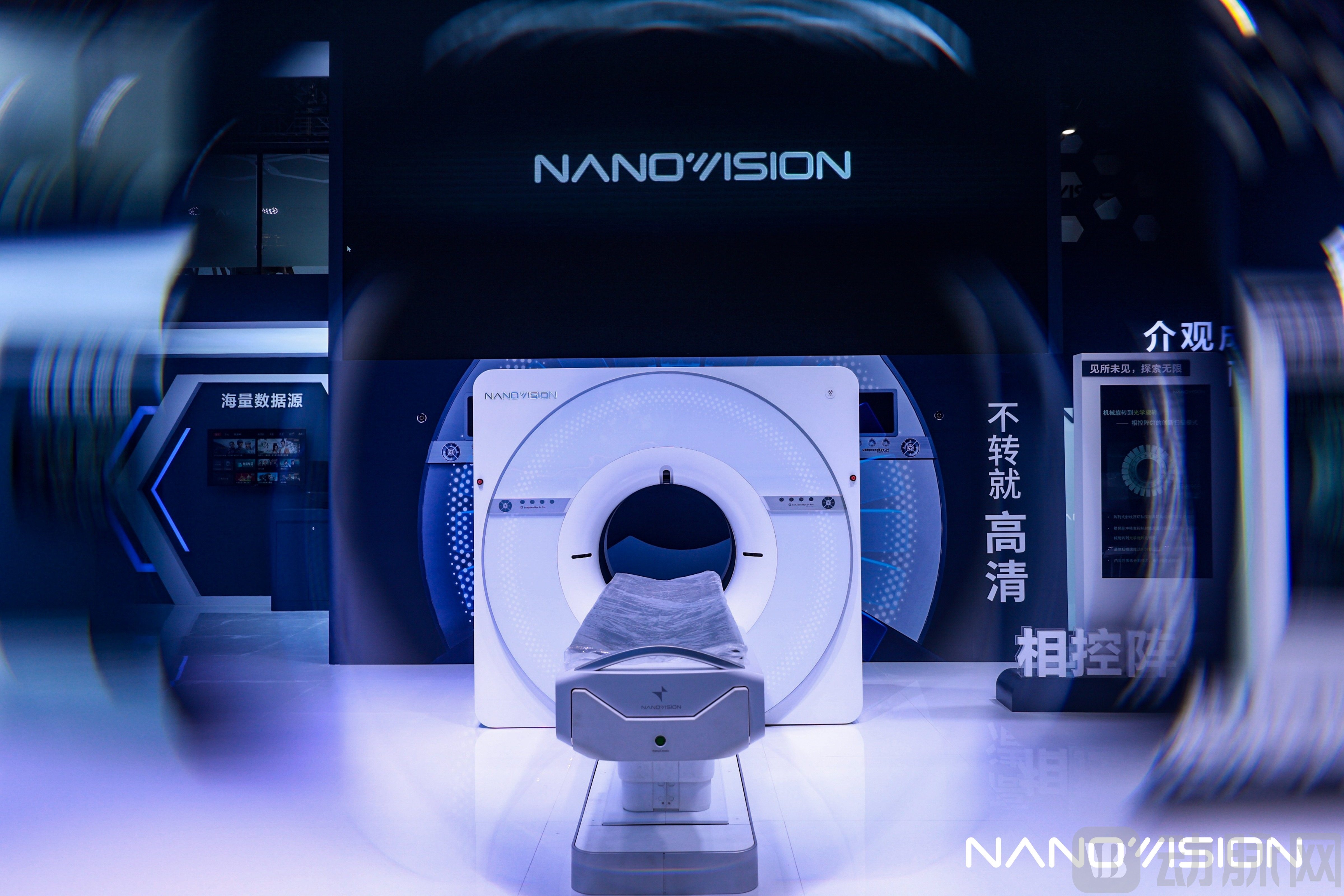
On the first day of CMEF 2025, Nanovision unveiled its phased-array CT system, “Compound Eye 24,” at the event.
The transition from “mechanical rotation” imaging to “optical rotation” imaging may appear straightforward, but in reality, it necessitates a comprehensive reconstruction of CT principles, architecture, materials, manufacturing processes, and other aspects.
Taking the X-ray tube as an example, spiral CT scanners require larger anode target surfaces and higher heat capacity to achieve greater imaging efficiency and quality. This necessitates additional space or more advanced technologies for heat dissipation, resulting in a continuous increase in the size of X-ray tubes as technology advances.
In contrast, phased-array CT employs an “array” dual-ring structure composed of an “array-type integrated X-ray source ring,” formed by multiple sets of X-ray tube–high-voltage generator units, and a “detector ring,” formed by a full-ring detector array, thereby effectively controlling the size of the aforementioned core components.
During scanning, phased-array CT relies on radiofrequency pulses to precisely control the arrayed X-ray sources for sequential pulsed exposure, creating a “distributed thermal capacity.” This is equivalent to dispersing the heat generated by a single X-ray source across 24 sources for shared dissipation. Each X-ray source generates heat for only 1/24 of the time, with the remaining time dedicated to cooling, ensuring that the cooling duration significantly exceeds the heating duration.
Under this operating mode, the cooling rate and heat generation rate of the X-ray tube anode are nearly equal, allowing the tube to cool promptly even under maximum load conditions. Consequently, there is no need to pursue extreme heat dissipation efficiency and thermal capacity for the anode, thereby avoiding the engineering challenges associated with high-power rotating anodes.
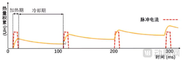 Schematic Diagram of Heat Accumulation in a Small-Array X-ray Tube for Phased-Array CT (Time Axis Magnified 5,000 Times)
Revisiting Detectors. NanoVision Imaging has found that existing photon-counting detectors fail to keep up with the count rate during sudden surges in photon flux, leading to signal distortion and compromised image quality.
To address this issue, NanoVision has developed the world’s first photon-flux detector, achieving ultra-micro-pixelation with a maximum spatial resolution of 25 LP/cm at an MTF of 10%.
In terms of detector materials, Nanovision Imaging utilizes eutectic materials with microscopically ordered structures. Through advanced growth processes, two crystalline phases with distinct refractive indices are integrated into a single eutectic material. Due to the difference in refractive indices, under X-ray irradiation, while the scintillator crystalline phase converts incident X-rays into visible light, the visible light propagates directionally along the crystal growth direction via total internal reflection at the interface between the scintillator crystalline phase and the matrix crystalline phase. This approach eliminates the need for physical segmentation of detector micro-units using titanium dioxide, thereby removing the concept of "rows" from Nanovision Imaging’s phased-array CT systems.
Furthermore, to achieve complete control over its supply chain, Nanovision has independently developed key components such as phased-array CT chips and narrow-pulse high-voltage generators, thereby realizing truly end-to-end in-house R&D and manufacturing across the entire supply chain in the field of ultra-high-end medical imaging equipment.
In addition to achieving technological breakthroughs, existing clinical trial evidence has also intuitively demonstrated the value of phased-array CT.
In the diagnosis of pulmonary nodules, images of nodules smaller than 6 mm generated by conventional CT typically lack sufficient precision and appear blurred. Therefore, international guidelines use 6 mm as the threshold, recommending clinical evaluation for pulmonary nodules with a diameter greater than 6 mm, and deferring further diagnosis until the nodules grow to a size that meets the capabilities of imaging equipment.
Images generated by phased-array CT exhibit virtually no artifacts, enabling comprehensive visualization of details in 4-mm nodules and even clear delineation of surrounding vasculature. This allows physicians to determine the benign or malignant nature of nodules at an earlier stage, thereby further reducing the difficulty and risks associated with surgical resection of malignant nodules. If this technology achieves widespread clinical adoption, it may reshape the clinical guidelines for lung cancer diagnosis.
Another piece of evidence supporting the clinical value of phased-array CT stems from the treatment of liver cancer. Currently, most physicians opt for surgical resection of hepatic tumor tissue; however, 70%–80% of patients experience recurrence within five years postoperatively.
This is because hepatocellular carcinoma tissues are typically rich in nourishing blood vessels that supply nutrients to the tumor. However, these vessels are too small to be clearly visualized by conventional contrast-enhanced CT. Consequently, during preoperative assessment, the extent of hepatectomy is often underestimated, leaving behind many infiltrating and microinfiltrating vessels, which subsequently leads to recurrence of hepatocellular carcinoma.
With higher-precision imaging, phased-array CT can clearly detect subtle clues that are often overlooked by conventional CT. Currently, NanoVision Imaging is collaborating with a leading Grade-A tertiary hospital on a research project funded by the National Natural Science Foundation of China. This innovative technology is expected to reshape the surgical planning process for liver cancer in the future, effectively improving patients’ five-year postoperative survival rate.
Despite its many advantages, NanoVision’s phased-array CT also has its limitations.
Currently, mainstream spiral CT scanners have a resolution of 512×512, with individual patient imaging data occupying approximately 200–300 MB. In contrast, the Phased-Array CT “Compound Eye 24” now achieves a resolution of 3072×3072, with nearly 10 million pixels per slice, resulting in approximately 20 GB of imaging data per patient.
For physicians, high-definition phased-array imaging undoubtedly reveals more detailed patient information, but it also multiplies their workload several-fold compared to the past. In China’s current healthcare environment, efficiency is an issue that medical institutions cannot afford to overlook.
To address the aforementioned challenges, NanoVision has partnered with multiple domestic AI imaging companies to leverage AI for pre-processing CT images, thereby enhancing physicians' efficiency in image interpretation.
It is worth noting that the medical images used for training by AI companies are almost exclusively 512×512 in resolution, and existing algorithms cannot effectively process higher-resolution images. Therefore, NanoVision’s partners are developing AI algorithms tailored specifically for phased-array CT.
For them, developing algorithms capable of processing high-quality medical images is both a challenge and an opportunity.
From traditional spiral CT to next-generation ultra-high-end CT systems such as photon-counting CT and phased-array CT, the resolution of medical images generated by these new devices has surpassed conventional limits. To build competitiveness in this new era, medical imaging AI companies must secure higher-quality data to train next-generation AI models.
Thus, as imaging AI companies empower NanoVision’s phased-array CT, NanoVision also opens up new possibilities for these AI firms, helping them gain a competitive edge in the next generation of digital and intelligent healthcare competition.
Reviewing the nearly five decades of CT development, although core technologies have undergone several leaps, they have still failed to break through the millimeter-level barrier in macroscopic imaging. Consequently, physicians must still rely on observing anatomical structures and morphological changes of organs and tissues to make diagnoses.
The emergence of phased-array CT has now made it possible for physicians to precisely examine tissue and cellular information.
According to NanoVision Imaging, the phased-array CT system “Compound Eye 24” has completed clinical trials and is expected to receive regulatory approval within this year. The higher-resolution “Compound Eye 36” is currently under development, with the potential to achieve micrometer-level imaging within a few years.
In the past, lacking support from domestically produced ultra-high-end imaging equipment, radiologists were largely confined to retrospective studies. Today, with breakthroughs in phased-array CT technology, we hope to see more Chinese physicians transition from retrospective to prospective research, thereby driving medical imaging to advance continuously from macroscopic toward mesoscopic and even microscopic levels.