United Imaging Intelligence Unveils Groundbreaking Chest CT 'One-Scan Multi-Check' AI Agent Capable of Detecting 73 Thoracic Abnormalities

UNITED IMAGING
Artificial Intelligence Medical Product Developer
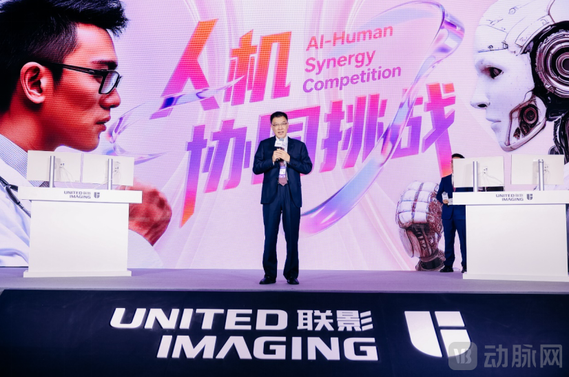
During the World Artificial Intelligence Conference, a special “showdown” is underway: Led by Zeng Mengsu, Director of the Department of Radiology and Director of the Department of Diagnostic Radiology at Zhongshan Hospital, Fudan University (hereinafter referred to as “Zhongshan Hospital”), three physicians completed imaging diagnosis and report writing with the assistance of an intelligent agent for multi-task chest CT analysis, while another three physicians relied on their clinical experience to compete.
Among the three on-site cases, the AI agent demonstrated significant advantages in diagnosing complex conditions. Physicians in the non-AI-assisted group had to manually drag the cursor across images, scrutinize each tomographic slice line by line, and draft reports by hand, taking eight minutes to complete both image interpretation and report writing. In contrast, physicians in the AI-assisted group leveraged the intelligent agent to instantly detect thoracic abnormalities such as pulmonary nodules and coronary artery calcification with a single click. They only needed to verify each finding individually to generate the report, resulting in a 25% overall improvement in efficiency.
It is important to note that UNITED IMAGING’s “Single Scan, Multiple Assessments” intelligent agent for chest imaging differs fundamentally from previous single-disease AI solutions. It is also not merely an application that simply stacks multiple single-disease small models and repackages them as a “Single Scan, Multiple Assessments” solution, as commonly seen in the market.
In the past, radiologists relied solely on their visual acuity and years of accumulated experience to meticulously identify lesions during image interpretation. The advent of medical AI has alleviated some of this burden; however, most existing AI solutions are single-disease, narrow-scope models capable of assisting in the diagnosis of only one condition at a time. This is akin to using specific keys for corresponding locks: a dedicated key is required to unlock the “nodule lock” for detecting pulmonary nodules, while a different key is needed to open the “pneumonia lock” for diagnosing pneumonia. When patients present with multiple concurrent conditions, physicians must frequently switch between these tools, which not only increases operational workload but also prolongs patient waiting times.
More critically, in current AI applications, image interpretation and report writing are disjointed: imaging data and diagnostic content cannot be correlated in real time, key information requires repetitive transfer, resulting in cumbersome workflows that easily disrupt clinical reasoning.
In response to the aforementioned challenges, in early 2024, the Department of Radiology at Zhongshan Hospital and United Imaging Intelligence jointly designated non-contrast chest CT as the inaugural application scenario for “single-scan, multi-condition screening,” targeting 73 types of thoracic abnormalities and aggregating over 400,000 chest CT imaging datasets.
Throughout the collaboration, radiology experts from Zhongshan Hospital were deeply involved in the entire process of algorithm optimization and product design. They provided improvement recommendations based on actual clinical needs, driving continuous product iterations to better align with frontline diagnostic and treatment workflows.
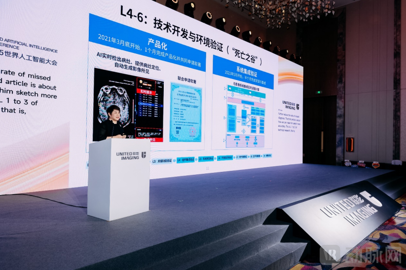
After more than a year of intensive R&D efforts, the two parties have successfully developed the world’s first AI agent for multi-condition screening via a single chest CT scan. Leveraging non-contrast chest CT images, the system can automatically detect 73 common thoracic abnormalities, including pulmonary nodules, fractures, emphysema, and aortic dilation, achieving an average AUC of 94% and setting a new industry benchmark for diagnostic accuracy. Furthermore, this AI agent not only enables automated generation of reports from imaging data but also supports voice-based report entry by physicians, pioneering a new experience in radiological image interpretation and report writing.

Physicians in the AI-assisted group are drafting imaging reports.
Overall, the practical experiences of frontline physicians intuitively validate the value of human-AI collaboration. Physicians in the AI-assisted group stated, “The multi-detection intelligent agent for chest scans acts as a ‘super assistant,’ capable of accurately screening all lesion information and enabling real-time synchronization between images and reports. This transforms report writing into report review, thereby facilitating the auditing process for senior physicians and clinicians.”
Zeng Mengsu stated after the match, “This game was not about ‘human versus machine,’ but rather a deep exploration of ‘human-machine collaboration.’”
He believes that AI not only improves work efficiency and reduces errors, but more importantly, lowers labor costs, enabling junior doctors paired with AI to achieve a high level of diagnostic accuracy. “In the future, I believe AI will further free our hands and make work easier.”
However, he also emphasized that this places higher demands on physicians: every diagnostic report must be drafted with greater meticulousness to ensure clear understanding by clinicians, thereby genuinely facilitating subsequent patient care and treatment.
Zeng Mengsu, Director of the Department of Radiology and Director of the Department of Diagnostic Radiology at Zhongshan Hospital, Fudan University
In the practice of top-tier hospitals, the application of AI agents has extended to broader healthcare scenarios.
To explore the ultimate potential of AI in healthcare, UNITED IMAGING has partnered with leading hospitals such as Zhongshan Hospital Affiliated to Fudan University and Sun Yat-sen University Cancer Center, collaboratively developing a systematic suite of intelligent agents that cover multiple medical scenarios, including imaging diagnosis, medical record documentation, patient consultation, and surgical procedures.
“Since 2018, we have partnered with United Imaging as an industry-medical alliance dedicated to conquering cancer, engaging in deep collaboration in fields such as precision radiotherapy and artificial intelligence. This partnership has yielded significant achievements, including online adaptive radiotherapy and AI for metastatic tumors,” said Sun Ying, Deputy Director of Sun Yat-sen University Cancer Center (hereinafter referred to as SYSUCC).
Sun Ying, Deputy Director of Sun Yat-sen University Cancer Center
"Diagnosing metastatic tumors in the brain, liver, bone, and other sites accounts for 70% of the workload in our hospital's radiology department. Taking brain metastases as an example, their incidence is relatively high. To detect minute metastatic foci at the earliest stage, 1-mm thin-slice reconstruction is required after routine MRI scanning, generating hundreds of MR images. Physicians must meticulously screen these images to identify occult lesions, making the task extremely challenging."
To this end, we collaborated with the United Imaging Intelligence team for over a year to develop an AI application capable of intelligently detecting brain metastases, automatically displaying lesion information, and automatically generating imaging findings. Building on this foundation, we have also launched an AI application for bone metastases.
“Currently, these two AI systems have been deployed and applied in over 400 hospitals across China, marking the expansion of this solution from Sun Yat-sen University Cancer Center to a national scale, thereby enabling more cancer patients to benefit from more efficient and precise medical services.”
Furthermore, Sun Ying shared insights into the collaborative exploration of AI agent applications across various healthcare scenarios, including patient consultations and hospital information management.
According to Sun Ying, patients often face numerous inconveniences when seeking medical care. For instance, they are required to carry bulky paper-based medical records, which is not only cumbersome but also prone to loss. Furthermore, due to difficulties in sharing medical data across different hospitals, test results are often not mutually recognized. This forces patients to undergo redundant examinations, thereby increasing their financial burden.
Based on this current landscape, UNITED IMAGING has collaborated with the Sun Yat-sen University Cancer Center to co-create a smart consultation room solution leveraging the “SYSUCC” expertise, thereby deeply empowering patient consultation scenarios with AI.
During the pre-consultation phase, patients can describe their personal information and symptoms in advance through interactive dialogues via digital humans or text-based conversations, and independently upload prior examination reports.
Empowered by this AI, physicians can conveniently review patients’ out-of-hospital medical histories and examination data prior to consultations, while also receiving assistance in generating various subsequent medical record documents, thereby significantly enhancing the patient care experience.
In medical record documentation, the two parties have jointly developed an electronic medical record (EMR) agent capable of real-time transcription of doctor-patient conversations into text and automatic generation of standardized medical records.
For patients from other regions or hospitals, the AI agent can automatically retrieve external hospital reports entered during the pre-consultation phase, providing physicians with a comprehensive basis for clinical judgment and ensuring more complete and accurate medical records.
Shache County, located in Kashgar Prefecture in southern Xinjiang, comprises 493 villages. With a permanent resident population of approximately one million, it is indisputably the most populous county in Xinjiang.
However, due to its remote location and mountainous terrain, Shache is free from the hustle and bustle of urban life, but it also lacks the comprehensive medical resources available in cities.
According to statistics, the total number of licensed physicians in Shache County is fewer than 900. At the meeting, Dr. Cai Sipeng, Director at the People’s Hospital of Shache County, stated that his radiology department, staffed by only 19 radiologists, issues over 910,000 imaging diagnostic reports annually, struggling to cope with a heavy workload throughout the year.
It is nearly impossible to bridge the shortage of medical resources in such a remote border town within a short period. Here, AI has provided them with new approaches to solving this problem.
The AI transformation at the People's Hospital of Shache County began with the national policy on building close-knit county-level medical consortia and support from Shanghai's aid to Xinjiang.
During this period, Shache People’s Hospital engaged in deep collaboration with United Imaging Group, progressively building systematic medical capabilities spanning high-end imaging equipment, healthcare informatics, and AI technology.
At the forum, Cai Sipeng shared his experience with AI during the event: “In the past, the quality of radiographic images at township health centers was inconsistent. Many technicians had not received professional training, resulting in an image qualification rate of only about 50%. This often required us to visit various health centers to provide on-site guidance.”
Today, township and village health centers under the jurisdiction of Shache County have gradually been equipped with United Imaging’s intelligent DR quality control system. This application enables real-time monitoring and guides technologists in patient positioning, informing them whether the acquired images meet diagnostic requirements. After a period of adjustment and optimization, our image qualification rate has exceeded 95%.
“Moreover, with the implementation of an information system, we can now review imaging quality from any township at any time, thereby addressing the incompleteness and subjectivity inherent in traditional spot checks, and eliminating the need for monthly rural visits and weekly supervisions.”
In addition to quality control in imaging, Shache County People's Hospital has implemented specialty AI solutions for post-imaging analysis in departments including Radiology, Orthopedics, Cardiology, Neurology, and Neurosurgery. These tools assist in diagnosing diseases affecting various anatomical structures, such as fractures, coronary arteries, head and neck vessels, pulmonary arteries, and the aorta, with the total number of applications exceeding 20.
This year, the hospital also jointly developed an AI system for tuberculosis with United Imaging Intelligence, which can rapidly detect and flag patients with suspected tuberculosis, thereby assisting physicians in conducting screening that is fast, effective, and cost-efficient.
In 2024, Shache County People’s Hospital reviewed one million chest X-rays, ultimately identifying over ten thousand high-risk individuals. With the assistance of AI for tuberculosis screening, they can finally shed this heavy “burden.”