Organoid Technology: Reshaping Drug Discovery as a 'Human-in-a-Dish' Platform
New drug development is a capital-intensive and high-risk systems engineering endeavor. It takes an average of 10–15 years and billions of dollars in investment for a new drug to move from the laboratory to the market, yet the ultimate success rate remains below 10%. The core bottleneck underlying this predicament is the lack of experimental models capable of accurately predicting how drugs will truly perform in the human body.
Traditional drug development primarily relies on two major tools: first,2D Cell Culture, which involves seeding human cells onto the bottom of culture dishes for adherent growth. Most of these cells are derived from tumors; after prolonged in vitro cultivation, their biological characteristics have significantly deviated from those of normal human cells. They fail to recapitulate the three-dimensional architecture of organs and lack the complex collaborative interactions among multiple cell types within tissues. Second,Animal Experiments, although drug effects can be observed at the level of intact organisms, species differences remain an insurmountable barrier. Many drugs that demonstrate efficacy in mouse models fail in human clinical trials; furthermore, some diseases unique to humans cannot be effectively replicated in animal models.
In this context, a revolutionary technology has emerged—OrganoidsThese “mini-organs,” cultivated from stem cells, typically measure only a few millimeters in diameter yet can spontaneously self-assemble into three-dimensional structures resembling real organs in culture dishes. They not only contain multiple functional cell types but can even perform certain organ functions. Organoids are regarded as a third approach, bridging the gap between simple cell cultures and complex animal models, thereby providing an experimental platform that highly recapitulates the human physiological environment for drug development.
The breakthrough in this technology began in the early 21st century. The team led by Mina Bissell at the Lawrence Berkeley National Laboratory in the United States pioneered the use of Matrigel (a hydrogel that mimics the extracellular matrix) for 3D cell culture, discovering that mammary epithelial cells could self-assemble into glandular structures and exhibit milk-secreting functions. In 2009, the team led by Hans Clevers at the Hubrecht Institute in the Netherlands achieved a milestone breakthrough: they successfully isolated single stem cells from mouse intestines and cultured three-dimensional structures containing all intestinal cell types in vitro, marking the birth of the world’s first true organoids derived from tissue stem cells.
Organoid construction primarily follows two technical routes, which ultimately converge on the same goal.
The first route is based onInduced Pluripotent Stem Cells (iPS Cells). This technology was invented in 2006 by Japanese scientist Shinya Yamanaka, who reprogrammed somatic cells (such as skin cells) into a pluripotent state similar to that of embryonic stem cells by introducing four key transcription factors, theoretically enabling their differentiation into any cell type in the human body.
Researchers precisely regulate culture conditions and apply specific growth factors in a temporal sequence to stimulate iPSCs, mimicking embryonic development and gradually guiding their differentiation into organoids of specific organs. This approach is particularly suitable for constructing organ models of the brain, retina, lungs, and other tissues from which it is difficult to directly isolate stem cells from adult sources.
The team led by Yoshiki Sasai at the RIKEN Center for Developmental Biology in Japan is a pioneer in this field. In 2011, they induced embryonic stem cells to aggregate into spheroids and discovered that these cells could spontaneously self-organize into optic cup structures, forming organoids containing all retinal cell layers. Subsequently, the team successfully constructed various neural system organoids, including those of the cerebral cortex and cerebellum, ushering in a new era of pluripotent stem cell-derived organoid research.
The second routeTissue stem cells are directly extracted from patient tissue samples for culture.Each organ has its own “repair crew,” namely tissue stem cells, which are responsible for replacing aging or damaged cells.
The 2009 breakthrough by Clevers’ team is a representative example of this approach. They isolated Lgr5-positive stem cells from intestinal crypts and demonstrated that, in a culture medium containing specific growth factors (such as EGF and Wnt signaling pathway activators), single stem cells could continuously proliferate and differentiate to form three-dimensional structures comprising various intestinal cell types, including stem cells, enterocytes, goblet cells, and Paneth cells. The greatest advantage of this method lies in its direct derivation from patients, thereby preserving individual genetic backgrounds and laying a solid foundation for personalized medicine.
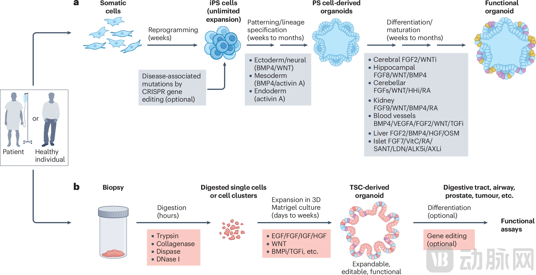
Figure: Human organoids derived from pluripotent stem cells and tissue stem cells (Source: Nat Rev Drug Discov)
Compared with traditional models, the core advantage of organoids lies in their high fidelity: they preserve the 3D architecture of tissues and the spatial relationships between cells, contain multiple functional cell types found in organs, can be cultured long-term and cryopreserved for biobanking, and are derived from human cells, thereby avoiding interspecies differences. When transplanted into immunodeficient mice, organoids derived from healthy tissues form normally functioning tissues, whereas traditional tumor cell lines often form tumors; this contrast clearly demonstrates that organoids more closely recapitulate normal physiological states.
One of the most promising applications of organoid technology lies in its ability to precisely recapitulate the pathological features of human diseases in vitro, thereby providing precise targets for drug development.

Figure: Different Types of Organoids in Disease Modeling
Cystic Fibrosis (CF)It is a paradigmatic example of the translational application of organoids. This genetic disorder is caused by mutations in the CFTR gene, leading to dysfunctional chloride channels and the production of viscous secretions in the patients’ lungs and intestines. In 2013, researchers at the University Medical Center Utrecht in the Netherlands developed an innovative assay: healthy intestinal organoids rapidly swell as water influx into the lumen occurs upon CFTR channel opening stimulated by forskolin (a compound that elevates intracellular cAMP levels), whereas organoids derived from cystic fibrosis (CF) patients fail to swell.
This “swelling assay” not only holds diagnostic value but, more critically, enables the prediction of drug efficacy. Multiple studies have confirmed that the response of organoids to CFTR modulators—such as ivacaftor, the lumacaftor/ivacaftor combination Orkambi, and the next-generation triple-combination therapy elexacaftor/tezacaftor/ivacaftor—is highly correlated with patients’ clinical outcomes, including improvements in lung function as measured by FEV1. This means that physicians can test which medication is most effective using a patient’s own intestinal organoids before prescribing, marking the realization of true personalized medicine in the field of cystic fibrosis (CF).
Hereditary Liver DiseasesAn ideal research model has also been identified. In 2015, Meritxell Huch’s team at the University of Cambridge established a long-term, stable culture system for human liver organoids. Using this platform to model alpha-1 antitrypsin deficiency—a genetic disorder causing liver and lung damage—patient-derived organoids recapitulated key pathological features such as protein aggregation and reduced secretion. Liver organoids modeling Wilson’s disease, a disorder of copper metabolism, exhibited abnormal sensitivity to copper handling, providing a powerful tool for studying defects in copper transport.
Following the outbreak of the COVID-19 pandemic in 2020, organoid technology hasVirology Researchrapidly demonstrated its unique value. A team from the Hubrecht Institute in the Netherlands discovered that ciliated cells expressing ACE2 and TMPRSS2 proteins in human airway organoids are the primary infection targets of SARS-CoV-2. More importantly, using an organoid biobank established via CRISPR gene-editing technology, they proved that the TMPRSS2 protein is a key host factor for the cellular entry of multiple coronaviruses, including SARS-CoV-2, SARS-CoV, and MERS-CoV. These viruses utilize this protein to cleave the viral spike protein, enabling direct fusion of the viral envelope with the cell membrane without requiring the endocytic pathway.
This finding strongly refutes the efficacy of a once widely used drug. In traditional 2D cell line experiments, hydroxychloroquine (a drug that blocks endocytic pathways) demonstrated antiviral effects, leading to its widespread prescription to patients during the early stages of the pandemic. However, in organoid models, hydroxychloroquine proved completely ineffective—because the virus does not enter cells via the endocytic pathway. This result is fully consistent with the subsequent failure of clinical trials, underscoring the value of organoids as a more reliable translational platform.
2016,Zika Virus (ZIKV)The outbreak of microcephaly in newborns in Brazil and other regions led the World Health Organization to declare it a Public Health Emergency of International Concern. Traditional research was severely limited by the inability to obtain brain tissue from infected human fetuses. At this juncture, multiple research teams independently turned their attention to brain organoids. Scientists at institutions such as Johns Hopkins University and Rockefeller University in the United States cultivated cerebral cortex organoids using induced pluripotent stem (iPS) cells. They discovered that the Zika virus specifically infects neural progenitor cells, causing these “brain builders” to undergo premature differentiation and death, thereby leading to impaired brain tissue development.
More excitingly, brain organoids have also been used forRapid Drug ScreeningResearchers tested thousands of compounds and found that the antidepressant hippeastrine hydrobromide could clear the virus from infected neural progenitor cells and rescue the microcephaly phenotype in organoids. A drug repurposing screen also revealed that the pan-caspase inhibitor emricasan could inhibit virus-induced apoptosis, protecting cortical neural progenitor cells, while the FDA Pregnancy Category B antiparasitic drug niclosamide could directly block viral replication. These efforts were completed within just a few months, demonstrating the rapid response capability of organoid platforms in addressing emerging infectious diseases.
Cancer organoids, especially patient-derived onesPatient-Derived Tumor Organoids (PDTO), reshaping the paradigm of oncology research and treatment. In 2011, Clevers’ team reported for the first time the establishment of long-term cultures of organoids from human colorectal cancer tissues. In 2015, they established the first patient-derived tumor organoid (PDTO) biobank for colorectal cancer, comprising dozens of tumor organoid lines from different patients. These organoids retained the histological features, genetic mutation profiles, and gene expression patterns of the primary tumors.
A key feature of PDTOs is their altered growth factor dependency. Organoids carrying the KRAS G12V mutation can grow without the addition of epidermal growth factor (EGF), whereas healthy intestinal organoids require EGF for survival. This difference can be used to distinguish between tumor and normal tissue growth, reflecting the autonomous growth capability acquired by tumor cells.
Drug sensitivity testing has demonstrated high clinical relevance. In a study on breast cancer patient-derived tumor organoids (PDTOs) published in *Cell* in 2018, researchers found that organoids with high HER2 expression were sensitive to afatinib, an irreversible ERBB family inhibitor, whereas organoids harboring BRCA1/2 mutations exhibited greater sensitivity to PARP inhibitors—findings that align perfectly with clinical treatment guidelines. In various other tumors, including pancreatic and gastric cancers, the response of PDTOs to chemotherapeutic agents has also been confirmed to correlate with patients’ actual therapeutic responses.
A prominent success story is MCLA-158 (petosemtamab), a bispecific antibody drug targeting EGFR and LGR5 that was developed entirely using colorectal cancer patient-derived tumor organoid (PDTO) lines and is currently in clinical trials. This marks the transition of organoids from auxiliary tools to a mainstream platform for drug development.
While drug efficacy is undoubtedly important, safety is often the key factor determining the success or failure of research and development. Many drugs are forced to be terminated due to severe adverse reactions during late-stage clinical trials or after market launch, resulting in significant losses. Organoids provide a warning system for drug safety assessment that more closely mimics human physiological conditions.

Figure: Organoid Systems in Drug Safety Evaluation
The liver is the primary organ for drug metabolism,Drug-Induced Liver Injury (DILI)It is the leading cause of failure during the development of candidate drugs, as well as the withdrawal or restricted use of marketed drugs; human liver organoids demonstrate unique value in this regard.
In 2019, a research team at the Korea Advanced Institute of Science and Technology (KAIST) developed functionally mature induced pluripotent stem cell (iPSC)-derived liver organoids capable of accurately recapitulating the injury patterns of known hepatotoxic drugs. For instance, troglitazone—an antidiabetic drug withdrawn from the market due to severe hepatotoxicity—induced dose-dependent cell death in these liver organoids at concentrations consistent with clinically relevant levels. The antibiotics trovafloxacin and levofloxacin also exhibited expected hepatotoxicity, with the organoids demonstrating significantly higher sensitivity than traditional 2D hepatocyte cultures.
A 2021 study published in Gastroenterology further advanced the field by establishing a high-throughput drug-induced liver injury (DILI) screening platform based on iPSC-derived liver organoids, which was used to test 238 drugs. This system not only accurately annotated the hepatotoxic potential of drugs but also identified genotype-specific susceptibilities: bosentan, an endothelin receptor antagonist known to cause cholestatic liver injury, exhibited differential toxic responses in organoids derived from different donors, highlighting the influence of genetic background on adverse drug reactions.
In addition to hepatocellular toxicity, organoids can also simulateBile Duct Injury. Researchers utilized intrahepatic cholangiocyte organoids to investigate chlorpromazine (an antipsychotic drug)-induced cholangiotoxicity, effectively recapitulating drug-induced impairments in bile secretion.
Many chemotherapy drugs damage the kidneys while killing cancer cells. Although kidney organoids derived from induced pluripotent stem (iPS) cells are not yet fully mature (lacking certain transporters and filtration functions), they are already capable of simulating drug-induced acute kidney injury.
In 2015, research teams at Harvard University and the University of Washington in the United States independently developed organoids containing multiple nephron structures, including glomerular podocytes, proximal tubules, loops of Henle, and distal tubules. These organoids exhibited dose-dependent apoptotic responses to classic nephrotoxic drugs such as cisplatin and gentamicin, and upregulated the expression of the kidney injury marker KIM-1, consistent with the molecular characteristics of clinical kidney injury.
Subsequent studies have introduced more advanced platforms. A study published in PNAS in 2023 combined kidney organoids with microfluidic chips, incorporating vascular endothelial cells and peripheral blood mononuclear cells (PBMCs) to construct an immune-infiltratedKidney Organoid-on-a-Chip. This system not only evaluates the toxicity of chemical drugs but also tests the side effects of immunotherapy—researchers treated the system with a T-cell bispecific antibody (TCB) targeting HLA-A2-presented WT1 peptides, successfully observing the selective killing of WT1-expressing cells, thereby simulating the target-mediated toxicity of biologics.
CardiotoxicityCardiac safety assessment is also a key focus. Studies using cardiac organoids have shown that the anticancer drug doxorubicin can cause decreased organoid viability, increased release of lactate dehydrogenase (LDH) and brain natriuretic peptide (BNP), apoptosis, fibrosis, and mitochondrial dysfunction—all of which are characteristic changes seen in clinical cardiomyopathy. In organoid models simulating myocardial infarction, doxorubicin exacerbated injury in hypoxic regions, disrupted calcium cycling, and promoted fibrosis in the infarct core.
Gastrointestinal ToxicityIn this regard, a study published in 2020 utilized human ileal organoids to validate 31 reference drugs, achieving a 90% accuracy rate in predicting drug-induced diarrhea. More importantly, co-culturing intestinal organoids with immune cells enables the assessment of off-target toxicity of biologics. Work reported in Nature Biomedical Engineering in 2023 demonstrated that patient-derived intestinal and tumor organoids supplemented with immune cells could capture tissue-specific off-target toxicity of T-cell bispecific antibodies (TCBs), successfully predicting adverse reactions observed in clinical settings—side effects that remained undetectable in conventional tissue models lacking immune components.
To more realistically modelAnthropomorphic Environment, ScienceExperts have developedOrgan-on-a-Chip(organoid-on-a-chip) technology. These platforms integrate organoids into microfluidic devices, simulating blood circulation through continuous media flow, and can even connect multiple organ chips to form a human-on-a-chip system.
A study published in Biofabrication in 2020 described a human-on-a-chip platform integrating seven tissue types—liver, heart, lung, vasculature, testis, colon, and brain—for screening drugs withdrawn from the market by the FDA. The system was capable of detecting ATP depletion and dysfunction in individual organs, demonstrating superior sensitivity compared to standalone 2D cell cultures.
Another study in 2021 constructedLiver-Heart Combined System, it was found that the antidepressant clomipramine, after being metabolized by hepatic cytochrome P450 enzymes into desmethylclomipramine, exerts toxicity on cardiac organoids, leading to reduced beating, disrupted calcium flux, and cell death—whereas this toxicity does not occur in the absence of liver tissue. This vividly demonstrates the importance of inter-organ interactions in drug metabolism and toxicity.
Despite its promising prospects, organoid technology still faces numerous challenges that limit its large-scale application in drug development.
Standardization and Reproducibilityis the primary challenge. Variations in culture protocols across different laboratories, genetic diversity of cell sources, and batch-to-batch differentiation variability all lead to inconsistencies in organoid function and drug responses, posing a severe challenge to drug screening that requires stringent quality control.
Insufficient MaturityThis limits certain application scenarios. For instance, neurons in iPSC-derived brain organoids more closely resemble a fetal state, making it difficult to fully recapitulate the late-stage pathological features of age-related neurodegenerative diseases such as Parkinson’s disease and Alzheimer’s disease.
Absence of Key Tissue ComponentsThis also limits the physiological relevance of the models. Most organoids lack components such as vascular systems, immune cells, and innervation, making them unable to recapitulate systemic inter-organ interactions, which is particularly pronounced when evaluating systemic drug effects.
Scalable and High-Throughput ApplicationsBottlenecks remain. The long culture cycle of organoids (ranging from weeks to months), high technical requirements, and complex data analysis limit their efficiency in scenarios requiring the screening of thousands of compounds.
To address these challenges, researchers are seeking breakthroughs from multiple directions.
Automation and Microengineering TechnologiesSignificantly enhanced standardization levels. Microcavity array systems and microfluidic printing technology can precisely control the quantity, size, and spatial distribution of organoids, ensuring batch-to-batch consistency. The OrganoPlate® system from Mimetas (Netherlands) and the Organ-on-a-Chip platform from Emulate (USA) have achieved automated control of culture conditions and full-process automation, substantially reducing variability associated with manual operations.
Bioengineering ModificationAddressing Functional Deficiencies. By co-culturing endothelial cells with organoids or using microfluidic chips to induce vascular ingrowth, researchers have successfully constructed vascularized organoids. A study published in Nature in 2024 achieved the simulation of immune-epithelial interactions through an immune cell co-culture system, which is crucial for evaluating immunotherapeutic drugs.
2D Organoid Monolayer CultureOpens new avenues for drug transport research. Unfolding 3D organoids into epithelial monolayers preserves cellular diversity and polarity while facilitating bilateral dosing and barrier function measurements, making this approach particularly suitable for absorption and metabolism studies.
Artificial Intelligence and High-Content ImagingAccelerating Data Analysis. Automated microscopy systems combined with computer vision algorithms enable real-time monitoring of organoid morphological changes and quantification of multidimensional parameters; machine learning models are being trained to predict drug toxicity and efficacy, promising a substantial increase in screening efficiency.
The global regulatory landscape is gradually opening its doors to organoid technology.
In 2022 and 2023, the U.S. Congress successively passed the FDA Modernization Act 2.0 and 3.0, explicitly stipulatingAnimal Testing No Longer Mandatory in Drug Development, allowing the use of new methods such as cell culture, organ-on-a-chip, and computational models to assess safety, marking a historic shift in U.S. drug regulatory philosophy since 1938.
In 2025, the FDA further announcedAnimal testing requirements in drug development, including for monoclonal antibodies, will be gradually phased out in the coming years., organoid toxicity testing is explicitly listed as one of the alternative methods. The European Medicines Agency has also introduced the “Safe Harbor Approach,” encouraging companies to submit research data based on new technologies such as organoids to accumulate evidence for formal qualification.
Currently, the application of organoids in the pharmaceutical industry is primarily concentrated in internal decision-making stages, such as early-stage drug screening and mechanistic studies, and has not yet entered regulatory submission processes on a large scale. However, pioneering cases have already emerged: in the field of cystic fibrosis, intestinal organoid swelling assays have been adopted by medical centers in countries like the Netherlands to guide clinical medication; in oncology, multiple clinical trials are underway to validate the predictive value of patient-derived tumor organoid (PDTO) drug sensitivity testing.
International organizations and industry alliances are also developing standardized frameworks. The Organisation for Economic Co-operation and Development (OECD) has provided validation guidelines, while the U.S. IQ MPS Consortium collaborates with the FDA to define quality control standards. A key consensus is that organoid validation should be use-case-driven, meaning it should focus on reliably predicting specific toxicity mechanisms rather than striving for comprehensiveness.
Looking ahead, organoid technology is poised to achieve breakthroughs across multiple dimensions.More Organ TypesOrganoids derived from organs such as the pancreas, thyroid, and prostate are rapidly advancing, providing indispensable tools for rare disease research. The large-scale application of personalized medicine is transitioning from scientific research to clinical practice. In the future, within 2–4 weeks after diagnosis, physicians may be able to test multiple therapeutic regimens using organoids to identify the optimal treatment, thereby significantly reducing the costs associated with trial-and-error approaches. As prospective clinical validation data accumulate, it is expected that within 5–10 years, certain organoid platforms will receive formal regulatory approval and become the standard alternative to animal testing.
In the long run, the ultimate goal of organoids isReplace Most Animal ExperimentsAlthough systems pharmacology still requires holistic biological assessment, human-derived organoids have demonstrated potential superior to animal models in areas such as local toxicity prediction, mechanistic studies, and early efficacy evaluation. If toxic or ineffective candidate drugs can be more accurately screened out during the preclinical stage, drug development costs are expected to decrease by 20–30%, and the timeline could be shortened by 1–2 years.
A more disruptive vision isIntegration of Digital Twins and Organoids, establishing multi-organ organoid systems and omics data-based computational models for each patient to constitute a personalized virtual body, in which drug efficacy and side effects can be predicted.
From tiny tissue structures grown in petri dishes to revolutionary tools that could transform the entire pharmaceutical industry, organoids are not intended to completely replace existing models but rather to add an option that more closely mimics the human body for drug development. For patients, this meansSafer New Drugs and More Precise Treatments; for the pharmaceutical industry, it representsHigher R&D Efficiency; for society as a whole, it symbolizesTech for Good, while reducing the use of animals, to make greater contributions to human health.
The organoid revolution is in full swing; as technologies mature and standards are established, organoids will ultimately evolve from novel laboratory tools into an indispensable standard component of drug development.