Fujian Medical University Union Hospital Transfers Capsule Endoscopy Mobility Assistance Device Patent for RMB 302,000
Recently, the First Affiliated Hospital of Fujian Medical University released a public notice on the transfer of patent rights, planning to transfer its held utility model patents through listed transactions.“An Auxiliary Device for Capsule Endoscope Movement”Transferred to Fujian Qianrun Medical Technology Co., Ltd., with a transfer amount ofRMB 302,000. According to the patent legal status, the change of patentee for this patent has been completed.
This utility model patent is byAssociate Professor Wei Jingjing and Professor Zhuang ZehaoCo-invention.Professor Zhuang ZehaoCurrently serving as the Director of the Endoscopy Center at the First Affiliated Hospital of Fujian Medical University, he has long been dedicated to clinical teaching. His recorded course, *Evidence-Based Clinical Practice*, has been designated as a textbook for the standardized residency training program in Fujian Province. The video publication *Clinical Skills Operation Tutorial*, for which he served as the chief advisor, was published by the People’s Medical Publishing House and received the Second Prize for Outstanding Achievements in Educational Technology from the Chinese Medical Association. The assignee, Fujian Qianrun Medical Technology Co., Ltd., is a startup focused on the development of mobile internet healthcare applications, committed to providing information service tools for clinical research and development.
This utility model patent falls within the technical field of medical devices. Its core structure comprises a flexible cannula closed at one end, a balloon body fixed to the outer side of the closed end of the cannula, and a water infusion channel and an air inflation channel running in parallel inside the cannula. By inflating the balloon with water, the movement speed of the capsule endoscope within the esophageal lumen can be precisely controlled, preventing it from passing through too quickly and thereby avoiding missed diagnoses. Meanwhile, the gastrointestinal lumen is distended via the air inflation channel to obtain a clear visual field, effectively addressing the challenges of poor controllability and inadequate visualization associated with traditional capsule endoscopy during esophageal examinations.
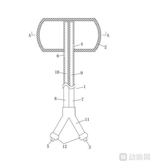
Image from the patent specification
Furthermore, the device is made of silicone material and equipped with scale markings and a three-way stopcock control valve, offering simple operation and high safety, making it easy for medical staff to master.
In the clinical application scenarios of capsule endoscopy, the examination of esophageal mucosal lesions has long faced multiple technical challenges, severely restricting diagnostic accuracy and clinical applicability. Capsule endoscopy, by virtue of itsNon-invasive Acquisition of Gastrointestinal Mucosal Imagesits unique advantages, has been widely applied in the diagnosis of small bowel diseases, and related technologies for the diagnosis of gastric and colonic diseases are also gradually becoming mature.
However, its application in the field of esophageal examination has always been limited. The core issue lies inThe capsule passes through the esophagus extremely rapidly after being swallowed.Clinical data indicate that the median transit time of existing capsules through the esophagus is only 2 seconds. Such a brief passage makes it difficult for healthcare professionals to conduct a comprehensive examination of the esophageal mucosa, leading to a high likelihood of missing potential lesions and thereby posing significant risks to early disease diagnosis.
To address this situation, two technological improvement directions have emerged within the industry; however, both exhibit significant drawbacks.
The first category is to enhance the capsule's own imaging capabilities,For instance, upgrading from a single-camera to a dual-camera system and employing high-speed imaging and storage technologies at 14–18 frames per second. Although dedicated esophageal capsule endoscopes have been successfully launched on the market, they fundamentally fail to address the core issue: imaging within the esophagus lacks targeting and selectivity, preventing focused observation of suspicious lesions. Furthermore, their transit relies entirely on esophageal peristalsis, lacking manual controllability—a limitation that also persists in magnetically controlled capsule endoscopy.
The second improvement strategy involves controlling the capsule's transit speed through external traction.For instance, the use of wire-controlled capsule endoscopes with fixed rods, traction devices utilizing rubber band applicators and traction wires, and control schemes employing four thin tethers. However, such approaches inevitably cause damage to the nasal and esophageal mucosa, significantly increasing patient discomfort during the examination and resulting in low clinical acceptance.
Furthermore, the clear visualization of detailed mucosal lesions in the gastrointestinal tract typically relies on an observation environment with adequate insufflation; however, no capsule endoscopes currently available on the market are equipped with an insufflation function. In clinical practice, although attempts have been made to distend the gastric lumen by administering gas-producing agents or having patients consume large volumes of water during the examination, the actual outcomes have been suboptimal: the gas production efficiency of these agents is inconsistent, while excessive water intake often leads to inadequate gastric distension, thereby compromising the clarity of the visual field and failing to provide reliable image support for diagnosis.
The compounding of these issues has created an urgent clinical need for a dedicated auxiliary device capable of both effectively controlling the movement speed of capsule endoscopes within the esophageal lumen and assisting in insufflation of the gastrointestinal tract, thereby driving the team’s research, development, and innovation in this technology.
This capsule endoscopy-assisted mobile device, with its innovative structural design and multifunctional integration, precisely addresses the bottlenecks of existing technologies, significantly enhancing its superiority in clinical applications. Its core advantages are mainly manifested in the following aspects:
In terms of core functionality, the device has achieved a dual breakthrough in "speed control + inflation," thoroughly addressing the critical shortcomings of traditional capsule endoscopy.It is inflated by injecting water into the balloon body through a water injection channel; the expanded balloon body assumes a cylindrical shape, with a maximum diameter of up to2cm, which can closely adhere to the entire circumference of the esophagus, effectively preventing the capsule endoscope from passing too rapidly through the cardia into the gastric cavity. Medical personnel can precisely control the movement trajectory of the capsule endoscope within the esophageal lumen by maneuvering the balloon body, enabling prolonged observation and repeated imaging of lesion sites. This significantly extends the effective observation time and fundamentally reduces the risk of missed diagnoses.
Meanwhile, the device’s independently configured gas insufflation channel can directly insufflate the gastrointestinal lumen, eliminating the need for gas-producing agents or large volumes of water. This enables rapid and stable gastric distension, providing capsule endoscopy with a clear and expansive field of view to ensure precise capture of mucosal lesion details, thereby filling the technical gap in current capsule endoscopy systems that lack insufflation capabilities.
In terms of structural design and safety, the device not only emphasizes practicality but also fully accounts for human-centered needs.The flexible cannula incorporates parallel, fixedly connected water-inflation and air-inflation catheters, both fabricated from soft, biocompatible silicone. Combined with a silicone-based lubricant coating on the balloon surface, this design minimizes friction and trauma to the oral and esophageal mucosa.
Compared with traditional external traction devices, this device significantly reduces patient discomfort during examination and improves clinical acceptance. The scale markings on the outer wall of the catheter help medical personnel to determine the position of the balloon in the body in real time.
Furthermore, the combination of three-way stopcocks and independent control valves enables mutually independent and controllable water and gas injection operations. This allows medical personnel to quickly master the operational essentials without extensive training, significantly enhancing operational convenience and clinical application efficiency.
In terms of clinical adaptability, the device demonstrates excellent flexible adaptation characteristics.Its length is meticulously designed to be 80 cm (including the balloon body), with the diameters of the water outlet and air outlet precisely set at 2 mm and 3.5 mm, respectively. The scientific matching between the catheter’s inner diameter and the channel caliber ensures that the water infusion rate and air inflation efficiency fully meet the requirements of clinical procedures.
Whether in high-end diagnostic and therapeutic settings with magnetic control capabilities or in conventional medical institutions lacking such equipment, this device achieves seamless adaptation: in magnetically controlled environments, it works synergistically with the magnetic field to enable more precise capsule navigation; whereas in non-magnetic environments, efficient visualization can be accomplished solely through the movement of the balloon body. This design fully meets the usage requirements across diverse tiers of healthcare scenarios, demonstrating broad potential for promotion and application.
The device features a simple and compact overall structure, eliminating complex and redundant component designs.Following ethylene oxide sterilization, its safety is assured, fully complying with the sterility standards for medical devices while significantly facilitating clinical storage and management. This feature further highlights the comprehensive advantages of the device in clinical applications.
Currently available and under-development capsule endoscopy locomotion assistance devices mainly focus on“Magnetic Field Drive”, “Mechanical Traction”Iterative Advancement of Two Major Technological Pathways Has Established a Diversified Product Portfolio.
Actively magnetically driven devices have become the mainstream trend in the current market, with their core advantage lying inPrecision Control. Among them,AnHan Technology's NaviCam® Magnetically Controlled Capsule Gastroscopy SystemThe product has successfully achieved mass production and been launched on the market. Leveraging active magnetic drive technology, this system enables flexible five-degree-of-freedom control of the capsule, including movement in superior-inferior, left-right, and anterior-posterior directions, as well as horizontal and vertical rotation. Its millimeter-level displacement and micro-angle rotation capabilities significantly enhance the precision of lesion observation. The system has obtained multiple international certifications and is widely used in clinical practice. Subsequently introduced smart modular products further integrate automated functions, substantially improving clinical workflow efficiency.
In addition,Anhan Technology has also made strategic moves in the field of mechanical traction devices.The company’s detachable tethered magnetically controlled capsule endoscopy (ds-MCE) system connects to the tail of the capsule via a detachable tether with a suction cup, enabling real-time, comprehensive visualization of the esophagus. After the examination, the detachable tether facilitates screening of the entire gastrointestinal tract, demonstrating high consistency in clinical diagnosis and good patient satisfaction. However, early-generation tethered capsules with fixed rods and rubber band deployment devices have been gradually replaced by detachable tether technology due to the risk of mucosal injury, fully underscoring the clinical emphasis on the core requirement of “safety and controllability.”
The technology transferred by the First Affiliated Hospital of Fujian Medical University stands as a paradigm for functionally integrated devices. Such devices extensively utilize biocompatible materials and feature optimized structural designs, prioritizing both operational convenience and patient comfort to meet the needs of healthcare scenarios at various levels. However, no commercial products based on this technology are currently available on the market. In the future, with continuous technological optimization and deeper market penetration, these devices are poised to play a significant role in the diagnosis of gastrointestinal diseases, injecting new vitality into improving clinical diagnostic standards and enhancing the patient care experience.