Southern Medical University to Spin Off PET Imaging Correction Technology Valued at RMB 1.52 Million with Vita (Xi'an) Medical
Recently, the National University Science Park of Southern Medical University released a public notice on the transformation of scientific and technological achievements, indicating that the university plans to“Positron Emission Tomography Imaging Correction Technology”The relevant patent achievements were capitalized as equity investment to jointly establish a new joint venture with Vita (Xi'an) Medical Technology Partnership (General Partnership), thereby promoting the industrialization of this technology. The valuation for this technology transfer was assessed by Guangzhou Zhirong Asset Appraisal Co., Ltd., atRMB 1.52 million.
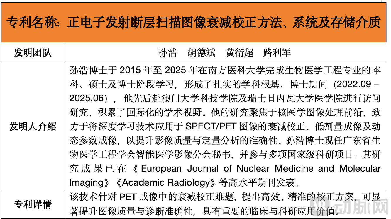
Patent Technology I
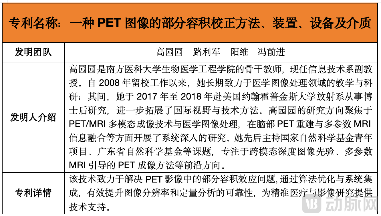
Patented Technology II
The Transferee of This TechnologyVita (Xi'an) Medical Technology Partnership, is an innovative enterprise focused on the research and development and industrialization of medical technology. The company is dedicated to the deep integration of medical imaging, artificial intelligence, and medical devices, promoting the transition of cutting-edge technologies from the laboratory to the market, and possesses strong capabilities in technological integration and commercial operation.
The two core patents included in this technology package are designed to significantly enhance the quality of positron emission tomography (PET) images through artificial intelligence and multimodal image fusion technology, together constituting an advanced intelligent PET image correction solution.
Positron Emission Tomography (PET) is a crucial functional imaging technique, playing a pivotal role in the early detection, precise staging, and therapeutic response assessment of tumors.
Its imaging principle isImages are generated by detecting photons produced from positron annihilation, based on the accumulation of radiotracers in metabolically active regions within the body (such as tumors).To achieve precise quantitative analysis of lesions, clinical practice often relies on semi-quantitative parameters such as the Standardized Uptake Value (SUV), the accuracy of which directly determines the reliability of diagnosis.
However, the quantitative accuracy of PET images is significantly affected by physical factors, among whichPhoton Attenuation EffectThis is particularly prominent. As photons traverse human tissue, they are absorbed or scattered, leading to attenuation of the signal reaching the detector. This results in reduced image contrast and the generation of artifacts, which severely distort the true radioactive distribution.
The current mainstream clinical correction method isAttenuation Correction Based on Co-registered CT Scanning.CT images can rapidly provide density distribution maps of various human tissues, thereby enabling the derivation of attenuation coefficients. However, this method has significant limitations: metal implants within the patient’s body cause severe streak artifacts in CT images, which are directly propagated into the generated attenuation maps and subsequently contaminate PET images; furthermore, slight patient movement between sequential CT and PET scans leads to spatial misalignment of the two image datasets, thereby introducing correction errors.
Another MRI-based correction method faces fundamental challenges:MRI images themselves reflect hydrogen proton density and relaxation times, but do not directly provide information on tissue attenuation of 511 keV photons. Therefore, complex image segmentation and tissue classification algorithms are required to indirectly estimate attenuation maps, a process that is cumbersome and difficult to ensure accuracy.
In addition to attenuation issues, PET images are also limited by the intrinsic spatial resolution of the detectors, resulting in significantPartial Volume Effect. This effect refers to the phenomenon where, due to the finite size of imaging units (voxels), a single voxel may simultaneously contain both high-uptake lesion tissue and low-uptake normal tissue, resulting in a signal that represents the average of the signals from these different tissues.
This leads to "dilution" of the signal from small lesions or lesion margins, resulting in blurred images and distortion of lesion size, shape, and uptake values, which severely impairs the detection of small lesions and accurate assessment of metabolic activity.
One current correction approach involves manually delineating or automatically segmenting the region of interest (ROI) containing the lesion on co-registered high-resolution CT or MRI images after image reconstruction, and then performing correction under the assumption of uniform radiotracer distribution within that region.
This method relies heavily on precise registration between PET images and anatomical images, as well as accurate segmentation of the anatomical images. Any registration deviation or segmentation error will directly translate into correction errors.
More importantly, the distribution of radioactivity in biological tissues is often non-uniform; the simplistic assumption of “regional homogeneity” does not align with actual conditions, thereby limiting improvements in correction efficacy.
In summary, existing clinical PET image correction schemes face core challenges in attenuation correction and partial volume correction, including artifact interference, data mismatch, inherent methodological limitations, and excessive reliance on the accuracy of registration and segmentation.
These limitations collectively constrain the accuracy and reliability of PET imaging in quantitative analysis and the diagnosis of subtle lesions.
Precisely because these critical bottlenecks have long constrained the accuracy and clinical utility of quantitative PET imaging, overcoming the limitations of traditional methods to achieve efficient and accurate image correction has become an urgent requirement for advancing molecular imaging.
The core advantage of this patented technology portfolio lies in itsIt adopts a cutting-edge deep learning architecture, fundamentally overturning the traditional paradigm of PET image correction methods that rely on single modality or manual intervention.Achieved smarter and more precise correction results.
To address the challenge of attenuation correction, the innovation of the first patent lies inAn intelligent correction model based on a three-dimensional conditional generative adversarial network (3D cGAN) was constructed.
This model does not directly rely on a specific type of anatomical image (such as CT or MRI) to derive attenuation maps; instead, it operates by learning the complex mapping relationships between large volumes of diverse, paired PET images and their corresponding high-quality attenuation correction maps. Its advancements are reflected in"Pre-training–Transfer Learning" Two-Stage Training StrategyOn.
The model is first pre-trained on one type of device or scanning condition (Type I images) to acquire a general understanding of the relationship between image structure and attenuation correction. Subsequently, transfer learning is employed to fine-tune the model using data from another condition (Type II images), enabling rapid adaptation to different PET scanners, tracers, or protocols.
This method ingeniously overcomes the fundamental obstacles of metal artifact propagation in traditional CTAC methods and the lack of attenuation information in PET/MR.
The model can ultimately generate high-precision attenuation correction maps directly from the PET images requiring correction, significantly reducing reliance on external anatomical images and the associated registration errors and artifact interference, thereby substantially improving the accuracy of quantitative parameters such as SUV.
With regard to partial volume effect correction, the advantage of the second patent lies in itsA Unique Dual-Channel Network Architecture Was Designed, this architecture comprises parallel PET reconstruction subnetworks and MRI reconstruction subnetworks.Its advancement lies in completely eliminating the tedious and error-prone step of precise segmentation of high-resolution MRI images, which is required in traditional methods.
During the training process, the MRI reconstruction subnetwork does not output segmented anatomical regions; instead, it focuses on extracting multi-level structural feature information (i.e., MRI information) from the training MRI images. These features are then subtly “injected” into the decoding process of the PET reconstruction subnetwork to guide and constrain the reconstruction of PET images.
This feature-level information fusion approach enables the model to fully leverage the detailed anatomical priors provided by MRI to recover details and true radiotracer distributions lost in PET images due to insufficient resolution, while avoiding the direct introduction of errors into the correction results caused by inaccurate MRI segmentation.
Ultimately, the model can directly output high-quality target PET images with significantly suppressed partial volume effects, without requiring any segmentation of the input first MRI image, thereby greatly enhancing the practicality and robustness of the method.
In summary,Two Patents Jointly Build a Complementary PET Image Enhancement SolutionThe first patent is dedicated to eliminating quantitative biases caused by photon attenuation, laying the foundation for precise quantification; the second patent focuses on restoring spatial details and true activity distribution in images, thereby enhancing the detection and delineation of small lesions.
Both approaches leverage advanced deep learning techniques to automatically learn correction patterns in a data-driven manner, significantly reducing reliance on manual operations, strict image registration, and precise anatomical segmentation. This not only promises to provide PET images with clearer contrast and more reliable quantification in clinical practice, thereby assisting physicians in making more accurate diagnoses, but also greatly promotes the development of PET imaging technology toward standardized and intelligent quantitative analysis.
In response to the sustained demand for safer, more precise, and smarter solutions in the medical imaging market, relevant technical teams are actively expanding multiple pipelines under development, leveraging their extensive expertise in multimodal image registration, feature fusion, and deep reconstruction.
In the international market,A research consortium comprising the Institute of Biomedical Engineering at Boğaziçi University, the Department of Nuclear Medicine at Istanbul University Faculty of Medicine, the Department of Nuclear Medicine at Koşuyolu Hospital of Yeditepe University, and the Department of Nuclear Medicine at Acıbadem Maslak Hospital has focused its collaborative efforts on the development and validation of partial volume effect (PVE) correction techniques in PET/CT imaging. By optimizing the calculation methods for recovery coefficients (RC), this initiative aims to enhance the accuracy of quantitative standardized uptake value (SUV) analysis for small lesions, thereby providing technical support for the precise diagnosis and therapeutic assessment of tumors and other diseases.
On the Research of Partial Volume Effect Correction Coefficients Based on Personalized Phantoms,The team achieved several key outcomes. By employing an Asymptotic Regression Model (ARM) to fit the relationship between Recovery Coefficient (RC) and sphere diameter, the team validated the stability of RC across different anatomical regions using heterogeneous data from two institutions. Furthermore, they demonstrated that this anthropomorphic phantom effectively obtains SUV correction values closer to clinical reality, thereby addressing the limitations of traditional NEMA phantoms, which fail to simulate human tissue heterogeneity and struggle to accurately measure subcentimeter lesions.
Currently, the related research findings have been published in the journal *BioMedical Engineering OnLine* and are in the public domain. Multi-center, multi-device clinical validation has been completed, demonstrating potential for clinical translation.
In China,A research team led by Dr. Hu Zhanli at the Lauterbur Biomedical Imaging Center, Shenzhen Institutes of Advanced Technology, Chinese Academy of Sciences, in collaboration with Sun Yat-sen University Cancer Center and Shanghai United Imaging Healthcare Co., Ltd., has developed a CT-free intelligent attenuation correction technology for whole-body PET imaging. This technology is primarily designed for the 2-meter total-body PET/CT system (uEXPLORER) launched by Shanghai United Imaging Healthcare.
Experimental results demonstrated that the attenuation-corrected PET images generated by this technique were highly consistent with ground-truth attenuation-corrected PET images, exhibiting superior image quality and enhanced contrast across various anatomical regions. Quantitative analysis yielded mean peak signal-to-noise ratio (PSNR) and structural similarity index measure (SSIM) values of 36.92 ± 5.49 dB (p < 0.01) and 0.980 ± 0.04 (p < 0.01), respectively. These findings have been published in European Radiology (IF = 5.9), a prestigious journal in the field of radiology. Currently, clinical data collection and experimental validation for this technology have been completed, and the results are publicly available.
Professor Gong Nanjie’s team from the School of Physics and Electronic Science at East China Normal University, the Shanghai Key Laboratory of Magnetic Resonance, and the Institute of Medical Magnetic Resonance and Molecular Imaging Technology collaborated with the Affiliated Hospital of Zunyi Medical University and the Second Affiliated Hospital of Chongqing Medical University to focus on the research and development as well as clinical validation of technologies related to PET image enhancement.
In terms of RaDynPET (an AI model for low-count PET image enhancement),The team has achieved multiple key results:
Clinical validation demonstrates that RaDynPET comprehensively outperforms other deep learning methods, including cGAN, cycleGAN, and RCAN, across both peak signal-to-noise ratio (PSNR) and structural similarity (SSIM) metrics, while maintaining stable performance across diverse medical institutions, patient characteristics, and scanning conditions;
Furthermore, the model demonstrates high performance in lesion detection, with a sensitivity of 95.45% and a specificity of 98.41%. It preserves the metabolic ratio between lesions and normal tissue, thereby ensuring the reliability of standardized uptake value (SUV) quantitative analysis. This makes it suitable for clinical scenarios that rely on metabolic quantification, such as differentiating between benign and malignant tumors and assessing treatment efficacy.
Based on the aforementioned technological landscape and industry dynamics, PET quantitative imaging technology is clearly evolving toward CT-free, low-dose, high-efficiency, and intelligent solutions. From domestic research institutions’ exploration of CT-free attenuation correction and low-count image enhancement to the international academic community’s ongoing efforts to standardize methods for addressing partial volume effects, technological iteration in this field is accelerating.
However, moving from algorithmic breakthroughs and clinical validation to becoming a widely adopted standardized tool still requires overcoming a series of hurdles, including data standardization, multi-center validation, and regulatory approval. In the future, the efficiency of clinical translation of core technologies and the ability to achieve commercial implementation will become key metrics for assessing the value of relevant stakeholders.