Kampto Neurotech Announces Breakthrough Wireless, High-Throughput Brain-Computer Interface with 65,536 Electrodes on a Single Chip
In the field of Brain-Computer Interface (BCI), achieving high-quality neural signal recording while minimizing surgical invasiveness has remained a significant challenge. Although traditional non-invasive techniques are safe, they offer limited signal resolution; conversely, invasive techniques provide clear signals but carry risks of tissue damage and infection.
On December 8, 2025, Professor Ken Shepard’s team at Columbia University, in collaboration with Stanford University, NewYork-Presbyterian Hospital, and other institutions, published a breakthrough study in the journal Nature Electronics.The team developed a novel wireless brain-computer interface named the “Bioelectronic Interface System to the Cortex (BISC).”
(Source: Nature Electronics)
This single-chip device integrates 65,536 recording electrodes, enabling the capture of cortical activity with ultra-high spatiotemporal resolution. By achieving full implantation and wireless power delivery, it paves a new path for high-throughput, minimally invasive brain-computer interface technology.
The successful development of the BISC system marks the formal entry of brain-computer interface (BCI) technology into a new era characterized by “single-chip, high-channel-count, and fully wireless” capabilities. It elegantly resolves the trade-offs among implant size, signal quality, and surgical trauma, opening a clear window into the inner workings of the brain with micron-level precision. As the technology matures and advances toward clinical translation, this tiny silicon chip holds promise to become a bridge connecting human thought with the digital world, offering renewed hope for countless patients afflicted by neurological disorders.
Electrocorticography (ECoG) is a technique that uses non-penetrating electrodes embedded in a flexible substrate to record electrical activity on the surface of the brain.Compared with penetrating electrodes, ECoG is located on the cortical surface, which can significantly reduce damage to brain tissue. At the same time, compared with scalp electroencephalogram (EEG), it can provide higher signal-to-noise ratio and bandwidth.
However, although existing high-resolution ECoG arrays can provide thousands of recording sites on a single substrate, their signal conditioning and data transmission still rely on discrete electronic components. Traditional wired connections not only limit the subject’s range of motion but also increase the risk of infection. Even current wireless solutions are often constrained by bulky electronic packaging or “canister” designs, which complicate surgical implantation and compromise long-term stability. More critically, this discrete design limits the number of recording channels and device scalability, making it difficult to meet the demands for detailed observation of the brain’s complex spatiotemporal dynamics.
To address these issues, the research team adopted a strategy of fully integrating electrodes and electronic circuits on the same chip.
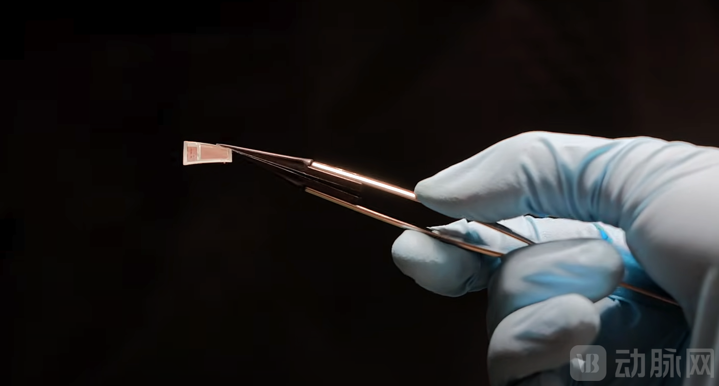
The core of the BISC system is a 12×12 mm complementary metal-oxide-semiconductor (CMOS) chip. Through post-processing techniques, the chip is thinned to less than 50 micrometers, granting it sufficient mechanical flexibility to conform closely to the curved surface of the cerebral cortex. This highly integrated design enables BISC to achieve a qualitative leap in volumetric efficiency—defined as the number of recording channels per unit implanted volume.
(Source: Columbia Engineering)
Data shows that BISC's volumetric efficiency is more than 400 times higher than that of the current state-of-the-art competitors.This single-chip architecture not only eliminates complex wiring but also significantly enhances signal quality and system integration by directly incorporating functions such as signal amplification, filtering, and analog-to-digital conversion into the pixel circuits beneath the electrodes.
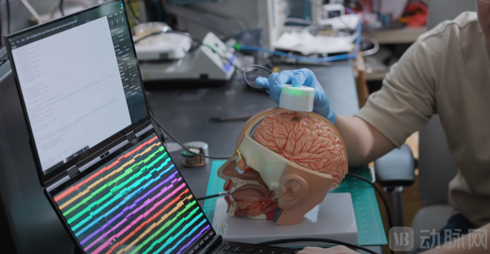
The design philosophy of the BISC system lies in achieving the ultimate integration of extreme miniaturization and high performance. It is not merely a recording device, but a comprehensive bidirectional neural interface platform. Through an external wearable “Relay Station,” the BISC enables wireless power delivery and high-bandwidth uplink data transmission at speeds of up to 108 Mbps. This wireless connection is based on Ultra-Wideband (UWB) impulse radio technology, which allows for the real-time transmission of massive volumes of neural data while ensuring low power consumption.
Moreover, this “paper-thin” device can be gently slid into the subdural space like a moistened tissue paper, eliminating the need for complex craniotomies or large-scale bone removal. This significantly reduces surgical risks and lays the foundation for future minimally invasive implantation.
The performance of the BISC system has been rigorously validated in multiple animal studies.The chip integrates 65,536 titanium nitride (TiN) electrodes. Although data bandwidth limitations prevent simultaneous recording from all electrodes, the system allows users to select 1,024 channels from these 65,000 electrodes for simultaneous recording at any given time.
In somatosensory evoked potential (SSEP) experiments using a porcine model, stimulation was applied to various locations on the snout of the pigs, and BISC successfully recorded the resulting cortical potential changes. t-distributed stochastic neighbor embedding (t-SNE) analysis revealed clearly separable clusters in the recorded signals, with a linear discriminant model achieving a classification accuracy of 97.8% ± 1.7%, demonstrating the system’s superior capability in capturing fine spatial distributions of tactile stimuli. Histological examination showed no significant pathological damage in the implantation area, confirming its favorable biocompatibility.
In non-human primate (NHP) models, the research team further demonstrated the potential of BISC in motor and visual decoding. During a motor task involving asynchronous reaching and grasping movements in macaques, BISC recorded neural activity from the primary motor cortex (M1) and the primary somatosensory cortex (S1). The decoder successfully predicted arm movement velocity using local field potential (LFP) signals, achieving a Pearson correlation coefficient of 0.53. Even more notably, long-term recordings lasting up to 64 days were conducted in the visual cortex. While presenting visual stimuli such as gratings, random-dot patterns, and natural images to the macaques, BISC not only accurately mapped the retinotopic organization of the primary visual cortex (V1) but also captured micron-scale “travelling waves.” These travelling waves represent coherent spatial propagation patterns of neural oscillations, which had previously been observed only at centimeter or millimeter scales.
(Source: Columbia Engineering)
Leveraging the high-density recording advantages of BISC, researchers discovered that these traveling waves in the γ band (30–90 Hz) carry rich information about the location of visual stimuli. By constructing decoding models incorporating convolutional neural networks (CNNs), Transformers, and hybrid architectures, they found that utilizing high-resolution traveling wave data significantly improves the decoding accuracy of visual stimulus locations (p < 0.01).
Furthermore, by leveraging the “Digital Twin” model—a predictive framework based on deep neural networks—researchers can generate “Maximally Exciting Images” (MEIs) for each recorded channel, thereby elucidating the hierarchical changes in visual feature selectivity of neurons across cortical areas V1 to V4. Area V1 exhibits a preference for oriented gratings, whereas areas V2 and V4 are sensitive to more complex color-opponent features. These findings fully demonstrate the robust capability of BISC in deciphering the brain’s fine-grained spatiotemporal dynamics.
The advent of BISC technology is not only a milestone in engineering but also brings new hope to basic neuroscience research and clinical treatment.
From a theoretical perspective, the unprecedented high spatiotemporal resolution provided by BISC enables scientists to observe dynamic patterns such as traveling waves in the cerebral cortex at the micrometer scale for the first time.This bridges the gap between microscopic neuronal activity and macroscopic brain region function, facilitating a deeper understanding of how the brain processes information through large-scale coordinated activity.
As Ken Shepard, Professor of Biomedical Engineering at Columbia University, stated, “We have integrated all functionalities onto a single silicon chip, demonstrating how brain-computer interfaces can become smaller, safer, and more powerful.” This ability to miniaturize computing power for implantation heralds a future in which medical implants are no longer constrained by size, but are as powerful and unobtrusive as high-performance computers.
In terms of clinical application, the minimally invasive nature of BISC makes it highly attractive.Traditional invasive electrodes often require penetration into brain tissue, which can easily trigger immune responses and glial scarring, leading to a decline in signal quality over time. In contrast, as a subdural implant, BISC avoids penetrative injury while positioning closer to neurons than epidural electrodes, thereby ensuring long-term, stable recordings with a high signal-to-noise ratio.
Its “slidable” implantation method means that in the future, implantation may be completed with only a tiny incision in the skull, significantly reducing surgical trauma and the risk of complications.This feature holds significant importance for the treatment of chronic neurological disorders, such as drug-resistant epilepsy, paralysis, aphasia, and visual restoration in the blind.Particularly for patients with epilepsy, the high-density array of BISC can more precisely localize the epileptogenic focus, providing a basis for precision treatment.
Currently, Kampto Neurotech, a startup based on BISC technology, has been established and is dedicated to translating this scientific achievement into clinical products.
Although the current external relay station still has room for size reduction and the system has not yet undergone human trials, its demonstrated scalability and modular design offer immense potential for future applications. The research team plans to cover larger brain regions by tiling multiple chips in the future and to meet the requirements of deep brain stimulation by connecting polyimide extenders.
With the deep integration of AI technology and brain-computer interfaces, the high-bandwidth, bidirectional interaction model represented by BISC will enable seamless connectivity between humans and AI systems. This advancement holds promise not only for treating diseases but also for enhancing human cognitive and perceptual capabilities, thereby redefining the future of human-computer interaction.