Intotobiotech Breaks Century-Old 2D Histopathology Blind Spot with Clinical-Grade Light-Sheet Microscopy, Ushering in a New Era of High-Precision 3D Pathological Diagnosis

INTOTO BIOTECH
Innovative Medical Device Developer
In the modern medical diagnostic system, pathology is referred to as the “foundation of medicine,” pathologists are thus known as the “doctors’ doctor,” and their diagnostic reports are regarded as the “gold standard” for disease diagnosis.
Traditional pathological diagnosis is, in essence, a process of continuous “subtraction.” The tissue sectioning preparation techniques it employs have been developed over more than a century: surgeons excise patient tissue samples 3–5 mm thick, which are then trimmed and embedded into paraffin blocks by the pathology department, before being sliced into 3–5 μm-thin sections for microscopic examination. This means that information from the tissue sample is progressively reduced at each step, so physicians actually observe only one-thousandth of the total information contained within the original tissue.
This classic workflow has long suffered from two major pain points. First, sections are prone to uneven thickness, irregularity, and even scratching during microtomy; more critically,Mainstream pathological diagnosis is limited to two-dimensional imaging, whereas human tissues are inherently three-dimensional structures. Within native tissues, cells exhibit complex volumetric features, projections, hierarchical organization, and spatial connectivity. Two-dimensional sections capture only arbitrary cross-sections, failing to reconstruct the spatial continuity of tissues, which results in the permanent loss of critical three-dimensional information during specimen preparation and increases the risk of missed or misdiagnosis.
In September 2025, a study from Harvard Medical School published in *Nature Methods* also pointed out that in traditional pathological sections, we almost never see a complete cell. The study calculated that in conventional sections with a thickness of only 4 to 5 micrometers, the proportion of intact nuclei is less than 5%.[1] In other words, traditional pathological images are actually pieced together by countless “broken cell fragments.” This “limited view” observation method is becoming a bottleneck for precision medicine today, when the demand for early cancer screening and targeted diagnosis is surging.
The key to breaking through lies in the observational leap from “two-dimensional fragments” to “three-dimensional panoramas.” Against this backdrop, INTOTO BIOTECH was established with the incubation and support of the National Innovation Center for High-Performance Medical Devices (hereinafter referred to as the “National Innovation Center”), dedicated to providing the industry with in situ imaging solutions for 3D spatial biology through next-generation light-sheet fluorescence microscopy technology.Its technological core lies in “no sectioning, panoramic viewing,” which involves non-destructive 3D imaging of intact tissues to clearly present the spatial architecture and interactions of cells, thereby fully preserving the original three-dimensional information of the tissue and filling a critical dimension missing in traditional pathological diagnosis.
"As one of the few high-end optical imaging enterprises in China with fully independent intellectual property rights and a 100% localized supply chain,"INTOTO BIOTECH has pioneered the advancement of 3D pathological imaging into clinical trials, becoming the first company in China and one of the earliest globally to do so.. Currently, INTOTO BIOTECH's technology platform has coveredLife Science Research, Clinical Precision DiagnosticswithInnovative Drug R&Dand other types of scenarios.
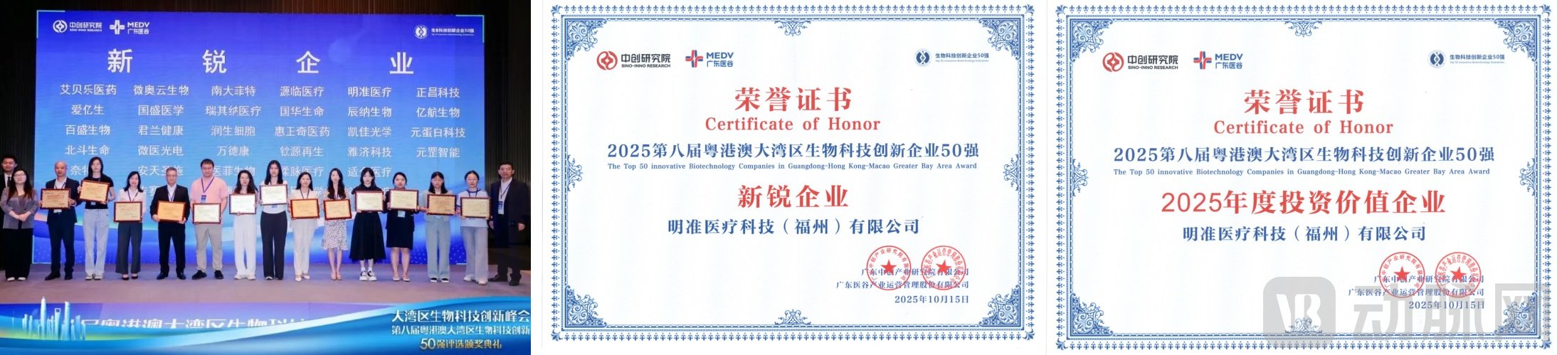
INTOTO BIOTECH Honored as a “Rising Star Enterprise” and “2025 Investment Value Enterprise” in the Top 50 Biotechnology Innovation Enterprises of the Guangdong-Hong Kong-Macao Greater Bay Area in 2025
To understand the core value of INTOTO BIOTECH, it is first necessary to answer two questions: What is light-sheet microscopy? And why has it been difficult to introduce light-sheet microscopy into clinical applications?
Light-Sheet Fluorescence Microscopy (LSFM) represents a significant technological breakthrough in the life sciences field in recent years. Its core principle involves tomographic scanning of specimens using an extremely thin beam of light, known as a "light sheet." During imaging, the light sheet is oriented perpendicular to the detection objective lens, exciting fluorescence exclusively within the focal plane. Rapid layer-by-layer scanning is then performed, and complete three-dimensional images are reconstructed through computational algorithms. The core technology of INTOTO BIOTECH is based on this approach.
Compared with the point-by-point scanning or wide-field illumination of traditional microscopes, LSFM offers significant comprehensive advantages: rapid light-sheet imaging, low photodamage, and the ability to perform continuous 3D observation of tissues ranging from centimeter to submicron scales. Consequently, it is regarded as a major breakthrough in life science imaging over the past two decades and is currently the only high-end microscopy technique that simultaneously ensures non-destructive sample handling, high contrast, large field of view, deep penetration, and high-speed 3D imaging.
Leveraging these advantages, light-sheet microscopy has become a key tool in scientific research fields such as embryonic development, neuroscience, and oncology. However, its application in the clinical domain remains a blank slate. This is not due to a lack of clinical interest, but rather because the barriers to industrialization are too high:
First are the technological and engineering barriers.Early light-sheet techniques commonly suffered from poor axial resolution, with lateral-to-axial resolution ratios reaching as high as 1:5. This meant that when physicians attempted to view three-dimensionally reconstructed lesions from a lateral perspective, the images would appear stretched and distorted, leading to misrepresentation of diagnostic information. Second-generation technologies attempted to address this issue through multi-angle illumination and algorithmic fusion, but their heavy reliance on computational processing resulted in unstable outcomes. Third-generation technologies employed purely optical methods to compress light-sheet thickness; however, simulated imaging results often outperformed actual imaging performance.
Secondly, there is a lack of engineering implementation and ease of use.Most light-sheet microscopes available on the market are designed for research institutions, emphasizing flexibility and expandability. Their operation is extremely complex, requiring specialized researchers to perform tedious calibration and parameter adjustments. However, clinical settings demand “tool-grade” devices that are stable, accurate, user-friendly, and reproducible. The complexity of research-grade equipment significantly hinders its widespread adoption in high-throughput hospital environments.
Finally, there are supply chain and market access barriers.The global light-sheet microscopy market has long been monopolized by German and American companies, forming a dense patent cluster. In pursuit of rapid shipments, some domestic manufacturers have taken the shortcut of "purchasing foreign frames for retrofitting." While this approach saves effort, it leaves core components dependent on external suppliers, and the products are essentially "retrofitted machines," unable to pass strict medical device registration approvals.
It is precisely these insurmountable technical and industrialization barriers that have kept light-sheet microscopy, a cutting-edge technology, long excluded from clinical diagnostics.
Unlike many medical device companies that transition from scientific research to clinical applications, INTOTO BIOTECH has chosen a path from the outset to redefine its architecture itself and drive technological iteration based on clinical needs.
During the initial phase of its development, INTOTO BIOTECH received full support from the National Innovation Center, which not only provided an incubation environment for the translation of cutting-edge technologies but also facilitated rapid access to top-tier clinical resources and seasoned industrialization experts, ensuring that INTOTO BIOTECH possessed a solid “clinical DNA” from the outset.
Leveraging the National Innovation Center, INTOTO BIOTECH has also assembled a team that deeply integrates industry, academia, and research. The company’s founder, Dr. He Jiaye, studied under Professor Jan Huisken, the inventor of light-sheet microscopy. Building on this strong technical foundation, Dr. He has led his team to successfully engineer and commercialize original technologies developed in the laboratory. Notably, Professor Jan Huisken serves as INTOTO BIOTECH’s exclusive technical advisor, ensuring that the latest research outcomes from his laboratory are prioritized for productization by INTOTO BIOTECH, thereby safeguarding the company’s continuous innovation in core underlying technologies.
The company’s core team also includes a group of researchers with over a decade of expertise in optical technology, as well as “industry veterans” from leading listed medical device companies such as Mindray. Leveraging its robust technological foundation, INTOTO BIOTECH has built core competencies to overcome the barriers to clinical translation of light-sheet technology through full-stack independent R&D—
Eliminate depth blind spots through isotropic optical resolution.INTOTO BIOTECH is one of the few manufacturers in the industry capable of achieving “optically isotropic resolution,” meaning that lateral and axial resolutions are nearly isotropic, thereby resolving the long-standing issue of poor axial resolution in early light-sheet microscopy technologies. More importantly, INTOTO BIOTECH achieves this through pure optical methods rather than algorithmic corrections. This core technology, grounded in fundamental optics, ensures the authenticity and stability of three-dimensional data, providing a reliable and accurate data foundation for clinical diagnosis.
Ultimate engineering enables "foolproof" one-click operation.To meet the high-load, fast-paced demands of clinical settings, INTOTO BIOTECH has adopted a unique open-top inverted structure and a pan-medium imaging chamber design, with built-in fully automated calibration processes and resolution matching mechanisms. Physicians need only place the sample and define the region of interest to initiate one-click scanning and imaging. Coupled with a clean and intuitive user interface, this significantly reduces operational complexity and maintenance costs. This ultimate ease-of-use design is the key guarantee enabling its products to truly integrate into clinical practice.
Ultra-wide resolution coverage and large-sample imaging empower all scenarios from research to clinical practice.INTOTO BIOTECH’s light-sheet microscope features automatic objective lens switching, with lateral pure optical resolution smoothly ranging from 400 nm to 2 µm and axial resolution covering 500 nm–2 µm. A single instrument balances high-resolution detail with large-field-of-view overviews, offering industry-leading versatility in imaging scenarios. Meanwhile, its standard imaging field supports a 2 cm × 2 cm cross-section and an imaging depth of 3–5 mm, fully meeting the requirements for clinical samples. Furthermore, the system accommodates the needs of large-sample, large-area imaging in scientific research, covering a broad spectrum of applications from clinical practice to research.
Core Components and Supply Chain 100% Domestically Produced; Next-Generation Light-Sheet Microscopy Imaging Capability ReservedUnlike “retrofitting vendors,” INTOTO BIOTECH has independently developed its underlying architecture from the ground up, achieving 100% localization of core components and supply chains. Through joint development with domestic suppliers, INTOTO BIOTECH has not only resolved issues related to cost and lead times but is also actively developing next-generation core components such as cameras and objective lenses. This means that the supply chain for 3D pathology will no longer be constrained by foreign dependencies, empowering Chinese teams to define the next generation of light-sheet microscopy imaging technology.
Leveraging the aforementioned technological, industrial, and resource advantages, INTOTO BIOTECH has established a clear product portfolio tailored to two core scenarios: scientific research exploration and clinical diagnosis.
Pano One is a next-generation light-sheet microscopy system designed for life science research, primarily serving the scientific research market.This system employs inverted imaging and a fully automated refractive index matching system, supporting cross-scale high-resolution imaging. Characterized by isotropic resolution, low photodamage, and high imaging contrast, it is suitable for long-term in vivo studies, such as tracking embryonic development and analyzing the tumor microenvironment. Widely used in neuroscience, developmental biology, drug screening, and other fields, it is a microscopic imaging tool that balances a large field of view, high resolution, and in vivo compatibility. Pano One utilizes axial scanning technology to improve its axial resolution by sixfold compared to traditional light-sheet microscopy. Leveraging this purely optical approach, it achieves isotropic, high-speed imaging with submicron resolution for samples ranging from centimeter to submicron scales.
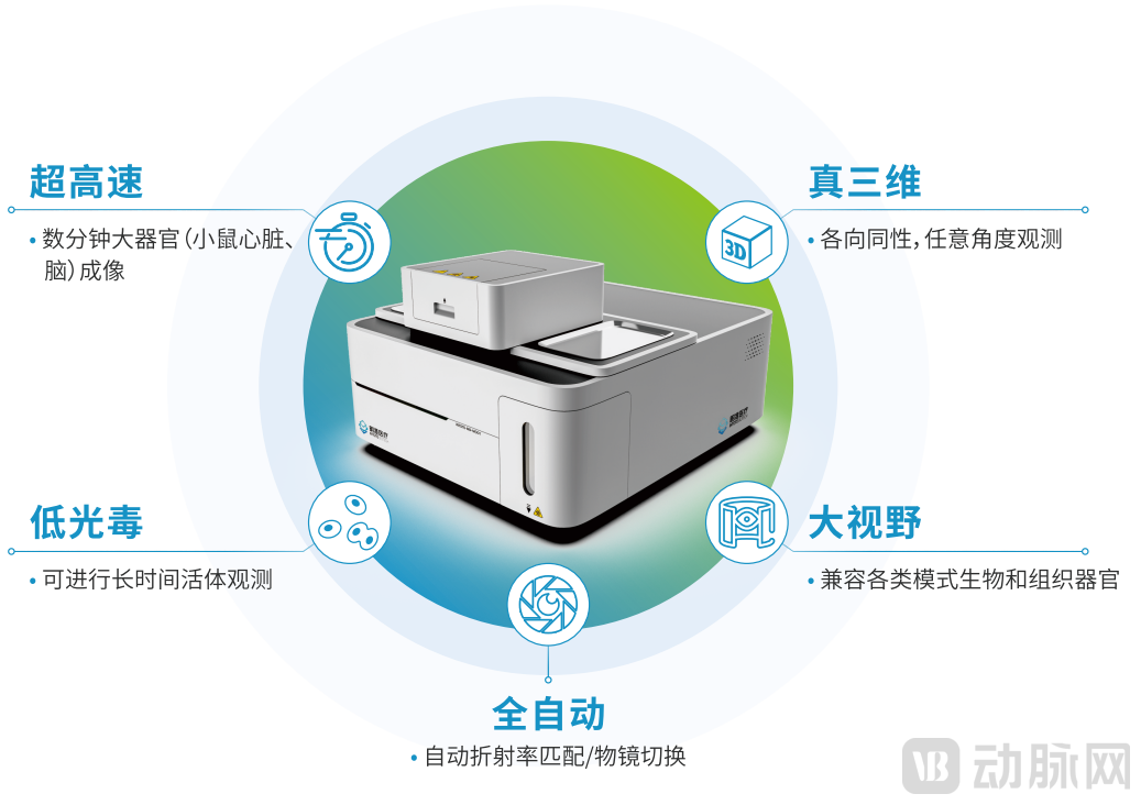
Pano One: The Next-Generation Light-Sheet Microscopy System
Patho One is a 3D non-sectioning histopathological imaging system designed for precision medicine.As the name suggests, it eliminates the need for physical sectioning and enables direct imaging of intact specimens, thereby preserving sample integrity and reversibility. The system supports 3D virtual H&E staining to reconstruct authentic tissue architecture and is applicable to various tissue types, including gastric cancer, colorectal cancer, and breast cancer. Furthermore, Patho One provides comprehensive information on 3D tissue structure and cellular heterogeneity, assisting physicians in accurately observing complex invasive structures.
As the first 3D non-slicing pathological imaging device in China, and potentially the first worldwide, to enter clinical trials, Patho One has completed imaging of nearly 1,000 human samples at multiple benchmark hospitals. Preliminary data indicate that 3D pathology can effectively improve the positive detection rate of cancer, holding promise to supplement and even revise existing clinical gold standards or guidelines.
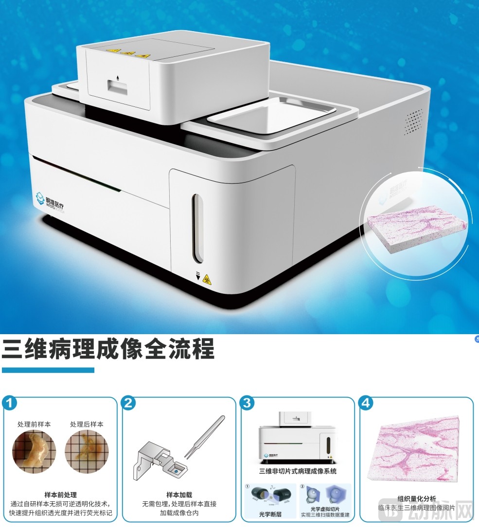
3D Non-Sectioning Histopathological Imaging System Patho One
Looking ahead, INTOTO BIOTECH is not content to remain merely an instrument manufacturer; instead, it harbors a grander vision for building a comprehensive ecosystem.
According to Dr. He Jiaye, founder of INTOTO BIOTECH, with the deep integration of computational optics and traditional imaging, the next revolutionary breakthrough in microscopy technology is highly likely to emerge in China. Chinese scientists have already reached the global forefront in fields such as super-resolution and photoacoustic imaging; however, international discourse power in the field of light-sheet microscopy remains predominantly held by German and American companies. The long-term goal of INTOTO BIOTECH is to become the “Chinese voice” in this domain.
To achieve this goal, INTOTO BIOTECH has formulated a clear "three-step" strategy:
Short-term: Deepen cultivation of the scientific research market,Market education for “3D pathology” is being conducted through technology promotion. It is reported that INTOTO BIOTECH is actively expanding its overseas presence, with its research-grade products expected to enter the international market in the second half of next year.
Mid-term: Layout the industrialization sector for new drug R&D,As a supplier of data acquisition equipment, we leverage the high-throughput capabilities of light-sheet microscopy to empower drug screening.
Long-term:With the approval of its medical device registration certificate, INTOTO BIOTECH willComprehensively Promote the Adoption of 3D Pathology in Hospitals, establish a comprehensive clinical solution encompassing imaging equipment, staining reagents, and data analysis, to build a three-dimensional spatial biology ecosystem.
The complexity of tissue is deeply rooted in its three-dimensional conformation, where micron-scale spatial variations are sufficient to render traditional two-dimensional sections “blind.” Whether for understanding the intricate architecture of normal tissues or precisely characterizing tumor heterogeneity, assessing therapeutic response, or evaluating disease risk, three-dimensional assessment is no longer merely an added advantage but a necessity for approaching biological truth. Piercing through a century of obscurity, INTOTO BIOTECH is using a beam of “light” to illuminate the uncharted territories of pathological diagnosis, providing a solid foundation and expanded possibilities for precision medicine.
References:
[1] <Highly multiplexed 3D profiling of cell states and immune niches in human tumors>,Nature Methods
[2] “A Complete Replacement for Confocal? Light-Sheet Microscopy May Disrupt the Paradigm of In Vivo 3D Imaging—An Interview with Dr. He Jiaye, Founder of INTOTO BIOTECH,” 3i Life Science Instrument Society
[3] "The New-Generation Light-Sheet Microscopy System PanoOne | Redefining Spatial Biology Research: A Complete Replacement for Confocal? Light-Sheet Microscopy May Disrupt the Paradigm of In Vivo 3D Imaging," Lab Cat