3-Minute Overview: AR-Guided Cataract Surgery Microscope System Files IPO Prospectus
The technological evolution of augmented reality (AR)-guided systems and methods for ophthalmic cataract surgery microscopes follows a core trajectory from optical visualization to digital imaging fusion, then to AR-precise navigation, and finally to AI-robot collaboration. Key milestones center on imaging upgrades, enhanced navigation precision, and optimized human-computer interaction, ultimately achieving dual improvements in surgical efficiency and safety. The following outlines the phased technological development timeline and the iteration of key technical methods:
I. Foundational Period (Mid-20th Century – Early 2000s): From Optical Microscopy to the Emergence of Digital Imaging
This phase centers on optical microscopy, gradually integrating digital technologies to establish the hardware and foundational imaging capabilities for AR guidance.
1. The Widespread Adoption of Optical Microscopy
In 1946, binocular surgical microscopes were introduced in ophthalmology to address the limitations of monocular vision, becoming a core device for cataract surgery. Subsequent optimizations, such as coaxial illumination and extended depth of field, improved the clarity of the surgical field; however, these systems relied solely on optical imaging and lacked digital overlay capabilities.
2. Preliminary Exploration of Digital Imaging
With the rise of computer image processing, surgical microscopes began to integrate CCD cameras, enabling digital acquisition of the surgical field, supporting image storage and basic analysis, and providing an image source for subsequent AR overlay, but without real-time guidance functionality.
Key Methods:Primarily based on optical magnification and digital acquisition, without spatial registration or virtual information overlay; used solely for postoperative review or teaching, not for intraoperative real-time guidance.
II. Early Technology Phase (Mid-2000s to Mid-2010s): Navigation Integration and AR Proof of Concept
Femtosecond laser-assisted technology has matured, with breakthroughs in digital navigation and spatial registration technologies; augmented reality (AR) is transitioning from the laboratory to clinical exploration, with its core achievement being the initial capability to overlay virtual information onto the surgical field.
1. Combination of Femtosecond Laser and Digital Navigation
In 2008, femtosecond laser-assisted cataract surgery was introduced for key steps such as corneal incisions and capsulorhexis, improving procedural precision. During the same period, navigation systems based on preoperative biometric data (e.g., IOL power, corneal topography) emerged, beginning to correlate preoperative planning data with the intraoperative field of view.
2. AR Proof of Concept and Preliminary Clinical Application
The research focuses on spatial registration algorithms, which overlay virtual markers (such as astigmatism axis and capsulorhexis boundaries) onto the microscope field of view through landmark or anatomical feature matching to assist surgeons in localization. Representative systems, such as the early Callisto eye, enable astigmatism marking and guidance for intraocular lens implantation.
Key Methods:Preoperative 3D reconstruction of CT/OCT data + intraoperative optical image registration, using rigid registration algorithms; AR information is primarily presented as simple lines/markers, with accuracy dependent on manual marking, resulting in limited real-time performance and adaptability.
III. Rapid Development Phase (Late 2010s – Early 2020s): Maturation of Digital Microscopy and AR-Precise Navigation
The widespread adoption of digital surgical microscopes, breakthroughs in 3D imaging, HDR, and real-time registration technologies have upgraded AR guidance from “auxiliary marking” to “full-process precision guidance,” enabling large-scale commercial application of these systems.
1. Digital Surgical Microscope and 3D Heads-Up Display
The launch of digital microscopes such as the Zeiss ARTEVO series and the Alcon NGenuity, which integrate high-resolution 3D imaging and HDR cameras, enables surgeons to perform “heads-up” procedures via 4K large screens, thereby reducing light-induced damage and improving ergonomic efficiency.
Key Developments:Achieve real-time intraoperative image enhancement (e.g., tissue detail enhancement), support dynamic registration of preoperative data with intraoperative images, and enable more precise AR markers (e.g., lens axial accuracy reaching 1°).
2. AI Assistance and Multimodal Data Fusion
Introduce AI algorithms (such as deep learning) to optimize spatial registration, enabling markerless automatic registration that accommodates positional changes caused by eye movement. By integrating multimodal data such as OCT and corneal topography, the AR layer provides richer anatomical information and surgical path guidance.
Key Methods:Dynamic registration (real-time tracking based on feature points/regions), multimodal data fusion, and AI-assisted surgical phase recognition enable AR information overlay throughout the entire process of capsulorhexis, nucleus fragmentation, and intraocular lens implantation, providing more targeted guidance.
IV. Technology Maturity Stage (2023–Present): AR + Robotics/Mixed Reality Collaboration, Intelligent Guidance Across the Entire Workflow
Centered on “precision, automation, and immersion,” the integration of AR with robotics and MR enables higher-precision and more natural human–computer interaction, driving the transformation of surgery toward a “data-driven” paradigm.
1. Integration of AR and Robotics
Represented by the Polaris robotic system, surgeons manipulate robotic arms using AR-processed 3D surgical field images to perform procedures such as capsulorhexis and phacoemulsification, reducing errors to 0.053 mm and enhancing operational safety through haptic feedback.
Equipment such as the ZEISS ARTEVO 800 deeply integrates AR navigation with 3D microscopy, enabling traceless marking and low-light surgery, while enhancing resolution and precision, and reducing surgical time.
2. Mixed Reality (MR) and Head-Mounted Display Applications
Head-mounted displays (e.g., Vision Pro) integrated with surgical microscopes provide an immersive AR view, supporting remote guidance and teaching. Surgeons can directly view overlaid virtual information through the head-mounted display without needing to fixate on screens, thereby enhancing operational freedom.
Key Methods:Real-time multimodal data closed-loop (preoperative planning → intraoperative AR guidance → robotic execution → postoperative evaluation), AI-driven phase-specific AR prompts (providing customized guidance for different surgical stages), and dual-mode interaction via head-mounted displays and large screens, accommodating the preferences of different surgeons.
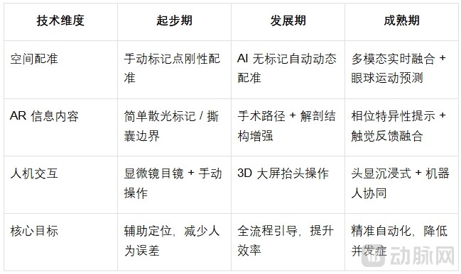
Core Technology Methodology Iteration Path:
V. Future Trends
AI Deep Empowerment:Real-time identification of surgical phases based on intraoperative video, dynamic adjustment of AR guidance strategies, and achievement of “adaptive guidance.”
Multimodal Fusion Upgrade:By integrating intraoperative OCT, fluorescence imaging, and other modalities, the AR layer presents richer physiological and pathological information to assist in decision-making for complex cases.
Lightweight and Universal:Miniaturization, low latency, and cost reduction of head-mounted displays are driving the widespread adoption of AR-guided procedures from tertiary hospitals to primary care institutions.