West China Hospital, Sichuan University Licenses Pressure Injury Detection Patent for RMB 720,000
Recently, West China Hospital of Sichuan University publicly announced a technological achievement transformation, planning to“A Pressure Injury Detection Method Based on Fractional-Order Composite Multiscale Sample Entropy”the patent was assigned to Sichuan Bojitai Medical Device Co., Ltd. via an exclusive license, with a transaction amount ofRMB 720,000. The inventor of this patent isJiang Yan and Her Team。
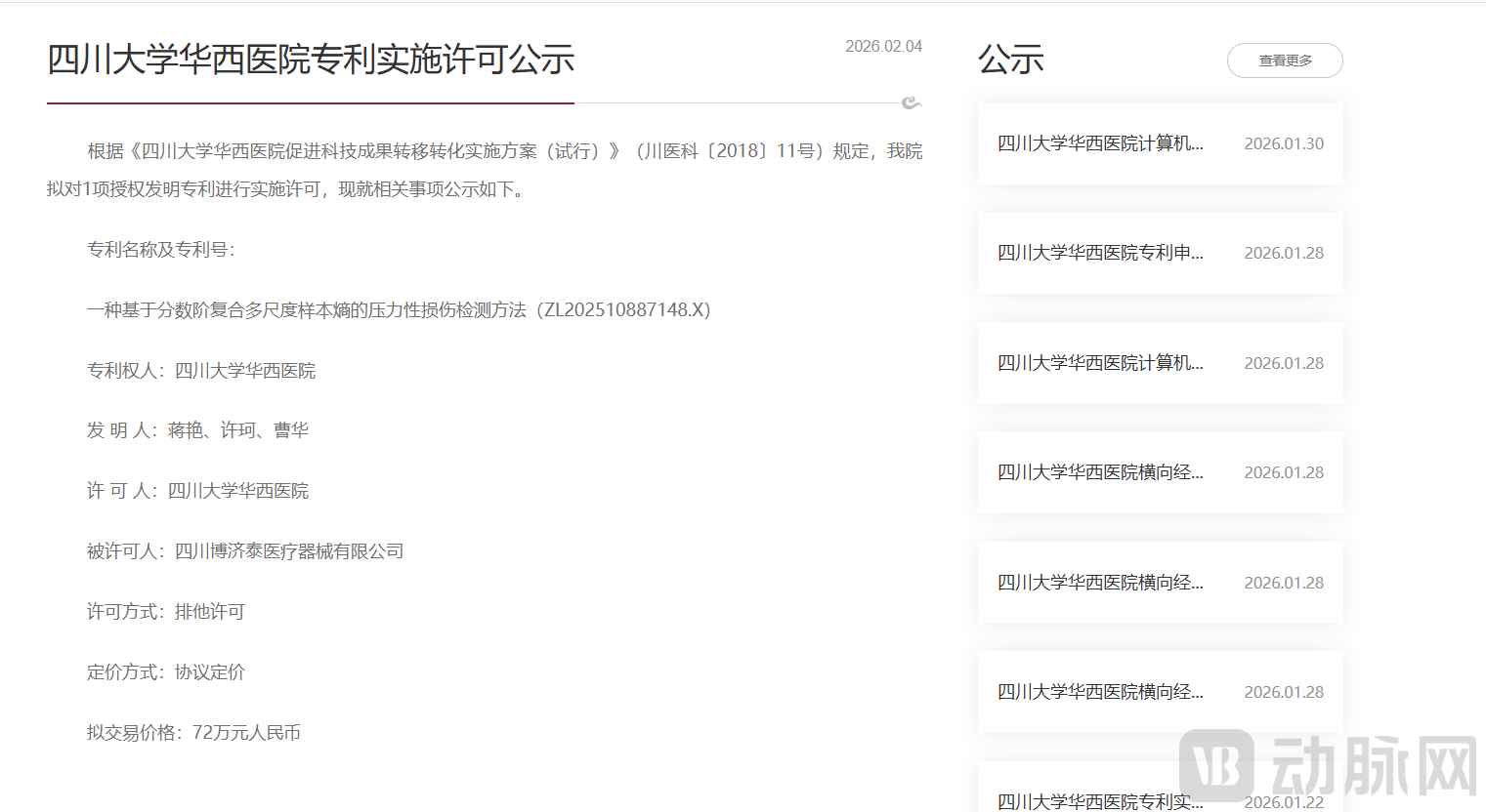
Image source: West China Hospital, Sichuan University Information Disclosure
This patent, throughAdaptive optical flow tracking, Eulerian video magnification, and other technologies, enabling precise staging and early identification of pressure injuries, overcoming the limitations of traditional static detection methods that are prone to misjudgment and have poor adaptability to individual differences, thus providing an intelligent solution for pressure injury screening in long-term bedridden patients.
Pressure injuries, also known as pressure ulcers, are a common complication in clinical nursing. Their pathogenesis is well-defined: prolonged pressure, shear forces, or friction on local tissues can impede blood circulation, leading to ischemia, hypoxia, and malnutrition of the skin and subcutaneous tissues. This ultimately results in progressive damage ranging from epidermal erythema to deep tissue necrosis. The condition is particularly prevalent among patients who are bedridden for extended periods, wheelchair users, or individuals with limited limb mobility due to disease. Bony prominences such as the sacrococcygeal region, heels, hips, and shoulders are typical sites prone to injury due to concentrated pressure. Clinical practice has shown thatEarly Precise Identification and Scientific Staging of Pressure Injuries, is the key to halting the progression of injury, reducing the complexity of treatment, and alleviating patient suffering; however, this very link faces intractable practical dilemmas.
The core pain points of clinical testing are concentrated in two aspects:1. Difficulty in Early Injury Identification, The typical presentation of a Stage I pressure injury is localized erythema; however, its intensity fluctuates cyclically with changes in subcutaneous microcirculation. This dynamic characteristic is easily overlooked during static observation, leading to misdiagnosis as transient hyperemia, which delays intervention and allows minor injuries to progress into deep ulcers;Second, staging assessment relies on subjective experience, variations in assessment criteria for injury severity among different healthcare professionals, coupled with a lack of objective quantitative indicators, can lead to staging discrepancies, directly compromising the precision and efficacy of treatment plans.
The technical limitations of existing detection methods further exacerbate this dilemma. Currently, mainstream clinical detection approaches are all based onStatic Image Recognitionas the core, making it difficult to meet the requirements for precise detection:Although the erythema threshold segmentation technique based on the HSV color model can locate suspected lesion areas, but it can only capture instantaneous static features, failing to reflect the dynamic evolution of injury and exhibiting insufficient sensitivity to early dynamic changes;Method for Quantifying Skin Texture Using Gray-Level Co-occurrence Matrix (GLCM), focusing on the analysis of surface roughness and uniformity of the skin, yet it struggles to distinguish the subtle differences between early-stage lesions and normal tissue, leading to a high risk of false-positive results;Near-Infrared Spectroscopy, Thermal Imaging TechnologyLocal ischemia is assessed based on temperature differences; however, detection stability is suboptimal due to significant influences from factors such as ambient lighting and variations in patient skin tone.
Even traditional image entropy methods incorporating temporal analysis exhibit significant limitations: they are highly sensitive to lighting conditions, leading to a marked decrease in detection specificity among patient populations with substantial skin tone variations. Multiscale entropy analysis, adopted in some studies, relies on fixed parameters and mean-based coarse-graining strategies. Consequently, it fails to effectively preserve high-frequency details of signals, such as transient fluctuations in erythematous areas, and exhibits insensitivity to subtle dynamic changes. Furthermore, its inability to adaptively adjust parameters to match inter-patient differences in skin characteristics ultimately restricts the accuracy of pressure injury staging, falling far short of clinical requirements for precise detection and real-time monitoring.
This patented technology is based onDynamic Signal Analysisas the core, integratingMedical Image Processing and Artificial Intelligence Algorithms, inDetection Logic, Feature Extraction, Model BuildingBreakthrough Innovations Across Three Key Dimensions, Completely Shattering the Technical Constraints of Traditional Static Testing.
At the detection logic level, a dual preprocessing mechanism of “video dynamic tracking + signal amplification” is innovatively adopted.Unlike traditional single-frame image analysis, this technique employs an adaptive optical flow field to dynamically track suspected injury regions: it calculates luminance variance based on illumination changes across video frames, determines the mean image gradient in conjunction with motion complexity, and dynamically adjusts adaptive weight coefficients to construct an objective function for optimizing luminance consistency error. This approach enables precise locking onto the movement trajectory of injured areas even under complex conditions such as illumination fluctuations and noise interference, ensuring that detection targets are not lost. Meanwhile, by processing the tracked videos using Eulerian Video Magnification, subtle skin micro-vibration signals can be effectively enhanced, capturing minute luminance changes imperceptible to the naked eye, thereby providing data support for early injury identification.
During the model construction process, we chooseRadial Basis Function (RBF) as the Kernel Function for Support Vector Machines (SVM), by optimizing the objective function for the hyperplane's normal vector and bias term, it maximizes the classification margin between different injury categories, thereby significantly enhancing the model's generalization capability. This kernel function can effectively handle high-dimensional nonlinear data, accommodating the complex classification requirements of pressure injuries across multiple stages, and ensuring precise differentiation between healthy skin and Stage I to IV injuries.
The multidimensional innovations of this technology can be directly translated into three core advantages, precisely aligning with the practical needs of clinical testing scenarios.
First, high accuracy in early identification.By dynamically tracking brightness changes induced by fluctuations in cutaneous microcirculation, it is possible to accurately differentiate early erythema associated with Stage I injury from transient hyperemia. This approach addresses the limitations of traditional static detection methods, which are prone to misdiagnosis and delayed intervention, thereby providing a scientific basis for timely management of mild injuries.
Second, it demonstrates strong detection robustness.By leveraging the dynamic parameter adjustment mechanism of adaptive optical flow fields and the noise suppression capability of fractional-order differentiation, this technology maintains stable performance across detection scenarios involving varying lighting conditions and diverse patient skin tones, significantly mitigating the impact of environmental factors and individual differences on detection results.
Third, a high degree of automation.From video preprocessing and feature extraction to staging assessment, the entire process requires no manual intervention. This not only minimizes diagnostic bias arising from clinicians’ subjective experience but also significantly reduces detection time, making it suitable for batch screening and routine monitoring of long-term bedridden patients, thereby effectively alleviating the workload of healthcare professionals.
In addition to West China Hospital’s patented technology for pressure injury detection based on fractional-order composite multiscale sample entropy, several mature products have already been implemented in clinical practice within the field of pressure injury detection. These competing products cover diverse technical approaches, including static image recognition, texture analysis, and thermal imaging, thereby providing a variety of solutions for clinical pressure injury detection.
ScarletredR&DScarletred® Vision Skin Imaging and AI Analysis SystemIts core functionality involves acquiring skin images through standardized mobile 2D and 3D imaging technologies, analyzing the hue, saturation, and brightness characteristics of skin erythema based on the HSV color space, segmenting erythematous regions using morphological processing algorithms, and automatically quantifying geometric parameters, color distribution, and texture features of the lesion areas to generate regulatory-compliant analysis reports. Its core value lies in achieving standardization and color calibration for skin imaging, effectively mitigating interference from factors such as lighting and viewing angles, thereby providing objective and traceable data support for the early identification of erythema in pressure injuries, suitable for both routine clinical monitoring and clinical trial data collection scenarios.
ArjoHuntleighLaunchedPressure Injury Assessment Tool(Pressure Injury Assessment Tool) Based on Gray-Level Co-occurrence Matrix (GLCM) technology, this tool quantifies core indicators such as skin texture roughness and uniformity to construct a texture feature database, thereby distinguishing textural differences between normal skin tissue and injured areas. The core value of this product lies in providing objective, quantitative texture data that serves as a standardized reference for assessing injury severity, making it suitable for monitoring and recording textural changes in moderate to severe injuries.
FLIR Systems, Inc.R&DFLIR ONE Pro Medical Edition Medical-Grade Thermal Imaging Device, captures thermal distribution images of the skin surface via a thermal imaging sensor, analyzes the temperature gradient differences between local tissues and surrounding normal skin, thereby indirectly reflecting local blood circulation status, and generates thermal distribution heatmaps and temperature differential data reports. The core value of this device lies in enabling non-contact detection to avoid secondary injury to fragile skin; meanwhile, it assists in assessing local ischemia through temperature signals, providing data support from a thermal perspective for early warning of injury risks.
These competing products, each leveraging their respective technological advantages, have formed a complementary landscape in clinical settings, meeting the testing needs of various scenarios. Whether for rapid screening, precise assessment, or risk early warning, they provide critical technical support for the clinical management of pressure injuries, driving the development of injury detection toward standardization and objectivity.