Shandong University Qilu Hospital and Jinan University License MR-to-CT Image Synthesis Patent for RMB 300,000
Recently, the invention patent jointly developed by Qilu Hospital of Shandong University and University of Jinan“Method and System for Generating CT Images from MR Images under Multi-Condition Constraints”Completion of the transformation of scientific and technological achievements, with the valuation of such achievements determined through negotiated pricing.300,000 yuanLicensed to Yibang (Beijing) Intelligent Technology Co., Ltd. for use; the individuals who contributed to the achievement includeZhang Menghua et al.Multiple R&D personnel.
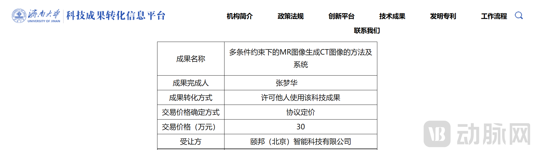
Image from the official website of University of Jinan
The core technologies involved in this transaction belong toMedical Image ProcessingIn this field, addressing the industry pain points of traditional MR-to-CT image synthesis techniques—namely, their excessive reliance on the quality and quantity of training samples and suboptimal synthesis results—we have innovatively designed a generator composed of a feature encoder, a cascaded cross-domain Transformer module, and a feature decoder. This design achieves, for the first time, feature selection and heterogeneous feature fusion of 3D and 2D features. Furthermore, by incorporating a degradation model, a gated fusion mechanism, and a self-attention mechanism to optimize model performance, the consistency between the synthesized CT images and the ground truth reaches85-90%, which can serve as a reference for clinical diagnosis. This approach overcomes the sample-dependency limitations of traditional methods at the technical level, significantly enhancing the quality and practicality of medical image synthesis.
CT, or computed tomography, as a core medical imaging diagnostic technology, relies onX-ray Beam TomographyBy integrating computer processing, it clearly visualizes internal human anatomical structures. Owing to its rapid imaging capabilities and rich detail, it has become a critical basis for clinical diagnosis, disease assessment, and formulation of treatment plans for various conditions, including tumors, cardiovascular and cerebrovascular diseases, and organic lesions. It is widely used and indispensable in the field of clinical medical diagnosis.
In current clinical diagnosis and treatment, it is often necessary to combineMR and CT ImagingConduct comprehensive diagnosis; in the absence of direct CT imaging, existing solutions primarily rely onMR-to-CT Image Synthesis Using Residual Transformer-Based Generative Adversarial Networks for Image Translation, this technique attempts to compensate for the limitations of traditional generative adversarial networks in capturing global information by incorporating deep feature extraction and feature enhancement modules within the generator, thereby achieving MR-to-CT image synthesis.
However, this existing technology has obvious technical drawbacks. The quality of its synthetic CT images depends entirely on the quality and quantity of training samples. In clinical practice, it is difficult to collect samples for rare cases and special conditions, which can easily lead toInsufficient sample size and variable sample quality... issues, which directly limit the accuracy of model training. The synthesized CT images are prone to structural deviations, blurred textures, and missing details, making it difficult to meet the precision requirements for clinical diagnostic imaging.
There is an urgent clinical demand for high-quality MR-to-CT image synthesis technology. On one hand, in clinical diagnosis, there is a need to rapidly and accurately synthesize CT images that meet diagnostic standards from MR images when direct CT scans are unavailable, thereby providing comprehensive imaging evidence for disease diagnosis. On the other hand, for conditions with limited sample availability, synthesis technologies must reduce excessive reliance on training datasets to ensure the stability and accuracy of the synthesized CT images. In this context, developing MR-to-CT techniques that overcome the bottleneck of sample dependency and generate high-quality CT images has become a key direction for meeting actual clinical diagnostic needs and improving the efficiency and precision of medical image diagnosis.
As an innovative achievement in the field of medical image processing, this patented technology has achieved multiple technical breakthroughs addressing the core pain points of traditional MR-to-CT conversion techniques. It combines significant technological innovation with clinical application advantages, with its core innovations and technical benefits reflected in four aspects:
1. Breakthrough in Core Technological Bottlenecks of Sample Dependence, Technological Innovation Introduces Reference to Similar Cases3D CT ImagesAs a constraint, it changes the traditional technology's complete reliance on the quality and quantity of training samples. Even in scenarios with insufficient samples for rare cases or special conditions, it can still ensure the quality of CT image synthesis, significantly improving the clinical adaptability and practicality of the technology.
Second, a pioneering cascaded cross-domain Transformer module is introduced to achieve efficient fusion of heterogeneous features., designing a Transformer attention mechanism with cascaded depth and height-width dimensions, achieving for the first time precise feature selection and heterogeneous feature fusion of 3D CT features with 2D MRI features. Through the synergistic interaction of multi-layer positional encoding, multi-head attention mechanisms, and multi-layer perceptrons, redundant features are effectively removed, thereby enhancing the comprehensiveness and accuracy of feature extraction.
3. Optimizing the Encoder-Decoder Architecture to Achieve Upgraded Feature Processing, the dual-branch feature encoder separately samples and encodes MR and reference CT images, with the last two sampling modules innovatively incorporating a degradation model that includes independent blurring and noising processes, thereby aligning feature extraction more closely with the actual characteristics of clinical imaging. The feature decoder combines gated fusion upsampling layers in the first two stages with self-attention upsampling layers in the latter two stages. The gated fusion mechanism enables intelligent integration of features across downsampling and upsampling paths, while the self-attention mechanism enhances the restoration of image details. This layer-by-layer optimization significantly improves the generator’s feature processing performance.
Fourth, a multi-dimensional loss function ensures high fidelity of the synthesized images., design a total loss function that combines perceptual loss, contextual loss, and structural loss to constrain the synthesized images from multiple dimensions, including high-level feature matching, local texture consistency, and image structural similarity, ultimately achieving consistency between the synthesized CT images and the ground truth.85-90%, which can meet the reference standards for clinical diagnosis and provide reliable technical support for clinical imaging applications.
In addition, this technology is also supported byEstablished the corresponding system architecture, includingImage Acquisition UnitandCT Image Generation Unit, and can be implemented on computer-readable storage media and various electronic devices. Its strong practical applicability and broad scenario adaptability have laid the foundation for large-scale clinical application.
AI-based synthetic medical imaging products that generate CT images from MR scans are poised for significant growth, driven by the dual advantages of digital transformation in healthcare and iterative advancements in AI technology. On the demand side, rising global needs for disease diagnosis, coupled with insufficient imaging resources at primary care levels and a scarcity of rare case samples, have created rigid application scenarios in fields such as tumor screening and the diagnosis of cardiovascular and cerebrovascular diseases. On the technology side, optimizations in AI algorithms are enhancing synthesis accuracy, meeting clinical reference standards, and accelerating commercial deployment.
United Imaging Healthcare “uAI Image Synthesis Module”As an AI software suite designed to complement United Imaging’s CT and MR systems, it is widely deployed in the radiology departments of tertiary hospitals across China. The solution addresses multi-site imaging conversion needs—including head, chest, and abdomen—and plays a supportive role in clinical scenarios such as preoperative tumor assessment and screening for cardiovascular and cerebrovascular diseases. Having received NMPA certification, it is compatible with United Imaging’s full range of proprietary imaging equipment and integrates seamlessly with hospital PACS systems; it has already been implemented in numerous hospitals nationwide. This product provides clinicians with rapid multimodal image fusion capabilities, reducing the need for repeat examinations and enhancing diagnostic efficiency in radiology departments. Particularly in emergency settings, it quickly delivers CT-level imaging references to support urgent diagnostic and therapeutic decision-making.
Infervision’s “Chest MR-CT Synthesis System” Focuses on Specialized Diagnosis of Thoracic Diseases, primarily used in scenarios such as lung cancer screening, tuberculosis diagnosis, and assessment of pulmonary infections, it addresses the need for CT image completion following routine chest MRI examinations. The product has been deployed in the respiratory and radiology departments of numerous hospitals across China, supports integration with mainstream PACS systems from major vendors, and does not require binding to specific hardware devices. It optimizes models specifically for the textural features and structural characteristics of chest images, providing precise support in pulmonary nodule detection and lesion delineation, thereby helping primary-care hospitals improve the accuracy of chest disease diagnosis.
Currently, China’s medical AI regulatory framework is gradually improving, and policies encouraging technological innovation are lowering barriers to market access and technology translation. Although both domestic and international brands have established a presence in the current market, existing products suffer from limitations such as reliance on large datasets and narrow application scenarios. Innovative technologies featuring cross-scenario adaptability, low data dependency, and high compatibility are poised to disrupt the competitive landscape. In the future, driven by the deepening adoption of precision medicine, growing demand for multi-modal image fusion, and increased penetration into primary healthcare markets, such products will evolve toward higher accuracy, broader adaptability, and lighter deployment. They will not only provide efficient and cost-effective imaging solutions for medical institutions at all levels but also play a significant role in emerging scenarios such as telemedicine and emergency diagnosis and treatment. The market size is expected to expand continuously, making this segment a core growth track in the field of medical imaging AI.