Intalight's DREAM OCT Powers Over 210 SCI Publications: Redefining Global Ophthalmic Research with Swept-Source Ultra-Widefield Imaging
Optical Coherence Tomography (OCT)Since its initial proposal in 1991, the technology has undergone more than three decades of evolution, progressing from time-domain to frequency-domain, and then to swept-source. Each leap has redefined the boundaries of what ophthalmologists can “see.” Now, as ultra-widefield swept-source OCT begins to illuminate the previously unseen peripheral retina and deep choroidal structures, a question arises:In this wave of technological transformation, who is truly driving the frontier?
This article attempts to address this question through a specific case study—Intalight SaiweiA self-developed swept-source ultra-widefield OCT/OCTA device—Ruyi Full-Eye OCT (DREAM OCT)Over the past few years, it has empowered academic communities both domestically and internationally to publish more than 210 papers in international journals. With nearly 1,000 units installed worldwide, its reach extends from top-tier ophthalmic centers in China, such as the Zhongshan Ophthalmic Center of Sun Yat-sen University, Beijing Tongren Hospital, and the Eye & ENT Hospital of Fudan University, to renowned global institutions including the Bascom Palmer Eye Institute in the United States and Moorfields Eye Hospital in the United Kingdom.
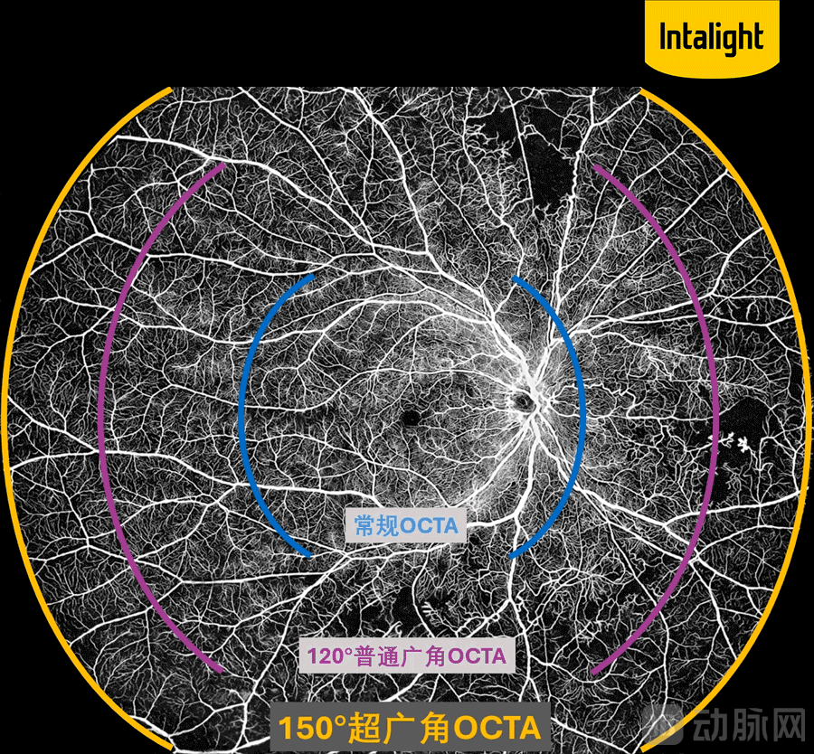
Over the past two decades, OCT technology has reshaped ophthalmic diagnosis as “optical biopsy.” However, traditional spectral-domain OCT (SD-OCT) has long been limited by“Insufficiently broad, deep, and clear vision”The advent of next-generation swept-source optical coherence tomography (SS-OCT) is systematically overcoming these bottlenecks. SS-OCT employs a long-wavelength swept laser at approximately 1050 nm, offering superior tissue penetration and faster scanning speeds compared to the conventional 840 nm wavelength. Furthermore, it incorporates non-contact, non-invasive optical coherence tomography angiography (OCTA), enabling physicians to visualize microvascular networks without the need for contrast agent injection. When SS-OCT is further integrated with ultra-widefield optical systems to form ultra-widefield SS-OCTA, the single-acquisition imaging field of view can be expanded from the traditional 30°–60° to 150° or even wider, as seen in the Ruyi Full-Eye OCT system. Additionally, the imaging depth has been extended from less than 3 mm to over 10 mm, progressively illuminating the previously existing "dark zones" and "blind spots."
This technological trend is profoundly reshaping the research paradigm in ophthalmology. From quantitative assessment of peripheral ischemic areas in diabetic retinopathy screening, to in vivo observation of the choroid and sclera for myopia control, and further to the discovery of early biomarkers for neurodegenerative diseases in the fundus, an increasing number of research directions previously hindered by equipment limitations are becoming feasible.
It is against this backdrop that a set of data from China has drawn attention.
As of early 2026, the Ruyi Whole-Eye OCT, independently developed by Intalight Saiwei, has cumulatively empowered global researchers to publishMore than 210 articlesInternational journal publications and nearly 1,000 installations worldwide. These figures alone may only tell part of the story. What deserves greater attention is: in which journals were these papers published, by which institutions, and what problems did they address?
1、Breadth of Institutional AdoptionIn China, all ophthalmic hospitals listed or nominated in the Fudan University-affiliated "Ranking of Specialty Reputation of Chinese Hospitals" have produced academic achievements based on the Ruyi Full-Eye OCT. This list includes the Zhongshan Ophthalmic Center of Sun Yat-sen University, Beijing Tongren Hospital, the Eye and ENT Hospital of Fudan University, the Second Affiliated Hospital of Zhejiang University School of Medicine, the Eye Hospital of Wenzhou Medical University, Shanghai Ninth People’s Hospital affiliated with Shanghai Jiao Tong University School of Medicine, Peking Union Medical College Hospital, and West China Hospital of Sichuan University, thereby encompassing nearly the entire core strength of ophthalmic academic research in China.
Internationally, multiple top-ranked global ophthalmology centers have adopted this device, with more than one institution making repeat purchases. These include the Bascom Palmer Eye Institute, which has ranked first in U.S. News ophthalmology rankings for 20 consecutive years; Moorfields Eye Hospital in the UK, the leading ophthalmic hospital in Europe with over 220 years of history; the Massachusetts Eye and Ear Infirmary affiliated with Harvard University; the Department of Ophthalmology at Stanford University School of Medicine; the Casey Eye Institute at Oregon Health & Science University; the Singapore National Eye Centre; Tokyo Medical and Dental University in Japan; and Kyung Hee University Medical Center in South Korea. In early 2026, the Memorial Sloan Kettering Cancer Center (MSKCC), renowned for its cancer research, also completed installation, utilizing the device for interdisciplinary research on tumor-related ocular lesions. This signifies that the application scenarios of the Ruyi Full-Eye OCT have extended beyond the traditional scope of ophthalmology.
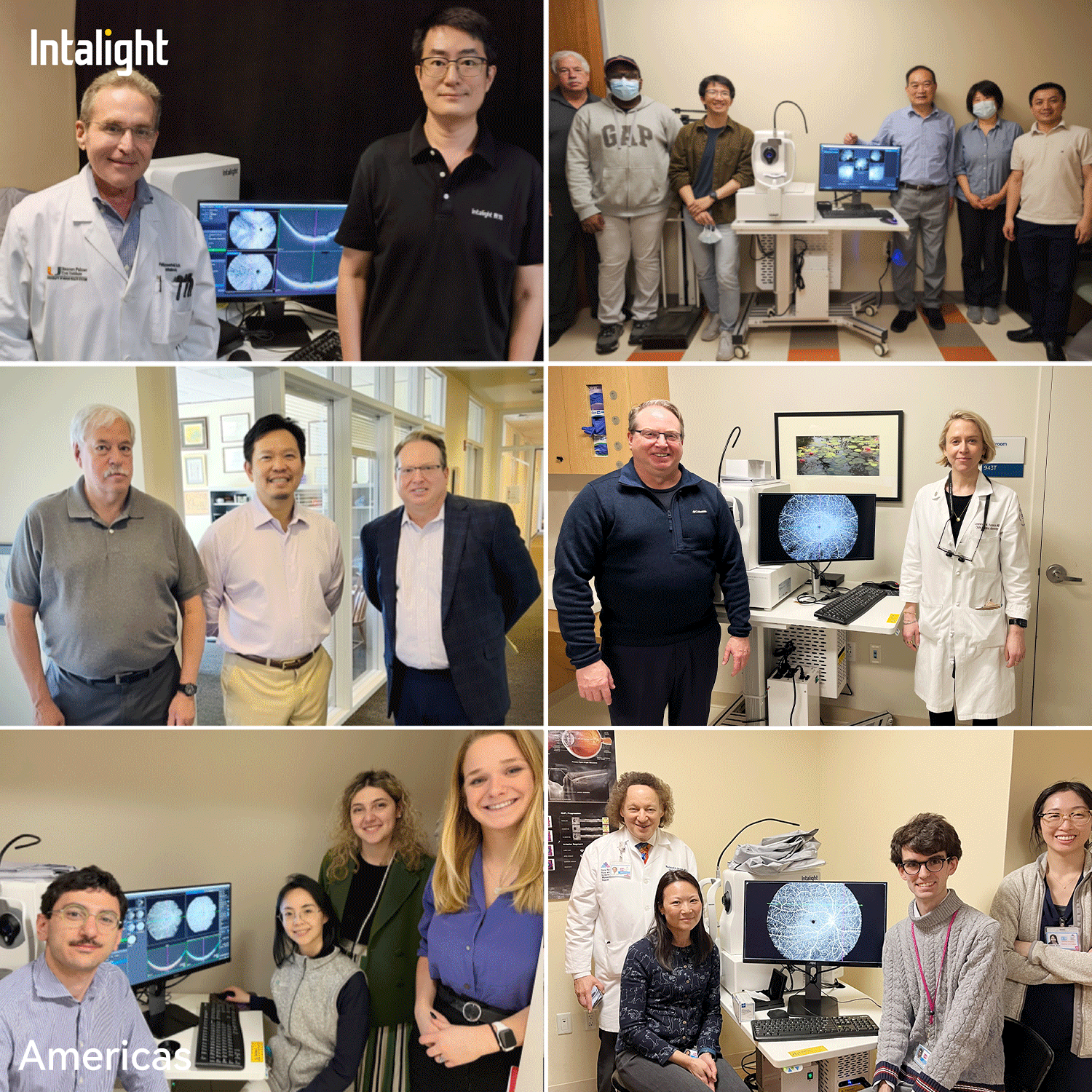
Group Photo of Installations at Some of the World’s Leading Eye Centers
2、Quality and Distribution of PapersAmong the more than 210 publications, approximately 85% of the studies relied directly on the device’s ultra-widefield imaging or deep choroidal imaging capabilities to achieve their core findings. In other words, these studies did not merely “incidentally use” the device; rather, the research was made possible precisely because it provided previously unattainable imaging perspectives. Of these, more than 20 articles were published in high-impact-factor journals, including Signal Transduction and Targeted Therapy (IF 40.8), Ophthalmology (IF 13.1), Alzheimer's & Dementia (IF 11.1), PNAS (IF 9.1), and JAMA Ophthalmology (IF 8.1).
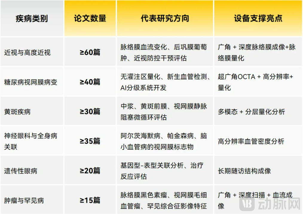
Figure: Ruyi Full-Eye OCT Empowers Renowned Experts at Top Global Hospitals to Publish 210+ SCI Papers
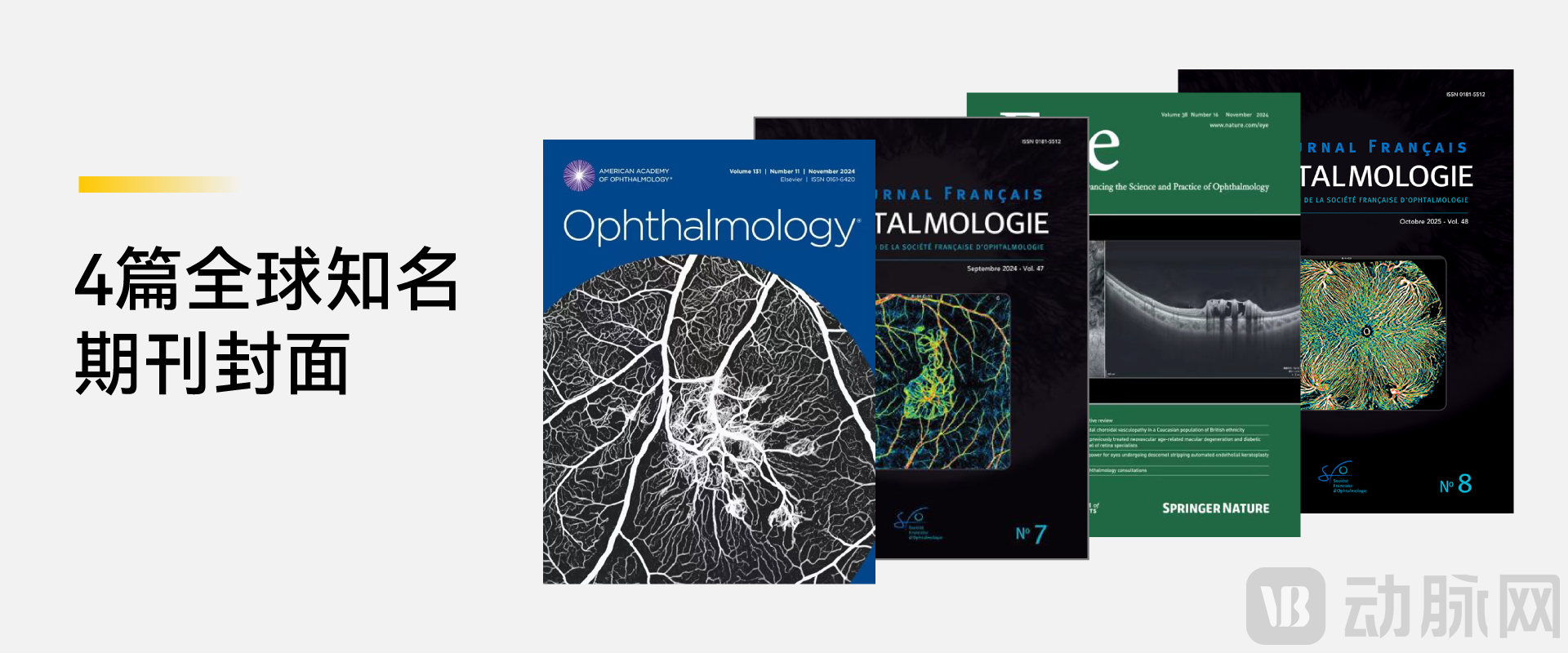
Furthermore, the key imaging provided by Ruyi Full-Eye OCTSelected four times as featured in *Ophthalmology*, *Eye*, and the *Journal Français d'Ophtalmologie*, among others.Cover images of international journals: In the field of ophthalmology, where image quality is a core competitive advantage, the selection of journal covers itself serves as an evaluation of imaging capabilities.
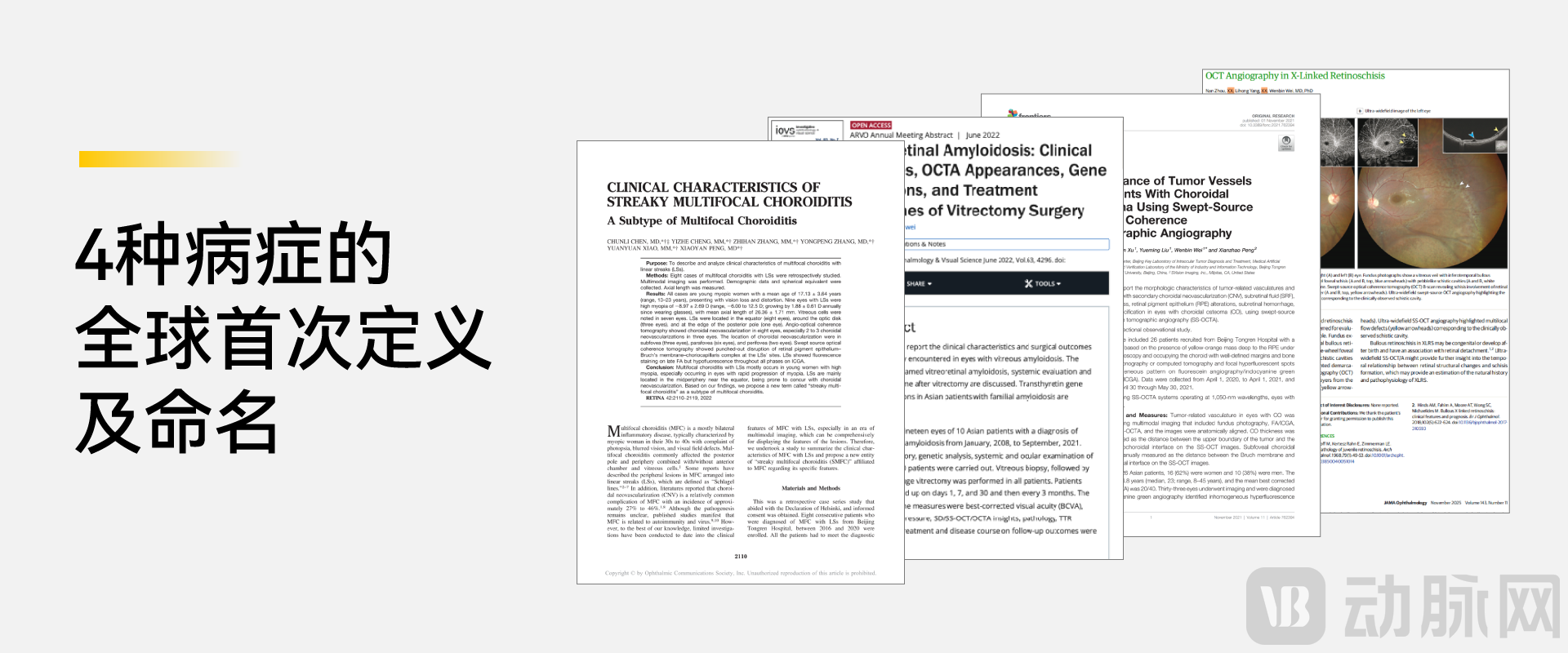
3、Pioneering Academic Contributions. Among more than 210 papers, one category of achievements stands out particularly. Leveraging the ultimate imaging capabilities of the Ruyi Full-Eye OCT,Chinese Scholars Complete the World’s First Definition and Naming of Imaging for Four Ophthalmic Diseases:The sea-fan vascular network in choroidal osteoma, a new subtype of striated multifocal choroiditis, the systemic imaging characteristics of vitreoretinal amyloidosis, and the cobblestone-like retinoschisis sign in X-linked retinoschisis. In the medical field, the ability to define and name a new sign signifies securing academic priority in that specific subfield. The common features of these achievements are:Their detection is highly dependent on previously unobservable imaging details revealed by ultra-widefield or deep choroidal imaging.
Why is this device capable of supporting such a high volume of academic output?The answer lies at the technical level. In contrast to the industry bottlenecks mentioned above, Ruyi Full-Eye OCT has achieved targeted breakthroughs in three key dimensions:
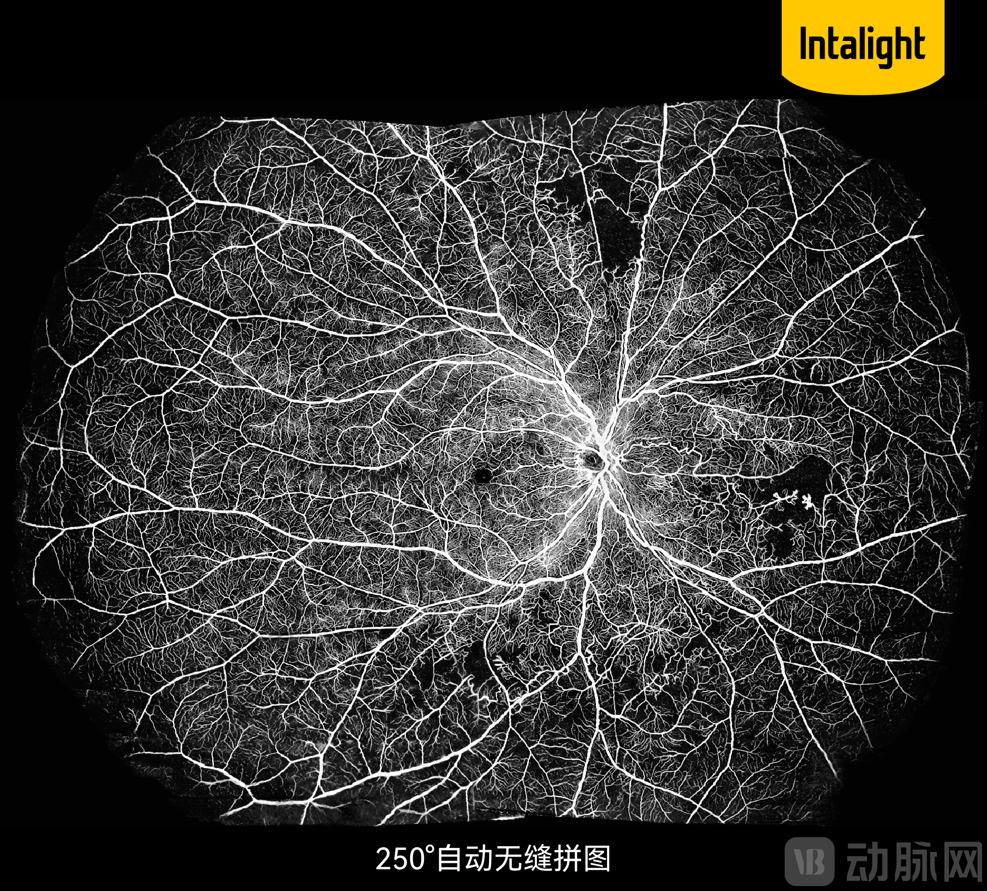
1、In terms of imaging breadthWith a single-scan range of 150° and automated montage covering up to 250°, this capability far exceeds the standard 30°–60° field of view of conventional SD-OCT, significantly improving the detection rate of peripheral retinal lesions and enabling researchers to perform near-panoramic in vivo observation of the retina for the first time.
2、In terms of imaging depth
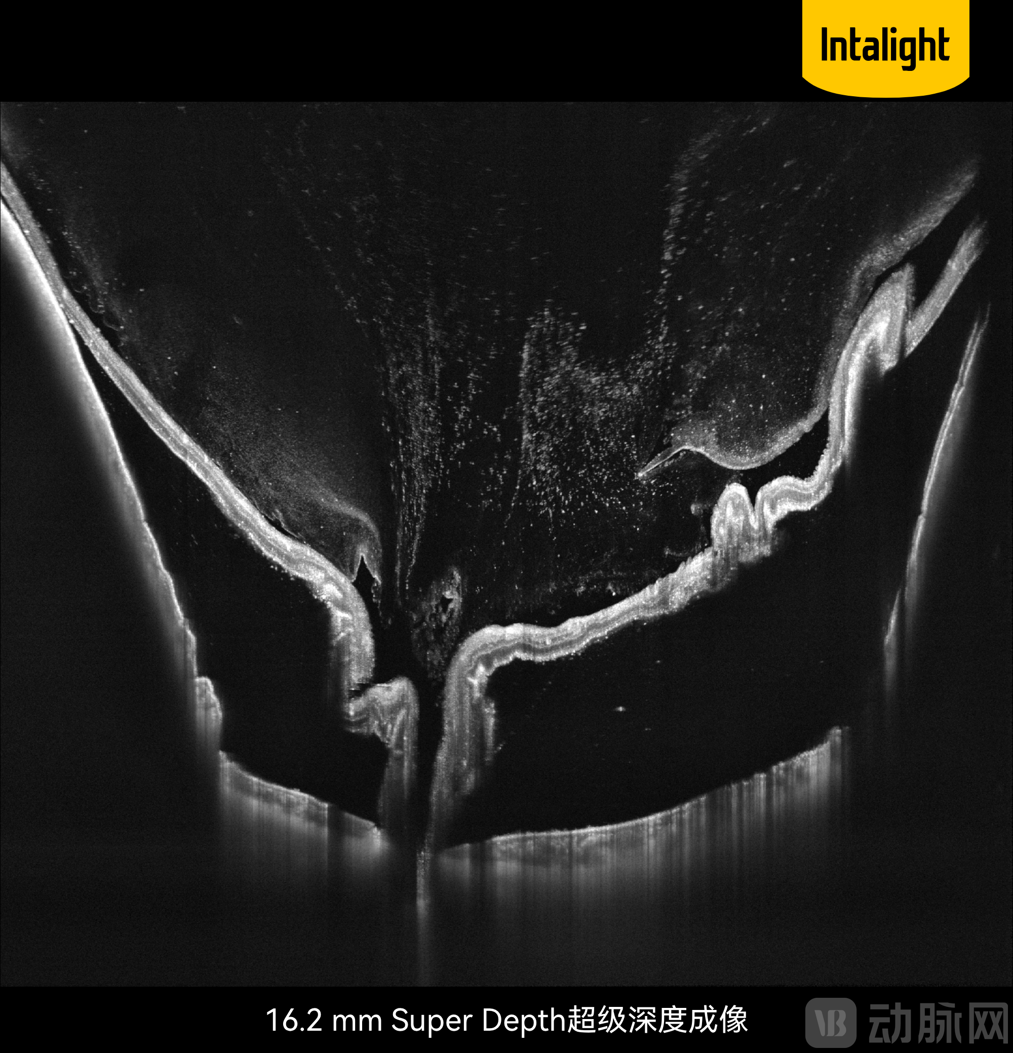
3、In terms of scanning speed, reaching 400,000 scans per second (400 kHz), which is 4 to 8 times faster than conventional SD-OCT. High-speed scanning not only reduces artifacts caused by eye movements but also enables large-area, high-resolution imaging within a clinically acceptable examination time.
4、Ruyi Multi-Lens Solution:Pioneering a multi-lens combination design, the system employs dedicated lenses for four distinct scenarios: fine imaging of the macula and optic disc, ultra-widefield fundus imaging, panoramic anterior segment imaging, and animal imaging. This approach avoids the performance compromises inherent in “one-lens-fits-all” solutions, ensuring greater satisfaction with every examination. Notably, ultra-widefield OCTA imaging can largely replace invasive fluorescein angiography (FFA), offering significant practical value in the management of chronic diseases requiring frequent follow-up.
These technical features collectively constitute research conditions that were previously unavailable, giving rise to academic breakthroughs in multiple directions: inMyopia Prevention and ControlIn this field, researchers have for the first time elucidated the negative correlation mechanism between scleral thickness and axial length at the in vivo level, providing a quantifiable imaging-based evaluation basis for interventions such as red-light therapy and orthokeratology lenses; inEarly Screening for Diabetic RetinopathyIn the field, ultra-widefield OCTA has significantly improved the detection sensitivity for peripheral and occult lesions, and its concordance with the gold standard FFA has been validated in multiple studies; inNeuro-ophthalmologyIn this direction, researchers are exploring the use of fundus indicators as early warning biomarkers for Alzheimer’s disease and cerebral small vessel disease, with related findings published in high-impact journals such as Alzheimer's & Dementia;Translational MedicineIn this field, Ruyi Full-Eye OCT has achieved in vivo dynamic monitoring of pathological processes in animal models for the first time, providing an indispensable imaging tool for preclinical research such as gene therapy and new drug evaluation.
The breakthroughs achieved by the Ruyi Full-Eye OCT across multiple core dimensions are attributable to the team behind it. Intalight established its R&D center in Silicon Valley in 2014 and was founded in Luoyang in 2015. Currently, its management headquarters is located in Shanghai, with R&D centers in Silicon Valley, Shanghai, and Luoyang. The core founding team includes Dr. Peng Xianzhao, Dr. Li Bing, Dr. Wang Zhengyu, and Dr. Wei Xing, all of whom are returnees from Silicon Valley with over 20 years of experience in semiconductor optics and precision equipment R&D. This cross-disciplinary background, which applies semiconductor-grade precision optical manufacturing to medical devices, is rare in China and explains how the Ruyi Full-Eye OCT has achieved differentiated advantages in optical system design.
Saiwei’s devices have also garnered attention from internationally renowned authorities in ophthalmology. Professor David Huang, an academician of the U.S. National Academy of Engineering and the inventor of OCT technology; Professor Philip Rosenfeld; Professor SriniVas Sadda, current President of the Association for Research in Vision and Ophthalmology (ARVO); and Professor Richard Spaide, among other globally distinguished retinal specialists, have all endorsed the Ruyi Whole-Eye OCT. In 2025, Saiwei established an Overseas Expert Advisory Board, chaired by Professor Philip Rosenfeld of the Bascom Palmer Eye Institute, bringing together eight international authorities from the United States, the United Kingdom, Japan, and Brazil to advance technological breakthroughs and clinical collaboration in ophthalmology worldwide.

Figure: Intalight Saiwei Overseas Expert Advisory Board
In terms of industry accolades, Saiwei’s swept-source OCT device was selected as one of the “Top Ten Advances in Chinese Ophthalmology” in 2021, following evaluation by a panel of experts including Professor Wang Ningli, an academician of the Chinese Academy of Engineering.“Internationally Leading”Scientific and Technological Achievement Evaluation, and Rated asNational-Level Specialized and Sophisticated “Little Giant” EnterprisesFurthermore, Saiwei was named the Global Best Fundus Imaging Equipment Brand of the Year by MedTech Outlook, a global medical technology magazine. In addition, Dr. Peng Xianzhao, the founder, collaborated with Professor Chen Youxin’s team at Peking Union Medical College Hospital to publish China’s first atlas of ocular diseases based on domestically produced OCT, titled Atlas of Swept-Source Optical Coherence Tomography and Angiography.
Ruyi Full-Eye OCT is currently Saiwei’s most representative product, though not its only one. Built upon its “AI + Optics” technology platform, Saiwei has developed the “Ruyi Series” product line, which covers multiple ophthalmic diagnostic scenarios. The Ruyi True-Color Camera employs an 8-light-source continuous-spectrum true-color imaging technology, enabling ultra-wide-angle imaging of 186° in a single capture with an optical resolution of 6.8 micrometers. The Ruyi Whole-Cornea Biometer combines swept-source technology with a proprietary telecentric optical system to perform one-stop precise measurements of parameters such as axial length and corneal curvature, as well as intraocular lens (IOL) power calculation.
Beyond diagnostic products, Saiwei is expanding into the therapeutic equipment sector, which presents higher technical barriers. Its high-end surgical microscopes and customized SMILE (Small Incision Lenticule Extraction) laser refractive surgery systems have entered the R&D pipeline. Meanwhile, the company is building an AI-enabled ophthalmic data platform called the “Ruyi Center,” aiming to break down data silos among high-end diagnostic devices and leverage intelligent analytics to assist physicians in making more efficient diagnosis and treatment decisions.
The company has obtained EU CE certification, and its overseas market expansion is underway. Saiwei summarizes its vision as“Meeting the Global Ophthalmology Community at Its Peak”. From a single OCT device to an integrated platform, the company is clearly strategizing for a broader vision.
The value of a medical imaging device is ultimately determined by whether it has transformed the way diseases are observed.
When the peripheral retina is no longer a diagnostic “blind spot,” when the choroidal microvascular network is visualized in its entirety for the first time, and when four disease signs are defined globally for the first time thanks to new imaging perspectives, these changes reflect a paradigm shift in imaging technology from “local sampling” to “whole-eye observation.” The more than 210 papers on Ruyi Whole-Eye OCT, in a sense, document certain facets of this transitional process.
OCT technology continues to evolve rapidly. For the ophthalmic academic community, what truly matters has never been the number of units sold for any given device, but rather whether it opens new observational windows for researchers. Based on existing academic outputs, Ruyi Whole-Eye OCT has indeed played a significant role in opening this window.