Sichuan University Announces Transfer of Fetal Brain MRI AI Patent for 178,000 RMB
Recently, the Industrial Technology Research Institute of Sichuan University released a public notice regarding the proposed transfer of scientific and technological achievements. This patent is an invention patent jointly held by West China Second University Hospital of Sichuan University and Sichuan University.“A Method, Device, Program Product, and Medium for Predicting Lateral Ventriculomegaly”, the proposed transaction price isRMB 178,000。
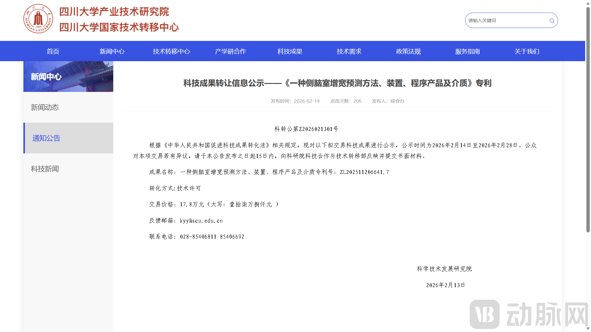
Image from the official website of the Industrial Technology Research Institute of Sichuan University
This patent focuses onIntelligent Analysis of Fetal Brain MRI, based onConstruction of a 3D nnUNet-Based Framework for Lateral Ventricle Segmentation and Adverse Prognosis Prediction, Innovative IntegrationTopological Loss Function, Cross-Modal Feature Alignment, Temporal Dynamic Modeling, and Multi-Model Ensemble Prognostic Risk Prediction Technology, breaking through the limitations of traditional 2D segmentation and single-sequence evaluation, achieving precise 3D segmentation of fetal lateral ventricles, classification of ventriculomegaly severity, and intelligent prediction of neurodevelopmental prognosis risk, thereby providing a high-precision, end-to-end technical solution for AI-assisted prenatal imaging diagnosis.
Fetal Lateral VentriculomegalyIt is an important early imaging marker of abnormal fetal central nervous system development, directly related to the neurodevelopmental outcomes after birth. It is a key indicator that must be closely monitored in prenatal screening and diagnosis. Its early precise identification, dynamic monitoring, and prognosis risk assessment have irreplaceable clinical value in reducing birth defects, guiding clinical interventions, and improving postpartum follow-up.
In current clinical practice,Prenatal Assessment of Fetal Brain StructureUltrasound remains the preferred and routine screening modality. Ultrasound offers the advantages of being non-invasive, real-time, repeatable, and highly safe, meeting the basic needs for structural observation; however, its inherentInsufficient resolution, limited tissue contrast, and poor visualization of deep intracranial structuressuch limitations make it difficult to provide stable and reliable diagnostic information when facing complex, subtle, and occult ventricular developmental abnormalities.Magnetic Resonance ImagingAs an important complementary imaging modality, it offers advantages such as higher spatial resolution, multi-sequence imaging, and superior soft-tissue contrast, enabling clearer visualization of the intricate anatomical structures of the fetal brain and thus serving as a key technique for evaluating complex cases.
However, most existing MRI-based methods and studies for lateral ventricle assessment remain at the stage ofTwo-Dimensional Analysis and Single-Sequence Application Level, there are widespread and significant technical limitations: on one hand, two-dimensional segmentation and measurement cannot fully reconstruct the three-dimensional spatial morphology and overall anatomical structure of the lateral ventricles, leading to deviations in the measurement of volume, width, and morphological parameters, making it difficult to objectively reflect the true extent of the pathology.
On the other hand, a single magnetic resonance imaging (MRI) sequence provides limited tissue information and fails to comprehensively characterize changes in tissue features under pathological conditions. Meanwhile, most existing techniques overlook the temporal dynamic evolution patterns of rapid ventricular development during the second trimester of fetal gestation. Static assessments are difficult to capture progressive abnormal changes, making it ineffective to judge the prognosis trend.
Overall, there is currently a lack of a comprehensive set of tools or frameworks in both clinical practice and scientific research that canAn Integrated Intelligent Prediction Framework Combining Precise 3D Structural Segmentation, Multimodal Image Fusion, and Temporal Developmental Dynamic Modeling, it is difficult to meet the core clinical needs for precise classification of lateral ventriculomegaly, assessment of progression trends, and early warning of adverse outcomes. There is an urgent need for more advanced, stable, and clinically integrated intelligent technologies to provide robust support for precise prenatal diagnosis and individualized treatment decision-making.
Core Innovations and Prominent Advantages of Intelligent Prediction Technology for Lateral Ventriculomegaly
To address the aforementioned clinical pain points and technical bottlenecks, this patent provides“Methods, Apparatuses, Program Products, and Media for Predicting Lateral Ventriculomegaly”Achieved systematic innovative breakthroughs and constructed an end-to-end intelligent solution ranging from image segmentation to prognostic assessment, with core advantages and innovations concentrated in four aspects:
First,Pioneering a fully automated segmentation architecture based on the "3D nnUNet network combined with topological loss function", abandoning traditional 2D segmentation approaches to achieve complete reconstruction and precise extraction of the 3D spatial structure of the lateral ventricles; topological constraints are applied to ensure continuous and smooth segmentation boundaries, significantly improving the accuracy and integrity of anatomical structure restoration, and fundamentally resolving issues of distortion in 2D measurements and incomplete morphological capture.
Second,Innovatively Introducing the “Cross-Modal Feature Alignment Mechanism”, supports feature fusion and spatial alignment of multi-sequence magnetic resonance images such as T1 and T2, breaking the limitations of single-sequence information. It enables optimization of feature consistency across different imaging modalities on the same anatomical structures, significantly enhancing the model's ability to represent and discriminate complex pathological conditions.
Third,"Construction of a 'Gestational Age Temporal Dynamic Modeling System'", targeting the temporal characteristics of rapid fetal brain development during the second trimester, this approach quantifies trends in ventricular morphological evolution through dynamic change maps. By integrating static structural analysis with dynamic developmental trajectories, it effectively captures progressive widening abnormalities, significantly enhancing the sensitivity for detecting early subtle lesions and trend-based anomalies.
Fourth,"Development of a Prognostic Risk Prediction Module Based on Multi-Model Integration", based on precise subtyping of lateral ventriculomegaly, integrates three-dimensional morphological features and temporal developmental indicators to assess the risk of adverse outcomes, forming a closed-loop system that progresses from structural classification to functional risk prediction. This system offers high diagnostic accuracy, clinical interpretability, and workflow compatibility, providing stable and reliable AI-assisted support for personalized prenatal interventions and long-term follow-up.
Overall, this technology is based onAI Imaging Analysiscentered on this, it addresses critical gaps in the precise assessment and prognostic prediction of prenatal lateral ventriculomegaly, combining technological advancement with clinical practicality.
Currently,AI-Assisted Diagnosis of Fetal Cranial Imaginghas become an important development direction in the fields of prenatal diagnosis and maternal and child health, with the overall market demonstrating a positive trend of continuous demand expansion, rapid technological iteration, and increasing penetration into lower-tier markets. From an industry-wide perspective,AI-Assisted Ultrasound Screening, MRI Image Post-Processing, and General Medical Image AnalysisRepresentative products have been progressively implemented across medical institutions at all levels, playing a positive role in enhancing examination standardization, optimizing image quality, and assisting physicians in interpretation. This fully validates the strong clinical demand for and widespread recognition of intelligent assessment tools for fetal cranial anatomy.
As the state places increasing emphasis on the prevention and control of birth defects and precision prenatal diagnosis, compounded by multiple favorable factors—including supportive policies for medical AI, gradual improvements in health insurance reimbursement, and continuous strengthening of primary care diagnostic capabilities—the fetal imaging AI sector is accelerating from the technology validation phase toward large-scale clinical application, with its future market space and development potential continuing to unfold.
AI-Powered Fetal Cranial Ultrasound Analysis Platform by AiYunJiYesGuangzhou Ai Yun Ji Information Technology Co., Ltd.This specialized AI-assisted diagnostic tool, launched for prenatal ultrasound scenarios, is also a representative product in the field of intelligent fetal cranial ultrasound analysis in China. The platform was jointly developed by the company and multiple Grade A tertiary hospitals as well as university research teams, withDeep LearningLeveraging core technologies, it focuses on real-time analysis and assisted interpretation of dynamic prenatal fetal cranial ultrasound images. It can automatically identify standard cranial planes, locate key structures, perform parameter measurements, and flag abnormalities related to the central nervous system, providing sonographers with standardized, workflow-integrated assistance. In terms of functional design, the platform aligns with the daily screening workflows of primary care and maternal and child health institutions, emphasizing real-time performance, ease of use, and quality control value. It helps improve examination standardization and structural recognition efficiency without altering existing inspection habits.
In terms of market application, this product is primarily targeted at ultrasound departments in hospitals at all levels, prenatal diagnosis centers, maternal and child health care institutions, and independent medical imaging facilities. It covers diverse scenarios such as routine prenatal screening, specialized fetal cranial ultrasound examinations, ultrasound quality control, and standardized residency training for physicians. Leveraging its strong ultrasound adaptability, moderate deployment threshold, and high integration with clinical workflows, the product has been implemented in medical institutions across multiple provinces and cities in China. It has become one of the more commonly used AI-assisted tools in the process of prenatal fetal cranial assessment, playing a positive role in promoting the standardization of fetal cranial examinations and enhancing screening capabilities at primary care levels, thereby reflecting the widespread clinical demand for intelligent assessment tools for fetal cranial structures.
Bracco AiMIFYis caused byBracco Imaging and AI Imaging Technology Companies Jointly DevelopofBrain MRI Contrast-Enhanced AI Software, and is also one of the representative tools in the field of post-processing for contrast-enhanced brain MRI on the international stage. This productAI AlgorithmsCentered on optimizing images for contrast-enhanced brain MRI, this solution intelligently enhances contrast enhancement effects under standard contrast agent protocols. It enables physicians to more clearly visualize subtle brain structures and lesion details. The product design adheres to routine clinical imaging protocols, is compatible with mainstream MRI equipment and existing workflows, does not alter physicians’ established operational habits, and seamlessly integrates into daily imaging diagnosis practices.
In terms of market application, AiMIFY has obtained access certifications for multiple major international markets by leveraging Bracco’s global commercialization channels and clinical resources. It is being promoted for use in scenarios such as radiology departments of general hospitals, neurology specialty centers, and diagnostic imaging centers, serving MRI diagnosis of adult brain lesions. The product is primarily used to enhance the display quality and detail recognizability of contrast-enhanced images, establishing stable application scenarios and a solid user base in the field of neuroimaging. As a mature AI tool for contrast-enhanced brain MRI, its deployment and widespread adoption reflect the extensive clinical demand for AI tools that improve brain MRI image quality, as well as the global healthcare market’s recognition and acceptance of specialized AI products for MRI post-processing.
From the perspective of technological development trends, the industry is transitioning from single-modality, two-dimensional measurement, and static assessment toMultimodal Fusion, 3D Precision Analysis, Dynamic Temporal Prediction, Full-Process Prognostic AssessmentUpgraded technical solutions featuring specialization, high precision, and end-to-end capabilities will better align with core clinical needs, thereby facilitating the establishment of stable application barriers and commercial advantages.
In the long term, as fetal MRI examinations become more widespread and AI-assisted diagnostic tools become standard in clinical practice, this patented technology can leverage its exclusive patent rights and technological originality to be rapidly transformed into standardized software products, integrated modules for imaging workstations, or specialized AI service systems. It will achieve broad implementation in scenarios such as prenatal screening, fetal medicine, and neurodevelopmental assessment. This not only provides clinicians with more scientific and objective evidence for decision support but also holds significant value in the prevention and control of birth defects and the intelligent upgrading of maternal and child healthcare. With broad market application prospects, it demonstrates strong potential for commercialization and sustainable long-term development.