Sichuan University to Transfer AI-Powered Cranio-Maxillofacial Surgery Technology Package for RMB 11.5 Million
Recently, Sichuan University released a public notice on the transformation of scientific and technological achievements, proposing to transfer“AI-Based Dental Arch Matching and Jawbone Segmentation Method, System, and Medium”transfer of related patent technology portfolios, with a total transfer fee of RMBRMB 11.5 million. The inventor of this patented technology isRon and His Team。
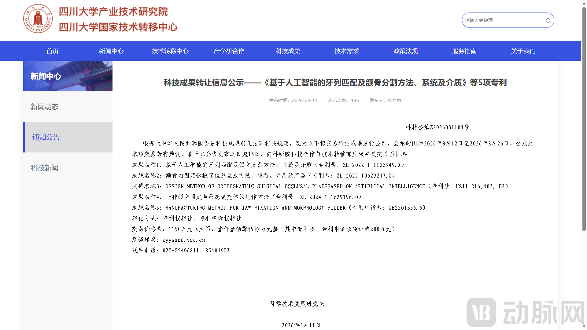
Image from the official website of Sichuan University
This setOral and Maxillofacial Surgery Technology PackageFirst, artificial intelligence is employed to achieve automatic dental arch matching and precise jawbone segmentation, providing an accurate digital model for surgical planning. Subsequently, customized PEEK material jawbone fixation and morphological fillers are fabricated to integrate bone segment fixation with bone contour augmentation, thereby making orthognathic surgery smarter, more precise, and more efficient.
Osteotomy and Bone Repositioning Surgeries for the Correction of Oral, Craniofacial, and Maxillofacial DeformitiesIt is the core approach for treating dentofacial deformities and improving facial aesthetics,Precision, Efficiency, and Postoperative Recovery Outcomes of SurgeryDirectly impacts the patient's treatment experience and facial aesthetics.
However, in current clinical oral and maxillofacial surgical settings, the inherent defects of traditional diagnosis, treatment, and restoration methods have led to a concentration of critical pain points: preoperative surgical design processes, such as jawbone segmentation and dental arch matching, rely entirely on manual operations by clinicians, lacking support from intelligent automation technologies; meanwhile, postoperative bone fragment fixation and morphological restoration employ traditional materials such as titanium plates and screws, with limited solution options. Consequently, oral and maxillofacial surgeries are significantly constrained by manual operational dependencies and the limitations of material properties.
Such scenarios pose multiple challenges to both healthcare professionals and patients:For patientsTraditional titanium plate and screw fixation has limitations in biocompatibility, easily causing adverse reactions such as metal allergies and inflammation. Additionally, metallic materials produce artifacts in imaging examinations, interfering with the assessment of postoperative recovery. Meanwhile, issues such as poor bone morphology and inadequate facial fullness often persist after osteotomy and distraction procedures, necessitating additional filler surgeries. This not only prolongs the treatment cycle but also increases surgical trauma and risks, potentially compromising facial aesthetic outcomes due to secondary interventions.
For healthcare professionalsPreoperative manual jaw segmentation and dental arch matching involve complex, highly repetitive workflows that consume an average of 40 minutes. This not only imposes a heavy workload but also encroaches on time critical for surgical plan optimization and preoperative assessment, resulting in wasted medical resources. Particularly in the diagnosis and treatment of complex dentofacial deformities, the accuracy of manual operations is susceptible to human factors, making it difficult to ensure consistency between model reconstruction and surgical design, thereby hindering precise surgical execution. Furthermore, postoperative fixation with titanium plates and screws, along with planning for subsequent auxiliary grafting procedures, further increases the burden on healthcare professionals and reduces overall diagnostic and therapeutic efficiency.
Currently, the mainstream surgical solutions for oral and maxillofacial surgery still rely on“Manual Preoperative Design + Titanium Fixation”Primarily, some hospitals have attempted to introduce simple computer modeling software for assisted design, but this approach is limited to basic model reconstruction and cannot achieve automatic dental arch matching or jawbone segmentation, thus still requiring substantial manual intervention. Meanwhile, restorative solutions employing novel materials are scarce and lack personalized custom design, making it difficult to adapt to the varying bone morphological characteristics of different patients. Furthermore, these solutions cannot simultaneously achieve both bone fragment fixation and morphological filling, necessitating the use of additional filler materials and resulting in cumbersome operational procedures.
Furthermore, existing solutionsA widespread lack of integrated solutions for “precise preoperative planning – personalized intraoperative repair – convenient postoperative assessment”, poor continuity between preoperative planning and intraoperative procedures, inadequate fit between the prosthesis and the patient’s bone tissue, and postoperative issues such as stress shielding and suboptimal bone morphology restoration due to material or procedural problems, thereby increasing the risk of postoperative complications. These pain points have created an urgent market demand forA Comprehensive Oral and Craniomaxillofacial Surgical Solution Integrating “Intelligent Preoperative Design + Personalized Material Reconstruction + Integrated Surgical Implementation”, filling the gap left by traditional approaches in “high efficiency + precision + multifunctional repair” and addressing the core challenges of oral and craniomaxillofacial surgery.
This intelligent digital technology package for oral and maxillofacial surgery, based onAI Algorithms and Personalized Material Innovationcentered on, it specifically addresses the industry pain points of traditional diagnosis and treatment—“manual inefficiency, limited repair capabilities, and hindered assessment”—demonstrating“Intelligent and Efficient, Precise and Multifunctional, Highly Biocompatible and Adaptable”Three Core Advantages: Delivering Superior Integrated Solutions for Surgeries Such as Correction of Dentofacial Deformities.
AI-Powered Intelligent Automation: Liberating Human Labor to Enhance Efficiency and Quality.Leveraging proprietary algorithms, we have developed an intelligent system for dental arch matching and jawbone segmentation. By solving objective functions under constrained motion, the system achieves automatic and precise alignment of digital dental arch models with cranial models. Furthermore, it enables automated segmentation of the mandibular condyle through threshold-based reconstruction, precise localization, and surface normal feature cropping. This innovation reduces the average processing time from 40 minutes of manual operation to just 2 minutes, completely eliminating reliance on physicians’ manual intervention.
This process not only significantly reduces the workload of medical staff, allowing physicians to focus on core tasks such as optimizing surgical plans and performing precise intraoperative maneuvers, but also eliminates human errors associated with manual operations. It ensures consistency between preoperative model reconstruction and surgical design, thereby enhancing overall surgical precision and diagnostic and treatment efficiency, while being suitable for various complex cases, including skeletal Class II and Class III malocclusions and asymmetric deformities.
Personalized custom design for integrated fixation and filling.This technology is based on three-dimensional CT imaging data of the bone morphology in the patient’s surgical area. Through three stages of refined design, Boolean operations, and edge trimming, combined with 3D printing technology, PEEK material implants and positioning guides are fabricated. The implants conform closely to the patient’s bone tissue, while the osteotomy lines and positioning holes on the guide enable precise intraoperative cutting and marking.
Unlike traditional titanium fixation materials, which can only achieve bone fragment fixation,Customized PEEK ImplantBone segment fixation following osteotomy and bone contour augmentation can be performed simultaneously during the same surgical procedure, eliminating the need for a separate grafting surgery. This approach significantly simplifies the surgical workflow, reduces surgical trauma and risks, while precisely filling bone contour defects to enhance facial volume and improve postoperative aesthetic outcomes of facial contours.
The Superiority of PEEK Material: Meeting the Needs of the Entire Clinical Workflow.The filler material adoptsPolyether Ether Ketone (PEEK) MaterialIts fabrication features excellent biocompatibility, avoiding adverse reactions such as metal allergy and inflammation, thereby fundamentally addressing the biocompatibility limitations of traditional titanium plates and screws. Meanwhile, the material’s elastic modulus is close to that of human bone, which effectively reduces stress shielding, better conforms to human physiological structures, and lowers the risk of postoperative complications.
Furthermore, PEEK materials produce no metal artifacts in imaging examinations and do not interfere with postoperative imaging assessments such as CT and X-rays, enabling physicians to monitor bone tissue recovery in real time. The accompanying positioning guides and implants can be used clinically immediately after sterilization. This technology is flexibly adaptable to various surgical sites, including the maxilla and mental region. By leveraging the flexible modeling capabilities of FreeForm Modeling Plus and CAD software, it achieves fully personalized customization based on individual patient differences, demonstrating strong clinical adaptability and versatility.
The digital restoration market in oral and maxillofacial surgery has taken shape.“AI-Assisted Design + Personalized Material Restoration”core development pattern, with domestic and international enterprises and research institutions focusing on“Enhanced Efficiency, Precise Adaptation, Integrated Functionality”Strategic layout centered on core demands, with products demonstrating differentiated competitiveness in algorithmic intelligence, material compatibility, and clinical adaptability, among whichAI Automation and 3D-Printed Personalizationis the mainstream R&D direction.
Fujian Zhongke Kangtai Material Technology Co., Ltd. “Patient-Matched Additively Manufactured Maxillofacial Prosthesis”, as the first laser 3D-printed maxillofacial bone defect repair implant approved for market launch in China, its core advantages lie in personalized anatomical fit and improved surgical efficiency. Fujian Provincial Medical Products Administration. The product utilizes Selective Laser Melting (SLM) technology to perform three-dimensional reconstruction and software design based on patient-specific maxillofacial anatomical data, enabling a complete match to the patient’s bone morphology. This eliminates the need for pre-bending of traditional implants prior to surgery, significantly reducing operative time. The product has received approval from the National Medical Products Administration.
Beichenxing Medical's 3D Digital Orthognathic Surgery Planning System Launched in China, focusing on the digital assisted design scenario for orthognathic surgery. Centered on 3D digital technology, this system provides preoperative planning support for procedures such as orthognathic surgery and genioplasty. It assists physicians in optimizing surgical plans through digital modeling. Although AI-based automatic matching and segmentation functions are not explicitly mentioned, its core direction in digital surgical design is highly aligned with the technology of Sichuan University. The product positioning emphasizes the visualization and convenience of clinical surgical planning, and it has currently showcased its clinical application scenarios on platforms such as Douyin.
In summary, this patented technology package has achievedIntelligent Preoperative Design, Integrated Intraoperative Repair, and Convenient Postoperative Assessment, addressing the core pain points of traditional diagnosis and treatment. This technology package featuresPrecision, Efficiency, and Biocompatibilityas its core advantage, providing clinicians with higher-quality integrated solutions and driving the digital and intelligent transformation of oral and maxillofacial surgery.
In the future, with the optimization of AI algorithms and advancements in material technology, such integrated intelligent diagnosis and treatment solutions are expected to further expand their application scenarios, reduce costs, become the mainstream diagnostic and therapeutic model in fields such as the correction of dentofacial deformities, and lead the high-quality development of the industry.