
In-Depth Analysis of Medical Evidence: DeepEvidence Supports Your Decision-Making
EditorpatientPress
Achieving a functional cure (clinical cure) is the most ideal goal for patients with chronic hepatitis B. With the continuous optimization of antiviral treatment strategies, clinical understanding of the nature of HBV infection is deepening. Previous assessments of viral burden primarily relied on serum HBsAg levels and hepatic tissue protein expression; however, these methods may not comprehensively reflect the true intrahepatic infection status. Accurate quantification of the intrahepatic viral load is of critical significance for the development of clinical cure strategies.
Recently, Gilead Sciences published a study in JHEP Reports showing that the intrahepatic HBV infection burden detected at the RNA level is approximately 3–7 times higher than that assessed at the protein level, suggesting that traditional protein-level assays significantly underestimate viral burden. The study also identified a previously unrecognized population of HBV-integrated cells, providing new insights and evidence for a deeper understanding of HBsAg clearance and clinical cure.
Research Methods
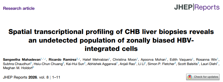
Using improved Visium spatial transcriptomics (ST) and single-nucleus RNA sequencing (snRNA-Seq), four commercially available HBV-positive liver biopsy samples and six clinical liver biopsy samples from the GS-US-174-0149 clinical trial were evaluated. Meanwhile, multiplex analysis targeting HBV viral proteins was performed.ImmunityMultiplex immunofluorescence (mIF) staining was performed, and chromogenic in situ hybridization (CISH) was used to detect HBV RNA and DNA. Machine learning methods were combined to annotate spatial transcriptomics (ST) data, distinguishing the presence of covalently closed circular DNA (cccDNA) from HBV integrated DNA (iDNA); deep learning algorithms were applied to classify based on mIF staining patterns of HBsAg and integrate this into the ST analysis framework.
Research Results
01 HBV infection load detected by RNA-based methods is significantly higher than that detected by protein-based methods
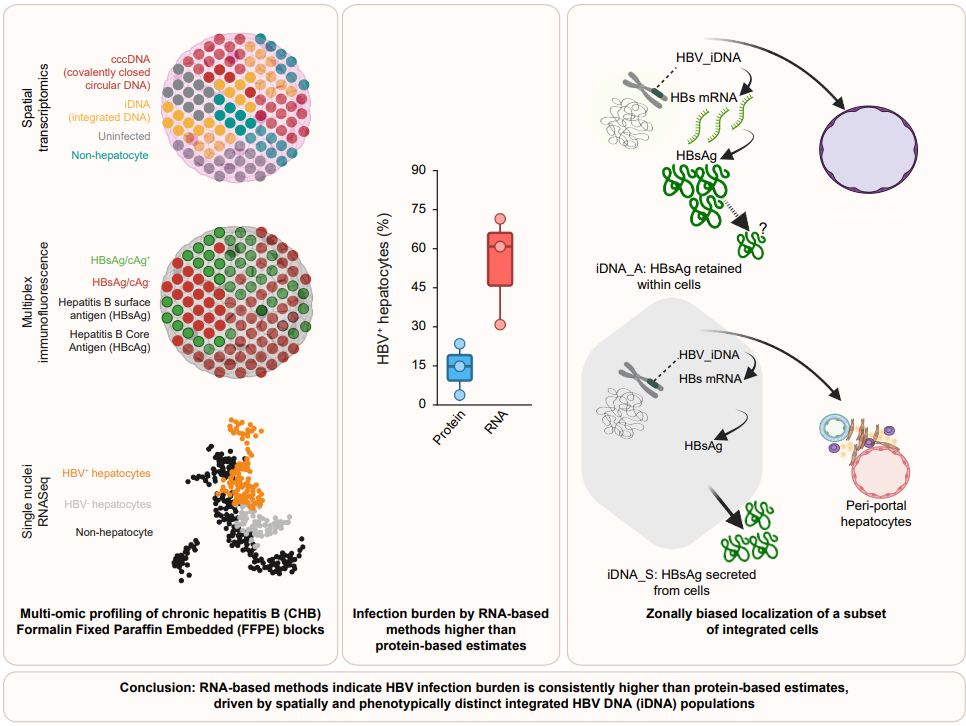
The intrahepatic HBV load reflected by HBs RNA-positive spots detected via ST was consistently higher than the estimates derived from mIF detection of HBsAg and/or HBcAg. Consistent with the ST results, snRNA-Seq revealed that the frequency of HBV-positive hepatocytes was 3–7 times higher than the quantification based on mIF.
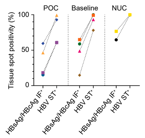
Figure 1. Comparison of the frequency of HBsAg-positive and/or HBcAg-positive tissue spots based on mIF detection with the frequency of iDNA- or cccDNA-positive tissue spots based on ST detection across all cohorts.
Figure 2. Comparison of spot-level HBs-P based on ST, spot-level HBsAg/HBcAg based on mIF, single-nucleus-level HBs-P based on snRNA-Seq, and single-cell-level HBsAg/HBcAg based on mIF
02 Comparison of mIF and ST results revealed a novel population of HBV-integrated cells
Traditional mIF identified two types of HBV-positive hepatocytes in tissue, indicative of cells containing cccDNA and iDNA, respectively. However, ST revealed a third hepatocyte population: positive for HBs RNA, but negative for both HBsAg and HBcAg by mIF, and also negative for HBc RNA/DNA (HBc-P) and antisense DNA (asDNA) strands by ST. This phenotype is consistent with the iDNA cell line PLC/PRF/5, suggesting it represents a subset of integrated hepatocytes previously unrecognized by protein-based detection methods.
03 HBs-P-Positive/HBsAg-Negative Hepatocytes Are Enriched in the Periportal Region of the Liver
HBsAg mIF staining-positive integrated cells are mainly localized in zone 3 around the central vein. The newly discovered population of HBsAg-negative but HBs RNA-positive integrated cells is significantly enriched in zone 1, the periportal region surrounding the portal vein. ST confirmed that there was no significant difference in HBs RNA levels between these two cell types within their respective enriched regions, indicating that the absence of protein staining is not due to reduced transcriptional activity.
Hepatic Health Insights
This study indicates that the quantity and types of intrahepatic HBV-infected cells may be more complex than traditionally recognized. RNA-based detection methods can identify a greater number of infected cells, suggesting that the burden of HBV infection may be underestimated. Furthermore, the presence of an integrated cell population that is HBsAg-negative but HBV RNA-positive suggests that a certain degree of viral reservoir may persist in the body even when HBsAg levels are low or after HBsAg clearance. This finding holds significant importance for understanding the implications of clinical cure and optimizing treatment strategies. Notably, the heterogeneity of viral distribution across different liver regions reaffirms that chronic hepatitis B treatment cannot be evaluated based on a single indicator. In the face of the complex mechanisms involving the coexistence of cccDNA and integrated DNA, the selection of treatment regimens should not only focus on reducing viral loads but also account for the modulation and long-term maintenance of immune function.
References:
Mahadevan S, Ramirez R, Mehrabian H, et al. Spatial transcriptional profiling of CHB liver biopsies reveals an undetected population of zonally biased HBV-integrated cells[J]. JHEP Rep. 2026, 8:101744.