Domestic 200,000 A-Scans/Sec Swept-Source OCT Launch Signals China's Rise in Ophthalmic Imaging

Intalight
High-end Ophthalmic Equipment Developer
In April 2018, the FDA approved IDx-DR, the first artificial intelligence-based medical imaging diagnostic product, which utilizes AI technology to analyze color fundus photographs for screening moderate or worse diabetic retinopathy. This groundbreaking approval ushered in the era of medical AI and spurred intensified research and development in ophthalmic AI.
Google DeepMind, a giant in the field of artificial intelligence, has also quietly entered the ophthalmology sector. In August 2018, DeepMind, in collaboration with Moorfields Eye Hospital in the UK, published an AI research study capable of accurately identifying 50 types of eye diseases, with diagnostic accuracy comparable to that of top ophthalmologists. Based on this framework, comprehensive identification of all eye diseases could be achieved in the future. Currently, DeepMind is working to gradually commercialize this technology.
IDx has achieved breakthrough success in color fundus photography, so why is DeepMind still holding tightly to this scenario?
The OCT technology favored by DeepMind, fully named Optical Coherence Tomography (OCT), is hailed as another major technological breakthrough following X-ray, CT, and MRI. It employs optics to image the microstructure of biological tissues, with resolution approaching that of histopathological sections, and is thus referred to as "optical biopsy" in the medical field.
Compared to color fundus photography, which provides two-dimensional imaging, a single optical coherence tomography (OCT) scan can generate hundreds to thousands of cross-sectional retinal images, representing an upgrade from “X-ray plain films” to “CT scans.” OCT provides precise information on lesion characteristics, thickness, and layered localization—details that are not visible to the naked eye—and is non-invasive and radiation-free, making it an essential imaging tool for ophthalmologists. Statistics indicate that by 2020, the annual number of ocular OCT examinations in the United States was projected to exceed 20 million.
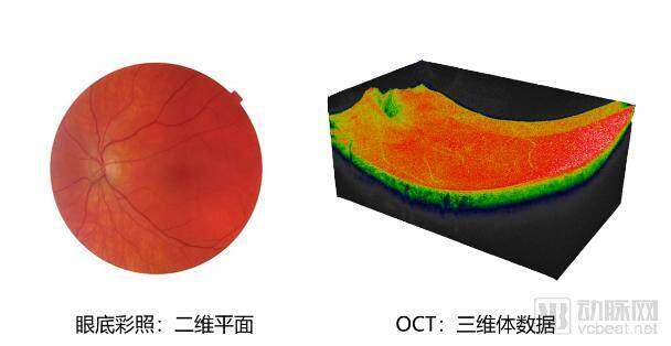
Comparison of Color Fundus Photography and OCT Data
Research indicates that, leveraging three-dimensional image data, AI-based analysis of OCT images can be applied to the screening and diagnosis of a broader range of diseases. However, there are two major challenges in developing artificial intelligence solutions based on OCT data: First, algorithmic analysis of 3D volumetric data is more complex than that of 2D planar data; second, AI companies incur substantial costs in acquiring raw OCT data.
In China, manufacturers of ophthalmic OCT devices rarely provide open data interfaces, and the market is virtually monopolized by imported equipment, making it even more difficult for AI companies to access raw data.
Can China’s “intelligent” manufacturing break the deadlock of import-dominated domestic OCT market and achieve more comprehensive AI-driven diagnosis in the field of ophthalmic OCT?
The scanning speed of OCT imaging directly determines the final image quality and resolution. OCT technology has evolved from the early time-domain laser technology with hundreds of scans per second, to the currently mainstream frequency-domain laser technology with tens of thousands of scans per second, and now to the most advanced swept-source laser technology with hundreds of thousands of scans per second. Currently, only two imported brands globally, Carl Zeiss from Germany and Topcon from Japan, have launched top-tier ophthalmic OCT systems based on swept-source laser technology, with a scanning speed of 100,000 scans per second. Among them, Japan’s Topcon Triton has obtained the Class II medical device registration certificate in China, while Zeiss’s Elite 9000 is sold only in Europe and America.
Recently, Intalight, a domestic company, launched the VG200, a high-end ophthalmic OCT device integrated with artificial intelligence, in Shanghai. This product significantly increases the scanning speed to 200,000 scans per second, akin to upgrading from a “64-slice CT” to a “128-slice CT.”
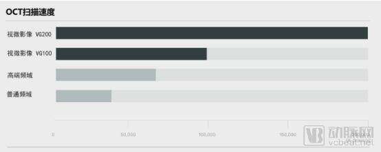
OCT Scan Speed Comparison
Meanwhile, to prevent physicians from missing ocular diseases during a single examination, Intalight has upgraded the field of view of its OCT products. Its newly released VG200 features a 56° scanning range and supports 16mm scans, offering an image acquisition area up to 33% larger than that of existing mainstream products. The en face imaging resolution is four times higher than that of top-tier international products, enabling simultaneous coverage of both the macular region and the optic nerve in the fundus, thereby facilitating higher-quality subsequent 3D reconstruction.
Dr. Peng Xianzhao, founder of Intalight, illustrated the importance of wide-field OCT using a personal anecdote: “A friend of mine, who is also one of our distributors, was highly interested in Intalight’s equipment and underwent an OCT examination using the VG200. The initial results showed a healthy choroid, but there were subtle signs of detachment at the peripheral retina. By adjusting the fixation light and changing the scanning angle to re-scan the affected area, we unexpectedly discovered a large-scale retinal detachment. Prior to this, he had never experienced any visual abnormalities, and routine examinations failed to capture the peripheral retina. Without this fortuitous discovery, he would likely have missed the opportunity for timely treatment.”
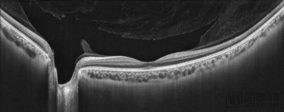
16mm HD Line Scan
Hardware breakthroughs provide the most robust support at the physical level. In an environment with scarce medical resources, physicians also rely on artificial intelligence to analyze this information, addressing challenges in 3D reconstruction and diagnosis.
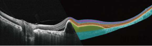
The eyeball exhibits a layered biological structure; however, the blurred boundary between the inferior border of the choroid and the sclera makes pathological layering of the choroid extremely challenging, rendering it difficult for traditional algorithms to classify images near these boundaries. To address this, Intalight employs its Deep Layer™ technology, based on deep learning, to identify various normal and abnormal retinal layers and assist physicians in three-dimensional reconstruction, significantly reducing the time required for image processing.
With each device deployed in hospitals, Intalight gains an effective data traffic entry point. The de-identified data collected through this portal features high quality, structured formats, and consistency, which are advantageous for training AI-assisted diagnostic products. As hardware deployment advances, Intalight’s AI solutions will demonstrate increasingly superior performance in assisted diagnosis.
Dr. Peng Xianzhao stated, “Most existing AI products are designed to identify a limited range of diseases and operate as external add-ons to imaging software or PACS systems, making it difficult to address issues of device universality. In contrast, our product is integrated directly into our proprietary imaging software. Leveraging high-quality, standardized data from our own devices, it will progressively achieve comprehensive coverage of more than 50 fundus diseases. This will constitute a truly clinically practical AI-assisted diagnostic tool.”
Standardized and comprehensive databases ensure the diagnostic quality of artificial intelligence, while also directly reducing the cost of using OCT technology, thereby facilitating its broader adoption in lower-tier markets.
“AI, as a groundbreaking technology, has an undeniable future; however, the industry still lacks a proven business model. For companies specializing in imaging software, profitability remains a significant challenge. At this stage, Intalight focuses its operations on hardware sales, ensuring survival while simultaneously deepening its AI capabilities and exploring potential commercial value.”
Therefore, following the launch of the VG200, Intalight will strengthen collaborations and promotional efforts with top-tier ophthalmology centers to raise awareness among ophthalmologists that China now offers OCT systems equipped with the most advanced swept-source technology, thereby aiming to increase hospital installations and expand data acquisition points.
In the realm of artificial intelligence, Intalight integrates identified structural and pathological data for model training. This approach not only provides AI-assisted disease diagnosis but also delivers quantitative metrics (such as size, volume, temporal changes, and clinical outcomes), forming a critical foundation for Intalight’s strategic collaborations with leading AI enterprises.
Artificial Intelligence Hierarchy
In early 2014, Dr. Xianzhao Peng, an optical systems expert with over a decade of R&D experience in Silicon Valley, and Dr. Bing Li, a medical image algorithm expert, founded an OCT startup in Silicon Valley. The company is dedicated to integrating cutting-edge technologies—including advanced optical design, imaging algorithms, artificial intelligence, and cloud platforms—to provide medical imaging solutions based on OCT technology for both physicians and patients.
Supported by national policies, the company relocated its headquarters to the Luoyang National University Science Park in 2015 and established an eight-character motto: “Regard the minute as significant; discern the big picture from small details,” adopting the name Intalight. In 2015, the company was selected for Luoyang City’s “Heluo Talents” Program and received tens of millions of yuan in financial support. It completed a Pre-A financing round worth tens of millions of yuan in 2018.
As the principal founder of the company, Dr. Peng Xianzhao specializes in research on leader systems and optical design, bringing 14 years of R&D experience from Silicon Valley. He earned his bachelor’s degree in Physics from Peking University in 1995 and received his Ph.D. in Spectroscopy from Oregon State University in the United States in 2002.
Dr. Li Bing, Co-founder of the team, oversees corporate management as well as software and algorithm research. In 1999, he entered the Department of Electronics at Peking University as the top science-stream scorer in the National College Entrance Examination (Gaokao) in Fuzhou City, and earned his Ph.D. in Image Processing from the University of Virginia in the United States in 2007. He holds copyrights for three medical atlas software suites and three U.S. patents, making significant contributions to R&D in image processing at Intalight.
Over the past five years, every technological breakthrough has been driven by Intalight’s deep understanding of clinical needs and its robust technical reserves. Today, the VG200’s performance rivals—and in some aspects even surpasses—that of the industry’s top-tier OCT systems. This year, Intalight will collaborate with leading Chinese ophthalmology experts to showcase its innovations at international ophthalmology exhibitions, bringing China’s advanced medical hardware technology to the world.
It is understood that Intalight will continue to develop cutting-edge technologies, such as 400 kHz swept-source OCT and swept-source biometers, to maintain the advancement of its hardware technology. Through seamless integration of software and hardware, the company aims to continuously enhance the core competitiveness of its products.