Digital Pathology Accelerates with 14 Funding Rounds in 2019 as AI Reshapes the Industry

Landing Med
Developer of Cervical Cancer Diagnostic Robots

Philips Medical Systems
Provider of Medical Imaging and Information Management Solutions

PathAI
Artificial Intelligence Medical Diagnosis Technology Developer

Thorough Images
AI Pathology Image Diagnosis Service Provider

Paige.AI
AI Cancer Diagnosis Technology R&D Provider

Knowledge Vision
Intelligent Decision-Making Platform Provider

Dipath
AI-Assisted Diagnostic Tool Developer

Proscia
Medical Imaging Diagnosis Supplier
On November 6, 2019, PathAI announced the completion of a $15 million Series B+ financing round, following a $60 million Series B financing round completed in April of the same year. The latest round was led by LabCorp, with participation from Merck Global Health Innovation Fund (Merck GHIF) and Bristol-Myers Squibb. The proceeds will be used to expand the clinical application of AI-driven diagnostic technologies and strengthen the company’s clinical development capabilities. PathAI will continue to focus on the field of cancer treatment, providing new therapies for patients.
PathAI, founded in 2016, specializes in developing AI-driven pathology research tools to enhance cancer diagnosis through artificial intelligence. Since its inception, PathAI has raised over $90 million in total funding. The AI-plus-pathology sector also experienced an investment boom in 2019; by November, the number of financing events had reached 14, representing a 27% increase compared to 2018.
November 6 may well be an auspicious day for the digital pathology industry. On this same day, Proscia, a representative enterprise in AI-powered digital pathology, secured CE marking for its Concentriq Dx solution in the European Union. Moreover, Paige.AI, a U.S.-based digital pathology startup, had its AI-driven cancer diagnosis platform granted the “Breakthrough Device” designation by the FDA earlier this March.
All of this indicates that digital pathology has recently entered a period of rapid growth. VCBeat (WeChat ID: Vcbeat) has also compiled the latest developments in this field.
From Pathology to Digital Pathology, and Then to AI + Digital Pathology
Pathology was described as the "foundation of medicine" by William Osler, the "Father of Modern Medicine," and pathologists are regarded as the "doctors' doctors." The critical value of pathology is self-evident, as the accuracy of its diagnoses directly impacts patients' health and fate.
# Digital PathologyDigital pathology refers to the application of computers and networking technologies in the field of pathology, integrating modern digital systems with traditional optical magnification devices. It utilizes automated microscopes or optical magnification systems to scan and acquire high-resolution digital images. These images are then automatically processed by computers through high-precision, multi-field, seamless stitching, yielding high-quality visual data for application across various domains of pathology.
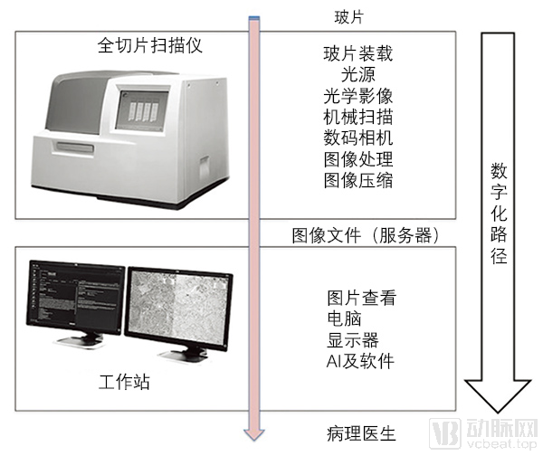
Image from *J Pathol Inform*
High-throughput automated digital pathology scanners can capture entire glass slides at magnifications comparable to those of microscopes, under either brightfield or fluorescence conditions. Digital slides can also be shared over networks using specialized digital pathology software. Automated image analysis tools are further available to assist in the interpretation and quantification of biomarker expression within tissue sections.
The history of digital pathology dates back 100 years, when specialized equipment was first used to capture microscopic images onto film. Telepathology, which involves transmitting microscopic images to remote devices, has also existed for nearly 50 years—in 1965, Prewitt and Mendelsohn scanned optical images under a microscope and reconstructed them digitally. However, at that time, digital microscopes cost as much as $300,000, and it took nearly a day to scan a single slide, making them practically unusable.
It was not until the past decade, with advancements in Whole Slide Imaging (WSI) technology, software interfaces, and network capabilities, that digital pathology began to undergo a true digital transformation, becoming fully integrated into pathological workflows. Compared to traditional methods, digital pathology provides pathologists with higher-clarity, consistent, and more user-friendly images, enabling them to rapidly participate in remote assessments and collaborations, thereby enhancing efficiency and productivity.
Whole Slide Imaging is a crucial component of digital pathology. Current software systems and methods for managing whole slide imaging data originate from the 1990s, with their initial motivation being the analysis, visualization, and querying of ultra-large-scale data obtained from satellite remote sensing. Using this software system, ultra-large-scale raw data collected by satellites can be associated with appropriate geospatial regions, spatially related data can be aggregated, and useful information can be generated.
Against this backdrop, Dr. Saltz’s team, then at the University of Maryland, College Park, developed the Active Data Repository (ADR) system, which optimizes storage to enable joint retrieval and processing of ultra-large-scale multidimensional data. This achievement received the National Science Foundation’s Grand Challenge Award.
At that time, Sal Pizzo, Chair of Pathology at Duke University, was considering the use of computers to replace microscopes in anatomic pathology. Following discussions with Dr. Pizzo, Dr. Saltz realized that this technology, originally used for processing Earth remote sensing data, could also be applied to handle the ultra-large datasets generated by whole-slide imaging in pathology. This insight laid the foundation for the subsequent development of virtual microscopy.
In 1996, when Dr. Saltz initiated his virtual microscopy research project, whole-slide scanners had not yet been developed. Imaging could only be achieved by stitching together individual fields of view captured via microscope scanning. Although James Bacus began designing a whole-slide imaging pathology scanner in 1994, it was not until 1997 that BLI (Bacus Laboratories Inc.), the company he founded, released the first whole-slide imaging pathology scanner.
In 2006, Olympus announced the acquisition of BLI, combining Olympus’s expertise in microscope hardware with BLI’s renowned whole-slide imaging and virtual microscopy technology to create an all-in-one imaging solution urgently needed by laboratories.
Another company worth mentioning is InterScope Technologies, which was co-founded in 1998 by Dr. Michael Becich and others in partnership with the University of Pittsburgh Medical Center. After several mergers and acquisitions, it was ultimately acquired by Carl Zeiss in 2007.
Modern whole-slide imaging systems have become a culmination of optics, robotics, and computing. Such devices typically include a microscope equipped with one or more objective lenses, a digital camera or robotic system for scanning to acquire microscopic images, a workstation including a display, and many other components.
Despite the rapid advancements in whole-slide imaging (WSI) technology, its adoption in clinical practice has been relatively slow. This situation is primarily attributed to a combination of technological, cultural, financial, and regulatory factors. In terms of regulation, the U.S. Food and Drug Administration (FDA) classifies AI-assisted diagnostic tools as Class III devices, meaning that such products must undergo rigorous clinical procedures before receiving approval.
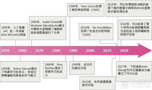
Image from *Nature*
It was not until April 2017 that Philips’ IntelliSite Pathology Solution, used for preliminary diagnosis in surgical pathology, became the first whole-slide imaging (WSI) system to receive FDA clearance. This milestone paved the way for other innovative pathology devices to obtain FDA approval. An increasing number of pathology institutions began considering equipment upgrades, and the rapidly growing market prompted more AI companies to explore the use of artificial intelligence to reshape digital pathology.
According to public reports, the first application of whole-slide digital imaging was in a 2018 study by Mukhopadhyay et al., which conducted the first large-scale blinded comparison of the diagnostic performance of whole-slide digital imaging and conventional microscopy. The study included specimens from 1,992 patients with various tumor types, which were blindly evaluated by 16 surgical pathologists using both whole-slide digital imaging and traditional microscopy. The results demonstrated that the primary diagnostic performance of whole-slide digital imaging was non-inferior to that of conventional microscopy-based methods.
Notably, whole-slide imaging technology has also facilitated the development of Vendor-Neutral Archives (VNA). This technology provides standardized interfaces and formats for the storage and archiving of medical images, even when these images originate from imaging systems of different brands.
This enables users to easily access PACS (Picture Archiving and Communication Systems) from different vendors via a VNA. In the past, PACS from different vendors had their own unique formats and interfaces, were incompatible with each other, and required cumbersome conversions for access.
Driven by commercial interests, various brands tend to build closed ecosystems using proprietary protocols and interfaces, which has led to the emergence of “data silos” within healthcare institutions.
Although VNAs are theoretically capable of supporting all medical image interfaces and formats, there remains a gap between this ideal and reality. Currently, no VNA can handle all image format types, interface with all imaging system protocols, and process all image formats. Meanwhile, the definition of VNA remains subject to debate; in some circles, VNA is considered equivalent to PACS.
In any case, as more and more enterprises begin to support VNA, its future is quite promising.
AI Will Reshape Digital Pathology
Despite significant advancements in digital pathology over the past decade, with substantial improvements in imaging speed, quality, and ease of use, final diagnoses still rely on the judgment of pathologists. This itself constitutes a major challenge.
According to relevant statistics, there are currently nearly 10,000 licensed pathologists in China. Based on the National Health Commission’s staffing requirement of one to two pathologists per 100 hospital beds, the shortage of pathologists amounts to as high as 90,000. Undoubtedly, each pathologist bears a workload five to ten times the normal level, making misdiagnoses and missed diagnoses inevitable.
This challenge is not unique to China; developed countries are also grappling with this issue. According to statistics from the article “Trends in the US and Canadian Pathologist Workforces From 2007 to 2017,” published in JAMA Network Open, the absolute number of pathologists in the United States declined from 15,568 in 2007 to 12,839 in 2017. The relative number also dropped sharply, with the number of pathologists per 100,000 people falling from 5.16 to 3.94. After adjusting for the annual number of new cancer cases, the diagnostic workload per pathologist in the United States increased by 41.73%, indicating a shortage of pathologists that is worsening over time.
The constraints on the development of pathology resources are not limited to the overwhelming workload; factors such as high work pressure, poor working conditions, low compensation, and lengthy training cycles have severely impacted the faculty responsible for pathology education. Globally, there is a "cliff-like" shortage of new entrants into the pathology workforce.
Pathology primarily relies on microscopic observation by the human eye. The identification of cancer cells is based on distinct similarities in their nuclei, cytoplasm, and cell membranes (such as color, density, size, and shape), as well as subtle variations in regional boundaries. Before the advent of digital slides, physicians were required to spend prolonged periods observing specimens under microscopes, resulting in an immense workload.
Digital slides are images composed of discrete pixels, allowing various features to be accurately calculated on a monitor. This technology has indeed greatly facilitated slide review for pathologists. Nevertheless, a qualified pathological glass slide contains at least 5,000 cells, and in some cases, tens of thousands. For pathologists, the task merely shifts from locating targets under a microscope to identifying them on a screen—a distinction without a significant difference.
The introduction of AI technology can significantly address this issue. Artificial intelligence, powered by deep learning, can process medical images in a rapid and standardized manner, highlight and render suspicious findings, and provide recommendations in structured language.
These tasks are highly labor-intensive and repetitive, whereas AI is not constrained by the nature of such work. Practice has demonstrated that with the assistance of AI, pathologists can not only enhance diagnostic efficiency and reduce workload, but also improve their working environment, ultimately lowering the rates of misdiagnosis and missed diagnosis.
The application of AI in digital pathology is not fundamentally different from its use in other medical imaging fields. It requires high-quality annotations by multiple pathology experts to ensure that the model can adequately learn the morphology of various pathological cells. Based on these patch-level annotations, the AI model first performs preliminary classification of samples, efficiently identifying those with higher densities of positive cells.
Subsequently, the AI model further performs precise identification of positive cells, ensuring accurate auxiliary interpretation results even in samples with low positive cell density. To ensure error-free results, the AI also selects a series of suspicious local fields of view for final review by pathologists.
In what areas can AI empower digital pathology?
Currently, artificial intelligence (AI) technology is widely applied in pathology. Quantitative analysis provides a series of quantitative indicators for pathological diagnosis, mitigating the unpredictable factors associated with physicians’ subjective experience and improving the accuracy of disease diagnosis. Beyond pathological diagnosis, early cancer screening for breast cancer, cervical cancer, and other malignancies, as well as the integration of pathology with new drug development, have become key focus areas for the deployment of digital pathology combined with AI.
Auxiliary Pathological Diagnosis
As previously mentioned, pathological diagnosis is the "gold standard" in disease diagnosis. It is particularly critical in the confirmation of tumors. It can be essentially stated that the diagnostic capability of the pathology department largely determines the overall level of diagnosis and treatment in a hospital. Typically, the initial diagnostic report for a disease is issued by the pathology department. Consequently, 80% of the treatment decisions made by physicians for patients are influenced by the pathology report.
Digital microscopy equipment can perform high-quality, fully digital scanning of entire pathology slides. The resulting digital slides enable AI-driven dynamic observation and assisted diagnosis, filtering out a large volume of negative data and reducing the slide review workload for pathologists. This allows them to devote more time to suspected positive cases and more complex diagnoses.
Patients suspected of having tumors, regardless of whether the specific symptoms present benign or malignant characteristics, can undergo digital pathology examination. In addition to tumors, certain special diseases such as tuberculosis, as well as specific allergic disorders like some forms of nephritis and nephropathy, can also be aided in diagnosis through digital pathology examination.
In this application area, nearly all domestic AI companies engaged in medical image data analysis have established a presence. Typical representatives include Thorough Images, DeepThinking, and Dipath.
Of course, do not overlook the major medical device manufacturers. Compared to standalone AI software systems designed specifically for analyzing pathological images, integrating hardware and software from the outset and embedding AI directly during image acquisition is clearly a superior approach. Traditional device giants, as well as companies with significant influence in the pathology field through their optical technologies—such as Leica Biosystems and Carl Zeiss—have all made corresponding strategic arrangements. In China, Fuyi Shares and Zhiying Medical are also representative players in this space.
New Drug Development
Pathological data can be utilized not only for diagnosis but also for analyzing the efficacy of new drugs, thereby facilitating drug development. Prior to the advent of AI, researchers could only roughly estimate the extent of lesions based on subjective judgment, making quantitative analysis of pathological slides impossible. The emergence of AI has enabled precise counting of tissue cells.
With the aid of AI, researchers can rapidly and accurately quantify the number, severity, and temporal changes of pathological cells in tissue sections over a given period, thereby observing the impact of new drugs on lesions in clinical trials. Furthermore, researchers can leverage AI to monitor histological and cellular changes in animal tissues following drug administration, thus providing more precise guidance for drug development.
Reveal Biosciences, PathAI, as well as China’s Knowledge Vision and Genome Wisdom Inc., are all representatives in this field.
Third-Party Medical Testing Center
Third-party pathology testing institutions are well-established in developed countries, whereas they remain an emerging sector in China. According to 2017 data, independent medical laboratories accounted for less than 5% of the total medical testing market size in China. In contrast, this proportion reached as high as 67% in Japan, 50% in Europe, and 35% in the United States. Data from LabCorp’s annual report indicates that in 2016, revenue in the U.S. clinical laboratory testing industry amounted to $8 billion.
For this reason, in August 2017, the National Health and Family Planning Commission approved five categories of medical institutions that could be independently established by third parties, including medical laboratories, pathology diagnosis centers, medical imaging diagnosis centers, hemodialysis centers, and hospice care centers. It also permitted investment from non-public sectors and allowed for chain-based and group-oriented operations. This move has made these institutions a key instrument for the state to implement tiered diagnosis and treatment systems and promote reforms in public hospitals, while also serving as a significant practical avenue for social capital to enter the healthcare industry.
The extensive recent application of artificial intelligence technologies and cloud pathology platforms is expected to enhance the medical capabilities of third-party medical testing centers, thereby accelerating the development of third-party pathology diagnostic centers. This model facilitates remote pathology diagnosis, enabling digital pathology to transition from a conceptual form of telemedicine to practical application, thus alleviating the current shortage of pathology testing resources in primary healthcare institutions.
Nowadays, multiple large enterprises in China have leveraged cloud platforms to deploy AI-based pathological diagnosis technologies to medical testing centers both domestically and internationally. Domestic remote pathological diagnosis platforms led by health authorities and medical consortia include the China Digital Pathology Remote Diagnosis and Quality Control Platform, the Henan Province Remote Pathology Consultation Platform, and the Ningbo Clinical Pathology Diagnosis Center.
Third-party clinical laboratories have also vigorously developed remote pathology diagnosis platforms. Institutions such as Southern Medical University, Guangzhou Huayin Medical Laboratory Center, KingMed Pathology Consultation Center, Dian Diagnostics Remote Pathology Consultation Center, Hengdao Medicine, and Wuhan Landing Medical High-Tech Co., Ltd. are all focused on this field.
Capital Markets Increasingly Recognize AI + Digital Pathology
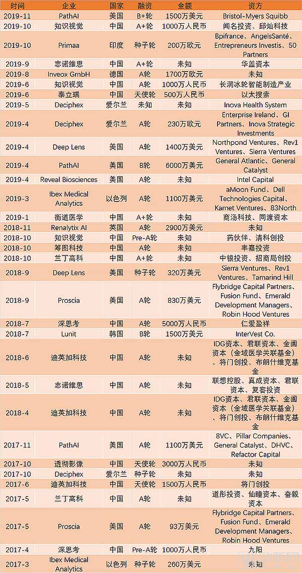
In recent years, an increasing number of enterprises have been combining AI with digital pathology. Although AI-powered digital pathology cannot compare with the booming field of AI-based medical imaging in terms of either funding amounts or rounds, it still demonstrates a gratifying upward trend. According to data compiled from the VCBeat knowledge base, there were a total of 33 financing events in this industry over the past three years, showing a year-on-year increase.
There were 8 and 11 financing events in this field in 2017 and 2018, respectively. By November 2019, there had already been 14 financing events in AI+pathology, highlighting the significant interest in this area.
Regulatory certification breakthroughs over the past two years should be one of the main reasons. In 2017, the FDA decided to reclassify AI-assisted diagnostic software systems combined with medical imaging from Class III medical devices to Class II medical devices, lowering the entry threshold and greatly promoting industry development.
In April 2017, Philips’ IntelliSite Pathology Solution became the first whole slide imaging (WSI) system to receive FDA clearance for primary diagnosis in surgical pathology.
In April 2019, Paige.AI, which had spun out of Memorial Sloan Kettering Cancer Center (MSKCC) and had been in operation for just over a year, bore fruit from its collaboration with MSKCC—Paige.AI’s AI-powered cancer diagnostic solution received the “Breakthrough Device” designation from the U.S. Food and Drug Administration (FDA). This marked the first time the FDA approved the use of AI for cancer diagnostic services.
Subsequently, in May 2019, Renalityix AI’s KidneyIntelX, an AI-based diagnostic system for kidney disease developed by a UK company, was granted Breakthrough Device designation by the FDA, becoming the first AI-driven diagnostic system for kidney disease on the market to receive this distinction.
In November 2019, Proscia’s Concentriq Dx solution for assisted diagnosis also received CE marking in the European Union.
It can be said that the growing number of approved AI-powered digital pathology devices has bolstered confidence in the capital market.
Although China has not yet approved any AI-assisted diagnostic solutions, regulatory authorities have been making intensive preparations in this regard. In November 2018, the Center for Medical Device Technical Evaluation of the National Medical Products Administration (NMPA) publicly solicited information from domestic and foreign manufacturers engaged in the production of artificial intelligence medical devices. The term “artificial intelligence medical devices” specifically refers to medical devices that employ “next-generation artificial intelligence technologies” in areas such as workflow optimization, data processing, and assisted diagnosis. Here, “next-generation artificial intelligence technologies” denote techniques that utilize data-driven approaches to train algorithms, represented by deep learning and neural networks.
Subsequently, in February 2019, the Center for Medical Device Evaluation of the National Medical Products Administration released the “Key Points for the Review of Deep Learning-Assisted Decision-Making Medical Device Software (Draft for Comments)” and solicited public opinions, signaling that the review standards for Class III AI medical devices were close to implementation and that policy bottlenecks hindering industry development were expected to be broken.
However, from the perspective of regulatory authorities, compared to individual physicians, AI algorithms have a much broader scope of potential harm to patients in cases of misdiagnosis, and may even trigger iatrogenic risks. Therefore, when artificial intelligence algorithms are applied in clinical practice, there is a greater need for systematic debugging, auditing, extensive simulation and validation, as well as prospective review. It is particularly essential to address the interpretability gap in AI decision-making processes—after all, black-box computations based solely on probabilistic clouds are not suitable for clinical application.
This issue has also seen improvement this year. In May 2019, the team led by Yang Lin, CEO of Dipath, published a paper titled “Pathologist-level Interpretable Whole-slide Cancer Diagnosis with Deep Learning” in Nature Machine Intelligence, proposing a solution for interpretable AI-based pathological diagnosis.
In the experiments described in the article, researchers employed AI technology to analyze and process pathological slides, while simultaneously providing the rationale for the AI analysis. This is the first monograph published in a Nature subsidiary journal that discusses the issue of AI interpretability in pathological image analysis.
This experiment offers new insights into the regulatory approval of artificial intelligence. Although current AI systems still lack genuine reasoning capabilities, this approach simulates the reasoning process by modularizing physicians' diagnostic steps. Furthermore, the text-matching procedure employed in the experiment adheres strictly to WHO standards. Consequently, unlike common segmentation methods generated through multi-sample deep learning, every step in this experiment allows AI to provide explicit rationale for its decisions.
Although this experiment still has some limitations, it provides a novel perspective for regulatory authorities on AI oversight. We believe that the impact of this experiment on the industry may soon be reflected in regulatory approvals.
Final Thoughts
With the advent of whole-slide imaging and the subsequent introduction of AI-assisted diagnosis, AI-based digital pathology technology is flourishing worldwide. Despite shortcomings in regulation, reimbursement, and deployment, healthcare professionals in the fields of pathology and oncology are showing growing interest in the development and use of these technologies.
Despite the challenges, the integration of AI with digital pathology has become an unstoppable trend. In recent years, healthcare institutions around the world have been digitizing, or are in the process of digitizing, the entire pathology workflow. The FDA clearance of Philips’ whole slide imaging system in 2017 marked a significant milestone indicating that digital pathology was on the verge of large-scale adoption.
Future pathology departments will first utilize digital slide scanners to convert all routine slides into digital formats, integrating them into daily workflows to enable AI-assisted primary diagnosis of digital slides. Furthermore, by leveraging the internet to establish regional pathology cloud platforms, these departments will employ artificial intelligence products for computer-aided pathological diagnosis, thereby further enhancing the diagnostic efficiency and accuracy of pathologists at primary care levels.
Therefore, AI methods will become key to analyzing and interpreting this vast amount of data, helping the ancient field of pathology revitalize in the process.
References:
Deep Tech: FDA Stumble? The World’s First AI Cancer Diagnostic “Breakthrough Device” Exposes Controversy Over Collaborations Between AI Startups and Top Cancer Centers
CCTV.com: China Faces a Shortage of 90,000 Pathologists: Long Training Periods, High Risks, and Low Pay Are the Current Reality
JAMA Network Open:Trends in the US and Canadian Pathologist Workforces From 2007 to 2017
jpathinformatics.org:Twenty Years of Digital Pathology: An Overview of the Road Travelled, What is on the Horizon, and the Emergence of Vendor-Neutral Archives
Nature:Artificial intelligence in digital pathology — new tools for diagnosis and precision oncology
Guangzhou Daily: AI Pathologist Sets New Global Benchmark for AI-Assisted Cervical Cancer Screening; Clinical Application Still Three to Five Years Away