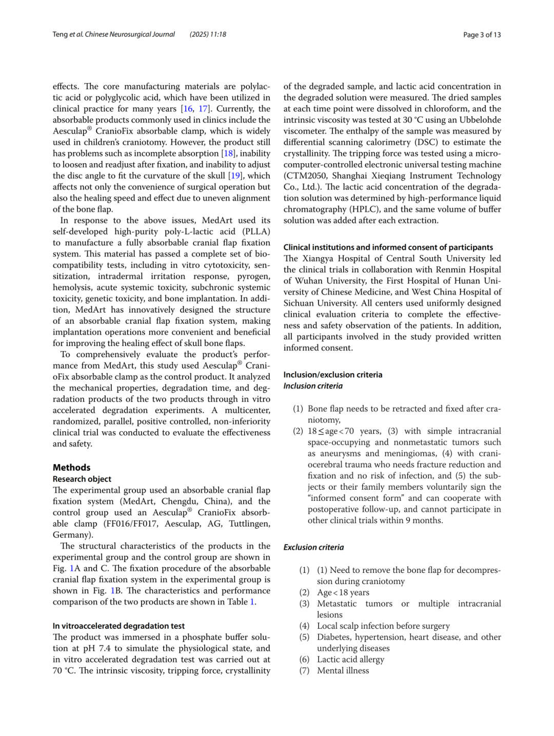
A Comparative Study of a Novel Absorbable Cranial Flap Fixation System and Aesculap CranioFix: In Vitro Experiments and Multicenter Clinical Trial

MEDART
Biomedical Materials Application Developer

Department of Neurosurgery, Xiangya Hospital, Central South UniversityProfessor Li Xuejun's TeamYuChinese Neurosurgical JournalThe results of a study titled “A comparative study of a novel absorbable cranial flap fixation system and Aesculap CranioFix” have been published online. This clinical study was led by Xiangya Hospital of Central South University and conducted across multiple centers, including West China Hospital of Sichuan University, Renmin Hospital of Wuhan University, and the First Affiliated Hospital of Hunan University of Chinese Medicine. The investigational product in the trial group is an absorbable cranial lock designed and developed by Chengdu Medart Medical Scientific Co., Ltd. (hereinafter referred to as “Medart”), which fills the domestic gap for similar devices. This study also represents the first head-to-head clinical comparison between a domestically produced absorbable neurosurgical internal fixation device and its imported counterpart. The study results showed:MEDART Absorbable Cranial Lock offers superior stability, prolonged maintenance of mechanical properties, enhanced cranial healing, and reduced bone flap displacement, ensuring highly reliable fixation and safety.Meanwhile, leveraging its self-developed biomedical material translation platform, MEDART has continuously developed China’s first absorbable craniomaxillofacial plate and screw system, establishing a product portfolio of absorbable internal fixation and reconstruction devices, with the aim of continually leading and promoting the upgraded application of cranial closure materials in neurosurgery.
Abstract
Background:Absorbable cranial locks have been widely used in neurosurgical procedures. However, these products present certain limitations, including incomplete degradability, inability to readjust after fixation, and non-adjustable angles of the lower fixation plate. To address these clinical challenges, Chengdu Medart Medical Scientific Co., Ltd. developed a fully absorbable cranial lock featuring enhanced ease of use and superior reduction outcomes, utilizing self-developed high-purity PLLA combined with an innovative structural design. This study thoroughly validated the safety and efficacy of the device through in vitro experiments and clinical trials.
Methods:In this study, the MEDART absorbable cranial fixation system was used in the test group, while a similar absorbable cranial fixation system was used in the control group. In vitro accelerated degradation tests were conducted to compare the trends in material and mechanical properties of the two products. A multicenter, randomized, parallel-group, active-controlled, non-inferiority clinical trial was carried out, with a 48-week postoperative follow-up period. The study compared the reduction in cranial suture distance, the qualified rate of bone flap displacement, trends in implant volume changes, and the incidence of postoperative adverse events between the two groups.
Results:In vitro accelerated degradation tests indicated that, in terms of the decline in intrinsic viscosity, the control group and the test group required 7 days and 14 days, respectively, to reach the test endpoint. Regarding the decline in mechanical properties, the control group and the test group lost clinical significance for secure fixation on day 3 and day 4 of degradation, respectively. In terms of degradation product release, the control group exhibited a burst release of lactic acid during the first 3–7 days, whereas the test group released lactic acid at a slow and constant rate. The clinical study randomly enrolled 90 patients, with 87 completing the surgery; the mean age was 50 years. For efficacy assessment, the reduction in the mean distance of cranial sutures was measured using three-dimensional reconstruction of cranial CT scans. At 1, 12, 24, and 48 weeks postoperatively, the mean suture distances in both groups were less than 2 mm. At 1, 6, 12, 24, and 48 weeks postoperatively, the qualified rate for bone flap displacement in the test group was 100%, whereas the control group had one unqualified case at 1 week postoperatively and two unqualified cases at 6, 12, 24, and 48 weeks postoperatively. Volume changes of the implants in both groups were analyzed using three-dimensional reconstruction technology based on CT data. At 48 weeks postoperatively, the residual volume in the test group remained close to 50% (approximately 48.8%), while the residual volume in the control group had dropped below 50% (approximately 43.9%) by 12 weeks postoperatively. Regarding safety, only one device-related adverse event occurred in the control group, with an incidence rate of 2.22%, manifesting as poor wound healing at the incision site.
Conclusion:In vitro experiments demonstrated that the test group exhibited excellent stability, with a longer duration of maintained mechanical properties and complete biodegradability in vivo. Clinical trials indicated that the test group significantly narrowed the cranial bone gaps after craniotomy, reduced bone flap displacement, promoted cranial bone healing, and provided highly reliable fixation efficacy and safety.
Clinical Trial Registration Information:Chinese Clinical Trial Registry, Registration No.: ChiCTR2500099674, Registration Date: March 27, 2025 (https://www.chictr.org.cn/showproj.html?proj=261082).
Keywords:Absorbable Cranial Lock, Multicenter Clinical Study, CT Three-Dimensional Reconstruction, Accelerated Degradation Test
Comparison of Material Properties

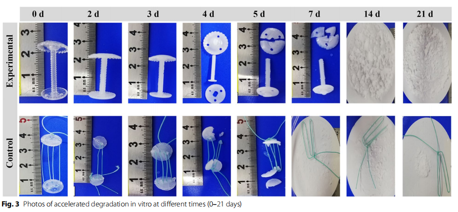
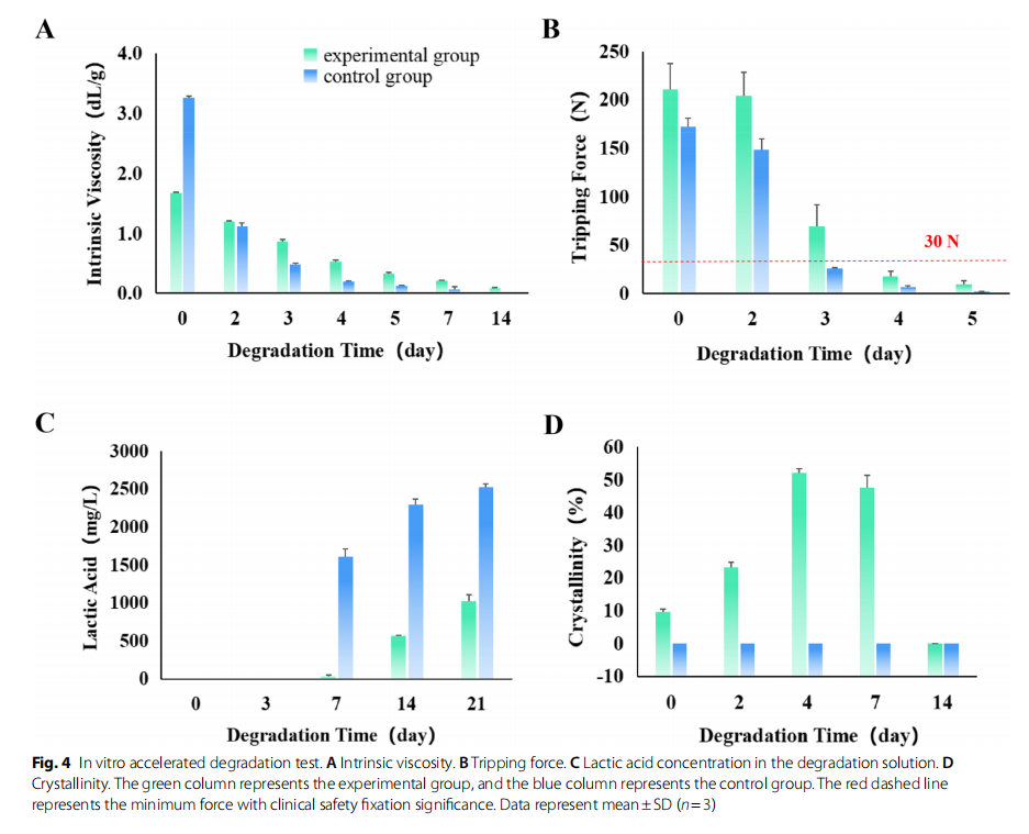
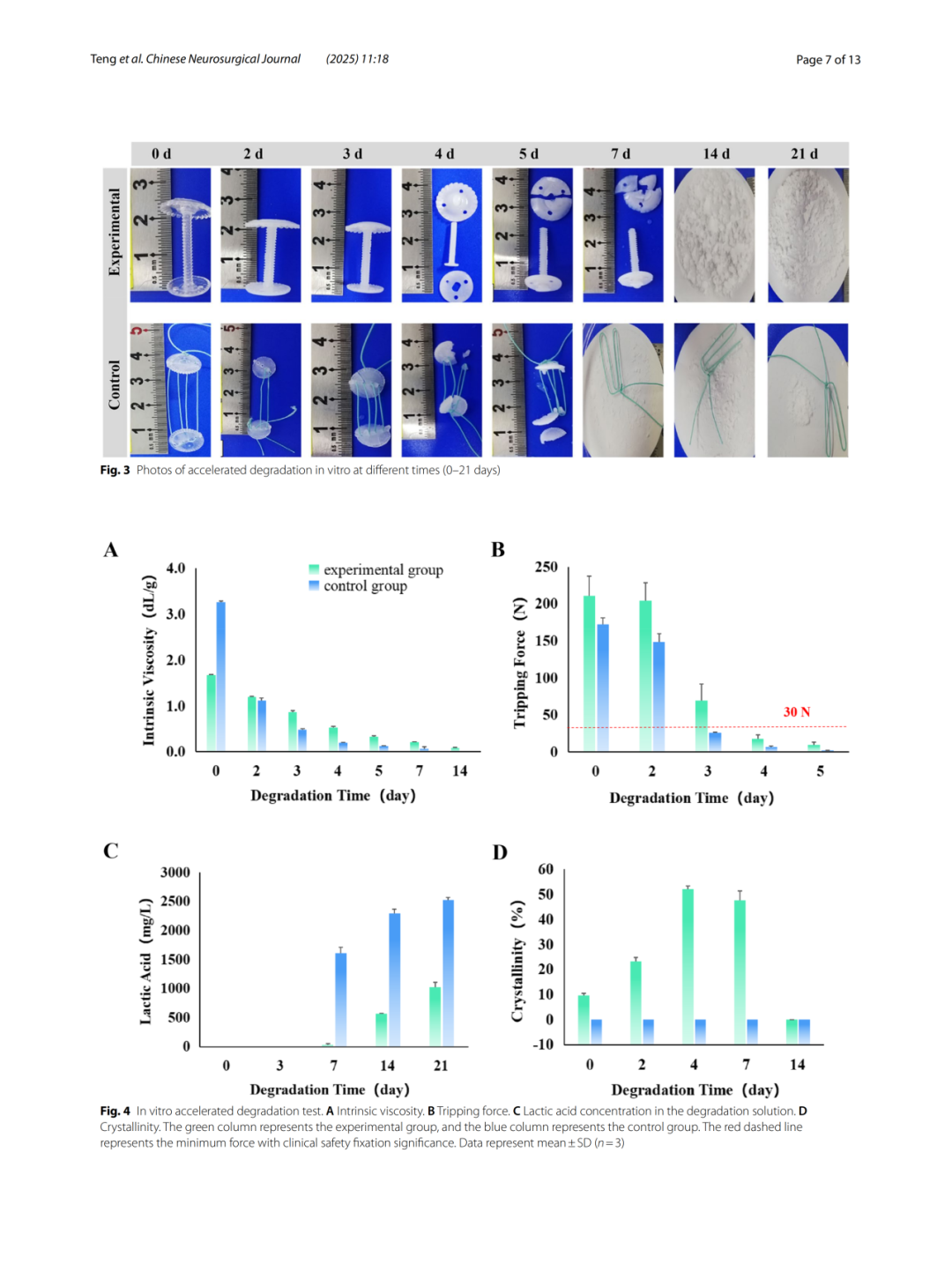
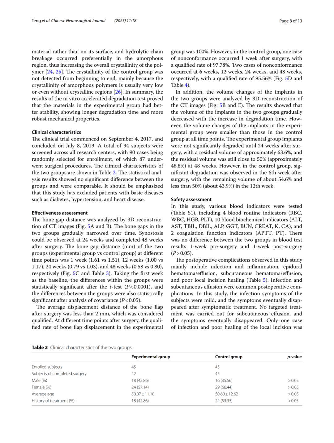
The in vitro accelerated degradation results of the two product groups are shown in Fig. 3. The results of the in vitro accelerated degradation test indicated that the trends in intrinsic viscosity changes were consistent between the test group and the control group. In the test group, the intrinsic viscosity decreased linearly during the first 5 days, at a slower rate than that observed in the control group. Using a decrease in intrinsic viscosity to below 0.1 dL/g as the endpoint, the control group reached this point in 7 days, whereas the test group required 14 days (Fig. 4A). Furthermore, the trends in pull-off force changes were consistent for both product groups. However, the test group demonstrated superior pull-off force compared to the control group during the initial steady state, at the onset of degradation, and at the end of degradation. Under simulated use conditions, the pull-off force for both groups decreased sharply on day 3. Based on the minimum force of 30 N required for clinically secure fixation, the control group and the test group lost their clinical significance for secure fixation on day 3 and day 4, respectively (Fig. 4B). The degradation product for both groups was lactic acid. The test group maintained a stable state for the first 7 days, releasing trace amounts of lactic acid; after day 7, it released lactic acid at a constant rate of 15.42 mg/day until the end of the test. In contrast, the control group exhibited a burst release of lactic acid between days 3 and 7, after which the release rate gradually decreased over time, with an average release rate of 40.09 mg/day (Fig. 4C). Regarding crystallinity, the crystallinity of the test group gradually increased during the first 4 days, reaching a maximum of 52.08 ± 1.25% on day 4; subsequently, the crystallinity gradually decreased, dropping to 0% after day 14, thereby becoming amorphous low-molecular-weight poly(L-lactic acid). The control group remained amorphous throughout the study (Fig. 4D).
Comparison of Efficacy

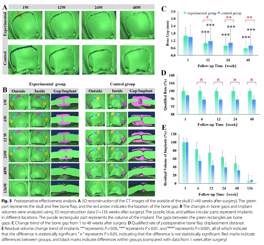
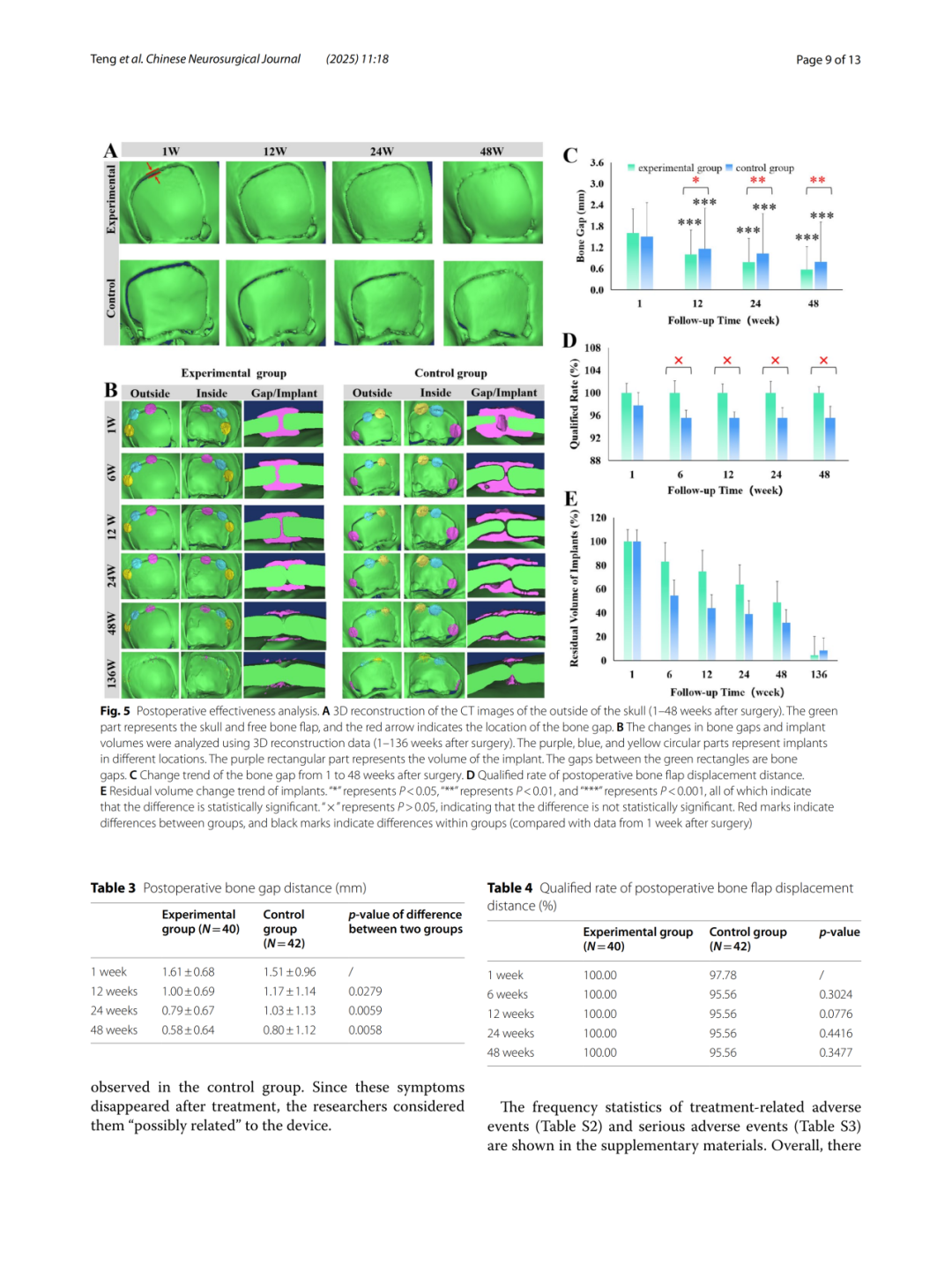
Postoperative cranial suture distances were analyzed using CT three-dimensional reconstruction data (Fig. 5A and 5B). In both groups, cranial sutures progressively narrowed over time postoperatively, achieving osseous fusion at 24 weeks and completing it by approximately 48 weeks. At various postoperative time points, the suture distances (mm) for the two groups (experimental group vs. control group) were as follows: 1 week (1.61 vs. 1.51), 12 weeks (1.00 vs. 1.17), 24 weeks (0.79 vs. 1.03), and 48 weeks (0.58 vs. 0.80) (Fig. 5C). Paired t-tests, with week 1 as the baseline, showed that the differences from baseline in each group were statistically significant (P < 0.0001). Analysis of covariance also revealed statistically significant differences between the two groups (P < 0.05).
A postoperative bone flap displacement of <2 mm was considered acceptable. At all postoperative time points, the qualification rate for bone flap displacement in the experimental group was 100%. In contrast, the control group had one unqualified case at 1 week postoperatively (qualification rate: 97.78%), and two unqualified cases at 6, 12, 24, and 48 weeks postoperatively (qualification rate: 95.56%) (Fig. 5D).
Furthermore, the volume changes of the implants in both groups were analyzed using CT three-dimensional reconstruction data (Fig. 5B and 5E). As shown in the figures, both groups exhibited the same trend of volume change in vivo, gradually decreasing with prolonged degradation time. The volume reduction in the experimental group at each postoperative time point was less than that in the control group. Significant degradation in the experimental group was observed only at 24 weeks postoperatively, with a remaining volume of approximately 63.6%; at 48 weeks postoperatively, the remaining volume was still close to 50% (approximately 48.8%). In contrast, significant degradation in the control group was observed as early as 6 weeks postoperatively, with a remaining volume of approximately 54.6%, and the remaining volume dropped below 50% (approximately 43.9%) by 12 weeks postoperatively.
Safety Comparison

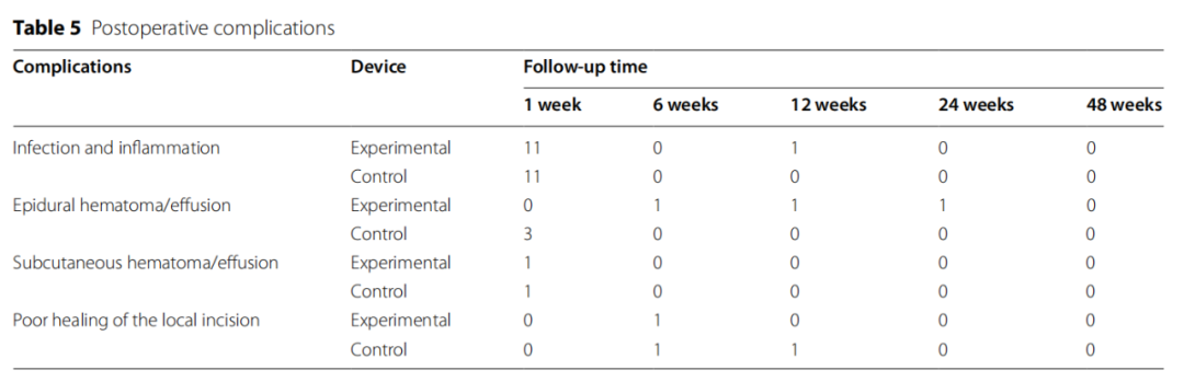
Postoperative complications observed in this study mainly included infection and inflammation, epidural hematoma/effusion, subcutaneous hematoma/effusion, and poor local incision healing (Table 5). Among these, infection and subcutaneous effusion are common postoperative complications. In this trial, all subjects with infection presented with mild symptoms, which ultimately resolved after symptomatic treatment. Subcutaneous effusions were not treated specifically, and the symptoms ultimately resolved. In the control group, one case experienced both infection and poor local incision healing. The infection resolved after treatment, and the poor local incision healing also ultimately resolved following targeted treatment. The investigator judged these events as "possibly related" to the device. No subjects in the experimental group experienced adverse events related to the investigational device. One adverse event possibly related to the device occurred in the control group, with an incidence rate of 2.22%, manifesting as poor healing at the incision site. No device defects that could lead to serious adverse events were identified in either group.
Conclusion

This study systematically evaluated the efficacy and safety of a novel absorbable cranial lock through in vitro accelerated degradation tests and multicenter clinical trials. The in vitro studies demonstrated that, compared with the control group, the test group exhibited superior stability, a longer degradation time, and enhanced mechanical properties. Clinical studies indicated that both the efficacy and safety endpoints of the test group were non-inferior to those of the control group. Furthermore, compared with the control group, the product in the test group offered more convenient surgical operation, more stable fixation, and a shorter cranial healing time. Major clinical risks were identified and controlled, with residual risks deemed acceptable. The success of the test product in terms of clinical efficacy and safety is primarily attributed to innovative structural design and optimized manufacturing materials. These findings hold significant importance for promoting the innovation and development of absorbable cranial locks.
Chengdu Medart Medical Scientific Co., Ltd. is a national high-tech enterprise dedicated to the clinical translation of biomaterials. Grounded in clinical needs, the company has designed and developed China’s first absorbable cranial lock, along with a series of novel cranial internal fixation devices, including an absorbable craniomaxillofacial plate and screw system. Located in the Western Park of Chengdu High-Tech Zone, the company has established a full-industry-chain biomaterial platform spanning from raw materials to finished products. It has deployed multiple industrialization systems for biomaterials, such as absorbable polyesters and collagen peptide-based hydrogels. Its material products are protected by more than 60 authorized patents domestically and internationally, and have obtained certifications from both the NMPA and the FDA. Adhering to the development philosophy of “integration of medicine and engineering,” the company aims to become a professional and reliable clinical partner.
Scan the QR code to view the full text

↓Swipe left or right to view the full content below.

BrainMed Hub AppNew Features “AI Q&A“Officially launched—your trusted assistant in clinical practice! Click here to explore.”Read the original text"Learn More!"

Click to Share

Add to Favorites

Give a like

Click "Like"