GE Healthcare Launches 'ZhiZhen AI+' Platform with Liver MRI AI Analysis for Clinical Use

GE Healthcare
Digital Solution Provider
May 25,GE HealthCarelaunched an all-new MRI artificial intelligence platform——"Zhizhen AI+", which also follows the release of "Smart & Simplefollowing the "AI 3.0" platform, another breakthrough in this field.
It is reported that, technologically, the "Zhizhen AI+ Platform" integrates GE MRI's rapid RF imaging chain, ensuring the acquisition of high-definition, high-quality imaging data from the source of data collection; in clinical applications, itEquipped with the "Liver Artificial Intelligence Analysis Platform" application—By GE Healthcare and its Edison partners “Shukun Technology”Joint research and development.
“ZhiZhen AI+” Platform
Both parties have expanded the application of AI technology in magnetic resonance imaging (MRI) to encompass image reading and the computer-aided diagnostic workflow, thereby enabling the automatic extraction of multiple hepatic lesions, automated description of lesion characteristics, and intelligent analysis of lesion pathology, among other capabilities.
Additionally,Leveraging the ultra-fast contrast-enhanced DISCO LAVA technology integrated into GE MRI systems, “Zhizhen AI+” increases the scanning speed of dynamic contrast-enhanced liver MRI by 60%, enabling precise acquisition of multiple hepatic arterial phases.
According to the MR clinical application team at GE Healthcare China, the application of DISCO LAVA technology enables the acquisition of six imaging phases during a single breath-hold, improving scanning speed and efficiency, allowing clinicians to capture multiple arterial phases, and ensuring the success rate of contrast-enhanced scans.The implementation of this technology serves as a prerequisite for the clinical deployment of AI-assisted analysis tools. It provides a precise imaging data foundation for post-imaging intelligent liver analysis applications, thereby driving the clinical implementation of AI tools.
Prior to this, GE Healthcare had iterated its MRI AI platform three times over a seven-year period:
In 2013, GE Healthcare launched the "AI 1.0" intelligent operation platform for MRI, enabling MRI systems to automatically identify anatomical structures, perform automatic positioning, and conduct automated continuous scanning;
In 2015, the "AI 2.0" intelligent post-processing platform was launched, enabling a one-click multi-modality fusion intelligent post-processing workflow at the MRI image post-processing stage;
In early 2020, the “ZhiJian AI 3.0” platform was released, achieving a breakthrough by implementing AI deep neural network algorithms at the core front-end stage of image acquisition. By eliminating artifacts at the imaging source and performing front-end image optimization, it doubled the imaging speed and increased the scan success rate by 20%.
Building upon the "IntelliSimple AI 3.0" platform, the "IntelliPrecise AI+" platform encompasses imaging chain AI, imaging platform AI, and clinical application AI, comprehensively integrating AI's digital technologies with MRI diagnosis to further unlock the potential of AI applications in the field of MRI technology.
The key is that these continuously innovating technologies within imaging equipment itself, while providing more comprehensive imaging information, can acquire sufficient high-quality data in a shorter timeframe, laying a solid foundation for the development and deployment of AI applications in post-imaging workflows and unlocking greater possibilities.
In fact, the onset and diagnosis of liver diseases currently face numerous challenges.
The first challenge lies in liver diseases. Data from the *2019 Annual Report of China Cancer Registry* indicate that in 2015, there were approximately 3.929 million new cancer cases in China, including approximately 365,000 new cases of liver cancer, which accounted for 50% of new cases globally. Liver cancer is the second leading cause of cancer-related death in China. Among individuals under the age of 60, liver cancer remains one of the most common and deadliest malignancies. (National Cancer Registry Center'sDataLinktypically lags behind by approximately 3 years)
The second is the complexity of diseases. Director Yang Zhenghan from the Department of Radiology at Beijing Friendship Hospital, Capital Medical University, and the National Clinical Research Center for Digestive Diseases, stated that liver diseases are highly diverse. Based on differences in etiology, pathogenesis, and pathological status, they are classified into focal and diffuse lesions. Hepatic focal lesions can be further subdivided into cystic or solid lesions, with dozens of common types. These focal lesions can be categorized as neoplastic or non-neoplastic, with neoplastic lesions further distinguished as benign or malignant. Consequently, both diagnosis and differential diagnosis are not only highly challenging but also time-consuming and labor-intensive.
Third, disease diagnosis remains highly challenging, and there is a shortage of specialized talent. Clinically, hepatic magnetic resonance imaging (MRI) can address the diagnostic challenges of the vast majority of liver diseases. However, as Director Yang Zhenghan noted: "There is a critical shortage of radiologists who are proficient in standardized MRI equipment operation and possess strong image interpretation skills. Given the inherent complexity of the liver, a single organ can manifest numerous pathologies. MRI yields extensive imaging information that requires rigorous study and clinical judgment to interpret. Therefore, the training and professional development of radiologists is a lengthy process."
In fact, while the integration of digital technologies such as AI into healthcare to assist diagnosis and enhance efficiency has been advancing for several years, progress in the magnetic resonance (MR) field has been relatively slower. This is closely tied to the ongoing development of MR technology, the complexity of imaging, and the massive volume of data involved.
Following a liver MRI scan, various imaging sequences and acquisition angles generate at least a thousand distinct images. This massive volume of information, along with AI algorithms, clinical diagnostic criteria, and diagnostic workflows, requires more precise source data and a more intelligent MRI platform foundation. Simultaneously, it also necessitates the development of AI algorithms that possess a deep understanding of clinical practice and diagnostic workflows.
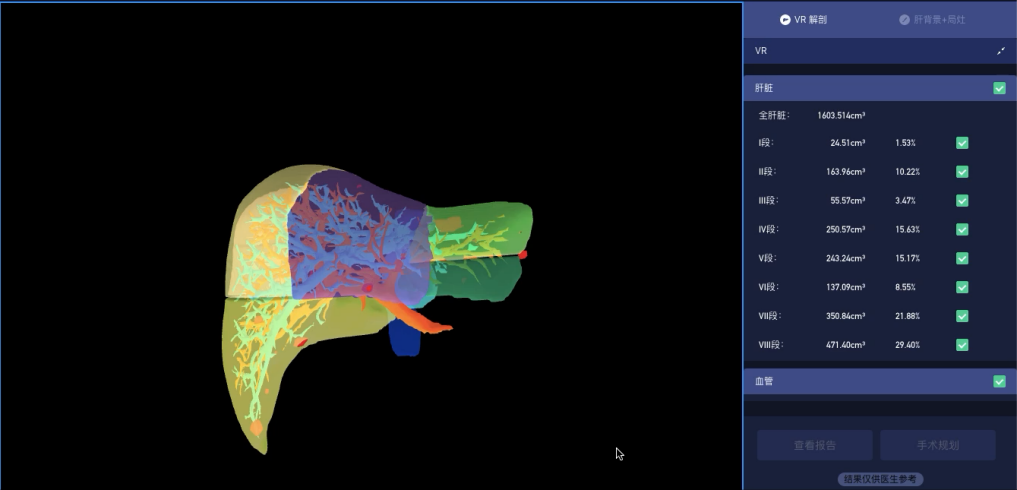
In this regard, the team stated that the aforementioned "Liver AI Analysis Platform" has implemented the "Post-Workflow AI" technological concept, extending the application of AI technology in MRI beyond the imaging acquisition phase into the image interpretation and diagnostic phases. Through one-click automatic sequence recognition, it performs lesion extraction and finding description, achieving intelligent analysis that "enables image interpretation and facilitates diagnosis"; the AI's capability to automatically extract anatomical features and lesion information has also injected new momentum into scientific research.
As the clinical partner in the development of this clinical AI-assisted system, Director Yang’s team at Beijing Friendship Hospital stated, "Liver lobar and segmental analysis is fundamentally based on the vasculature. Once the blood vessels are extracted, 3D reconstruction can be performed, providing a standardized model for subsequent procedures, whether for liver transplantation or hepatobiliary surgery. After the vascular structures are delineated, the non-vascular regions correspond to the lesions. We tested the currently trained model and found that the Dice coefficients for segmentation accuracy all exceed 0.9, with the highest surpassing 0.96, which adequately meets the requirements for routine clinical application."