Yiwei Technology Achieves Breakthrough in AI-Based Quantitative Fundus Analysis for Myopia Research in National Natural Science Foundation Project

EVsion
AI Detection Service Provider
On August 18, the research findings on myopic fundus changes supported by the National Natural Science Foundation of China were officially published in the leading international ophthalmology journal *TVST* (*Translational Vision Science & Technology*).This project was jointly completed by the team of Professor Wei Wenbin, Vice President of Beijing Tongren Hospital, Capital Medical University, in collaboration with the Aerospace Information Research Institute, Chinese Academy of Sciences, and EVsion.
This study employs AI-based quantitative fundus analysis technology to objectively characterize tessellated fundus changes and quantitatively calculate the fundus tessellated density index (fundus tessellated density, FTD).The research results indicate that,The mean tigroid patch density index of the fundus is significantly correlated with the subfoveal choroidal thickness in patients with myopia.
This research not only provides novel quantitative monitoring indicators for myopia-induced fundus changes, but also offers new research methodologies for elucidating the mechanisms underlying myopia progression in children and adolescents, controlling the progression of myopia, and reducing the incidence of blindness and high myopia.
Excerpted from the official website of the journal TVST
Tessellated fundus is one of the common features of myopic fundus changes, referring to a leopard-skin-like pattern visible in the fundus due to decreased pigmentation in the retinal pigment epithelium, increased interstitial tissue and pigmentation in the choroidal capillary layer, and the visibility of the large choroidal vessels and intervascular pigmented areas through the overlying retina.
In the 2011 population-based survey of the "Beijing Eye Study", a total of 3,468 participants were enrolled. Collected data included sex, age, fundus images, optical coherence tomography (OCT) images, axial length, and spherical equivalent. The study screened the results based on manually measured subfoveal choroidal thickness obtained from OCT images and the mean fundus tessellation density index derived from AI-based quantitative analysis. Ultimately, 3,074 participants were included in the final data analysis, comprising 1,733 females and 1,341 males.The results showed that tessellation density was significantly correlated with subfoveal choroidal thickness; the greater the average fundus tessellation density, the thinner the subfoveal choroidal thickness and the larger the peripapillary atrophic crescent.
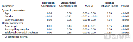
Multivariable Analysis of the Association Between Fundus Tessellation Density and Ocular and Systemic Parameters in the 2011 Beijing Eye Study
Relevant studies have shown that tessellated fundus changes are closely associated with the pathogenesis of blinding fundus diseases such as high myopia-related fundus changes, primary open-angle glaucoma, and age-related macular degeneration. However,Prior to the publication of this study, there was no reliable metric to objectively describe the degree of tessellated fundus changes. The tessellation density index proposed in this research provides a novel indicator for objectively quantifying the extent of tessellated fundus alterations, and also opens new avenues for future investigations into this condition.
Tessellated fundus is commonly associated with myopia. In this study, the tessellation density index extracted based on quantitative fundus analysis technology,This provides a novel monitoring indicator for myopia-induced fundus changes, representing a pioneering innovation in the research of tessellated fundus alterations. It offers new research tools and methodologies for elucidating the progression mechanisms of myopia, facilitating the control of myopia development, and reducing the incidence of high myopia and blindness.
In recent years, the prevalence of myopia in China has shown a rapid upward trend, and myopia has become a major public health issue affecting the eye health of the Chinese population, particularly adolescents. Epidemiological surveys indicate that there are nearly 700 million individuals with myopia in China, among whom over 70 million suffer from high myopia.
Numerous studies have shown that,With progressive axial elongation, in addition to causing myopia, a series of degenerative changes also occur in the ocular fundus, such as tilted optic disc, temporal peripapillary atrophy, tessellated fundus, posterior staphyloma, posterior pole retinal and choroidal lesions, and peripheral retinal changes. Meanwhile,Pathological myopia induced by high myopia is often accompanied by or leads to severe complications such as macular degeneration, cataracts, vitreous degeneration, retinal detachment, and retinoschisis. Identifying the "root cause" of this clinical challenge is particularly crucial.
In response, the state has attached great importance to this issue and has issued directives on multiple occasions: "Myopia among students in China is exhibiting a trend of high prevalence and younger onset, seriously affecting the physical and mental health of children. This is a major issue concerning the future of the nation and the people, which must be highly prioritized and cannot be allowed to go unchecked."
Based on this, EVsion, in collaboration with the ophthalmology team at Tongren Hospital and other partners, jointly conducted this research focusing on myopic fundus changes. The collaboration will continuously intensify targeted research efforts in this field and drive innovative scientific breakthroughs.
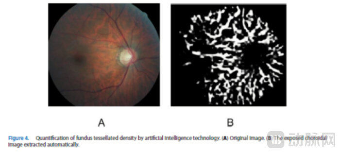
Artificial Intelligence Technology Quantifies Fundus Tessellation Density
( A ) Original image ( B ) Automatically extracted exposed choroid image
Based on proprietary visual computing technology, EVsion has pioneered quantitative fundus analysis, enabling precise digital annotation of key fundus structures such as blood vessels, nerves, and lesions., assisting experts in investigating the correlation between subtle ocular fundus changes and common diseases such as myopia, diabetes mellitus, cardiovascular and cerebrovascular diseases, kidney diseases, and neurological disorders, facilitating the understanding of disease onset and progression, and supporting etiological research, thereby enabling the formulation of rational treatment plans.
In the future, as population aging continues to intensify, the prevalence of fundus diseases will steadily rise accordingly. Whether conventional technologies can meet this challenge has become a critical question that warrants deep consideration within the medical field.
It is worth noting that both academia and industry are actively conducting innovative research and trials on myopic fundus changes, aiming to disrupt traditional treatment paradigms and gradually overcome the formidable challenges posed by highly complex, blinding fundus diseases. The recent innovative breakthrough by EVsion in myopia treatment, leveraging AI-based quantitative fundus analysis technology, holds considerable reference value and practical significance.
With its distinct advantages in image recognition, data analysis, and deep learning, artificial intelligence technology has sustained a rapid development momentum since entering the healthcare sector. In recent years, AI has yielded numerous achievements in the medical field. Bolstered by the introduction of supportive policies and the translation of scientific research into practical applications, AI-assisted medical image interpretation and diagnosis have made substantial progress. Although healthcare advancement is inherently a slow and incremental process, the era of large-scale deployment of AI medical applications may well be imminent.