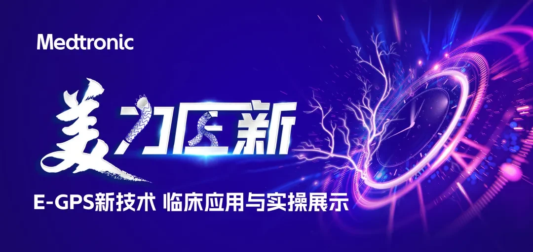
Medtronic Showcases E-GPS Technique for Complex Aortic Aneurysm Repair at VINNOVA 2026

Medtronic
Medical Device Manufacturer

PREFACE
May 22–24, 2026, co-hosted by the Professional Committee of Vascular Medicine, Chinese Research Hospital Association, the Vascular Devices Branch, China Association of Medical Devices Industry, the Capital Medical Science and Technology Innovation Center, and the Beijing Borui Vascular Health Public Welfare Foundation,“The 11th Vascular Innovation Conference”Held in Beijing. This conference brings together leading experts, entrepreneurs, engineers, investors, and researchers in vascular medicine from China and abroad, focusing on cutting-edge diagnostic and therapeutic technologies and evidence-based medical research in vascular diseases, providing strategic directions for industry pioneers, and jointly forging a new chapter in the advancement of the discipline.
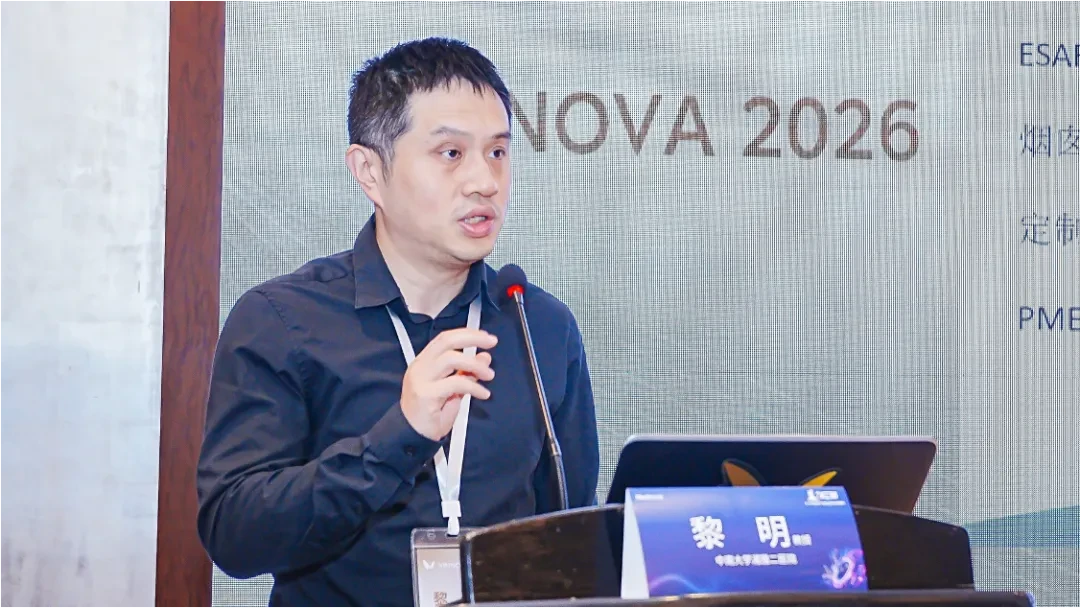
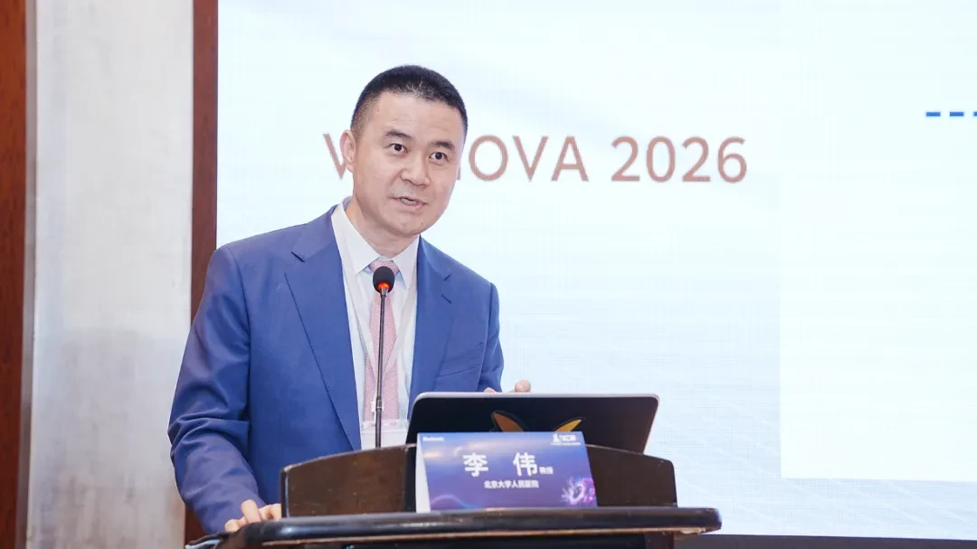
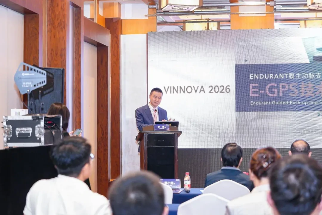
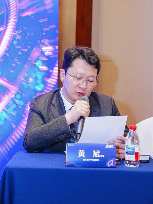
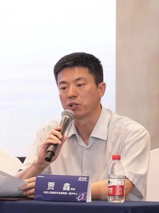
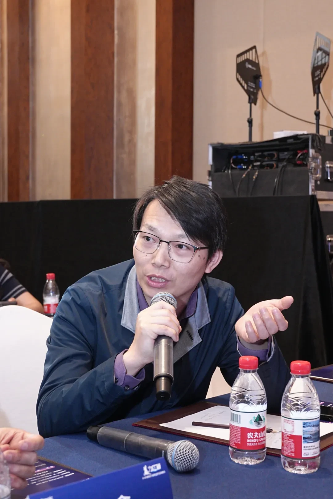
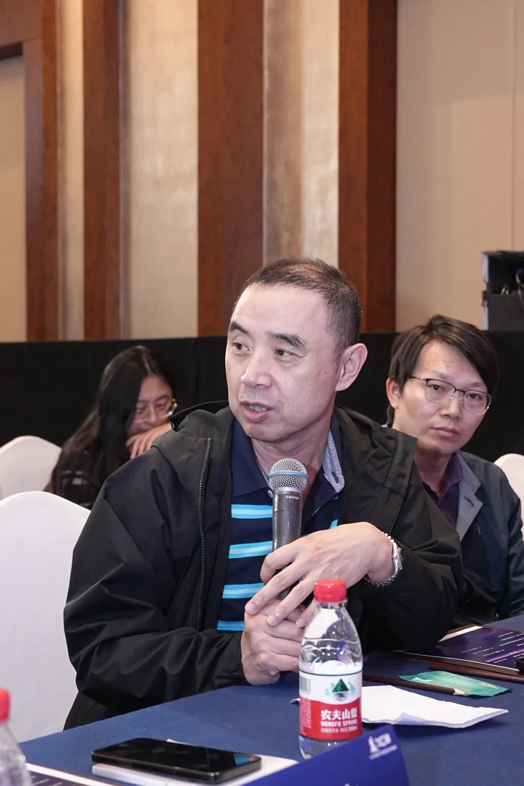
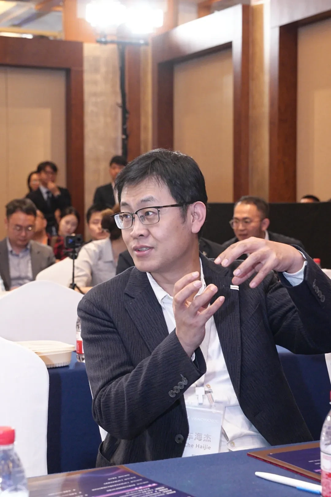
During the meeting,MedtronicIn collaboration with senior Chinese experts in the aortic field, jointly hosted on May 23「Craftsmanship & Innovation」Special Session: Clinical Application and Hands-on Demonstration of E-GPS New Technology。This event focuses on the challenges and clinical pain points in managing complex aneurysm necks during endovascular repair of abdominal aortic aneurysms, with a particular emphasis on the E-GPS technique optimized for the Endurant stent graft system. Through theoretical lectures, hands-on demonstrations using 3D-printed models, and discussions of challenging cases, the program aims to assist clinicians in achieving more precise stent positioning and more reliable proximal sealing under complex anatomical conditions such as short necks, severe angulation, and irregular necks. This ultimately reduces the incidence of Type Ia endoleaks and improves long-term clinical outcomes for patients with complex aneurysm necks.


Full-Process Simulation of the E-GPS Technical Workflow Using a 3D-Printed Model


Copyright Notice: This platform aims to assist healthcare professionals in staying informed of the latest developments in relevant disease areas. The information published on this platform does not imply endorsement of the descriptions or viewpoints presented, but is provided solely for informational purposes. Should any copyright issues arise, rights holders are kindly requested to contact us, and we will address the matter promptly. This information is intended exclusively for healthcare professionals for informational purposes and must not, under any circumstances, replace professional medical guidance or be construed as diagnostic or treatment advice. The platform and its authors assume no liability should this information be used for purposes other than informational reference.Partnership Contact Email:vascular@edoctor.work。